
Rabbit Anti-SPHK2 antibody
sphingosine kinase 2; SK 2; SPK 2; C76851; MGC102297; SPHK2_HUMAN.
View History [Clear]
Details
Product Name SPHK2 Chinese Name 鞘氨醇激酶2抗体 Alias sphingosine kinase 2; SK 2; SPK 2; C76851; MGC102297; SPHK2_HUMAN. literatures Research Area Cell biology Signal transduction Apoptosis Kinases and Phosphatases Cell differentiation Immunogen Species Rabbit Clonality Polyclonal React Species Human, Mouse, Rat, Applications WB=1:500-2000 ELISA=1:5000-10000 IHC-P=1:100-500 IHC-F=1:100-500 ICC=1:100 IF=1:100-500 (Paraffin sections need antigen repair)
not yet tested in other applications.
optimal dilutions/concentrations should be determined by the end user.Theoretical molecular weight 72 kDa Cellular localization The nucleus cytoplasmic The cell membrane Form Liquid Concentration 1mg/ml immunogen KLH conjugated synthetic peptide derived from human SPHK2: 101-200/654 Lsotype IgG Purification affinity purified by Protein A Buffer Solution 0.01M TBS(pH7.4) with 1% BSA, 0.03% Proclin300 and 50% Glycerol. Storage Shipped at 4℃. Store at -20 °C for one year. Avoid repeated freeze/thaw cycles. Attention This product as supplied is intended for research use only, not for use in human, therapeutic or diagnostic applications. PubMed PubMed Product Detail This gene encodes a kinase that phosphorylates sphingosine into sphingosine-1-phosphate, which is involved in cell differentiation, motility, and apoptosis. The encoded protein plays a role in maintaining cellular levels of sphingosine-1-phosphate. The gene product also enhances apoptosis in different cell types and suppresses cellular proliferation. In mast cells, the encoded protein is necessary for influx of calcium, protein kinase C activation, and cytokine production and degranulation. Alternative splicing results in multiple transcript variants. [provided by RefSeq].
Function:
Catalyzes the phosphorylation of sphingosine to form sphingosine 1-phosphate (SPP), a lipid mediator with both intra-and extracellular functions. Also acts on D-erythro-dihydrosphingosine, D-erythro-sphingosine and L-threo-dihydrosphingosine. Binds phosphoinositides.
Subcellular Location:
Isoform 1: Cytoplasm. Membrane. Isoform 2: Lysosome membrane.
Similarity:
Contains 1 DAGKc domain.
SWISS:
Q9NRA0
Gene ID:
56848
Database links:Entrez Gene: 56848 Human
Entrez Gene: 56632 Mouse
Omim: 607092 Human
SwissProt: Q9NRA0 Human
SwissProt: Q9JIA7 Mouse
Unigene: 528006 Human
Unigene: 24222 Mouse
Product Picture 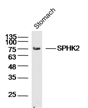 Sample:Stomach (Mouse) Lysate at 40 ug
Primary: Anti-SPHK2(SL2653R)at 1/300 dilution
Secondary: IRDye800CW Goat Anti-Rabbit IgG at 1/20000 dilution
Predicted band size: 72kD
Observed band size: 75kD
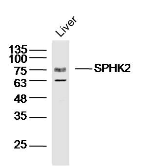 Sample:Liver (Mouse) Lysate at 40 ug
Primary: Anti-SPHK2(SL2653R)at 1/300 dilution
Secondary: IRDye800CW Goat Anti-Rabbit IgG at 1/20000 dilution
Predicted band size: 72kD
Observed band size: 75kD
 Sample:
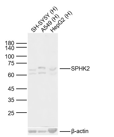
Lane 1: Human SH-SY5Y cell Lysates
Lane 2: Human A549 cell Lysates
Lane 3: Human HepG2 cell Lysates
Primary: Anti-SPHK2 (SL2653R) at 1/1000 dilution
Secondary: IRDye800CW Goat Anti-Rabbit IgG at 1/20000 dilution
Predicted band size: 72kDa
Observed band size: 72kDa
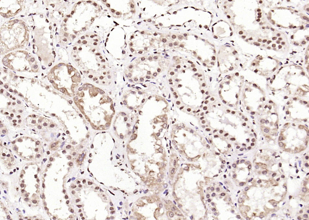 Paraformaldehyde-fixed, paraffin embedded (Human kidney); Antigen retrieval by boiling in sodium citrate buffer (pH6.0) for 15min; Block endogenous peroxidase by 3% hydrogen peroxide for 20 minutes; Blocking buffer (normal goat serum) at 37°C for 30min; Antibody incubation with (SPHK2) Polyclonal Antibody, Unconjugated (SL2653R) at 1:2000 overnight at 4°C, followed by operating according to SP Kit(Rabbit) (sp-0023) instructionsand DAB staining.
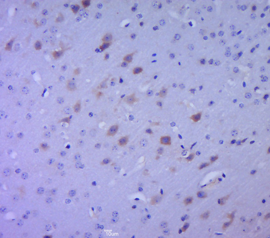 Paraformaldehyde-fixed, paraffin embedded (mouse brain tissue); Antigen retrieval by boiling in sodium citrate buffer (pH6.0) for 15min; Block endogenous peroxidase by 3% hydrogen peroxide for 20 minutes; Blocking buffer (normal goat serum) at 37°C for 30min; Antibody incubation with (SPHK2) Polyclonal Antibody, Unconjugated (SL2653R) at 1:400 overnight at 4°C, followed by a conjugated secondary (sp-0023) for 20 minutes and DAB staining.
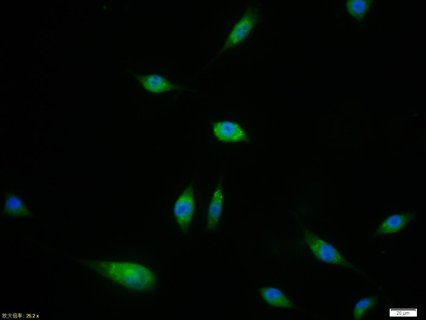 SH-SY5Y cell; 4% Paraformaldehyde-fixed; Triton X-100 at room temperature for 20 min; Blocking buffer (normal goat serum, C-0005) at 37°C for 20 min; Antibody incubation with (SPHK2) polyclonal Antibody, Unconjugated (SL2653R) 1:100, 90 minutes at 37°C; followed by a conjugated Goat Anti-Rabbit IgG antibody at 37°C for 90 minutes, DAPI (blue, C02-04002) was used to stain the cell nuclei.
Cartpieces
Totalgoods,subtotals:¥Checkout
Partial purchase records(bought amounts latest0)
No one bought this product
User Comment(Total0User Comment Num)
- No comment


 +86 571 56623320


