
Rabbit Anti-VPS4a antibody
hVPS4; SKD1; SKD1 homolog; SKD2; Vacuolar protein sorting 4 homolog A; vacuolar sorting protein 4; VPS4.
View History [Clear]
Details
Product Name VPS4a Chinese Name 液泡分选蛋白4抗体 Alias hVPS4; SKD1; SKD1 homolog; SKD2; Vacuolar protein sorting 4 homolog A; vacuolar sorting protein 4; VPS4. literatures Research Area Cell biology Cyclin Cell differentiation Immunogen Species Rabbit Clonality Polyclonal React Species Human, Mouse, (predicted: Rat, Dog, Pig, Rabbit, ) Applications WB=1:500-2000 ELISA=1:5000-10000 IHC-P=1:100-500 IHC-F=1:100-500 IF=1:100-500 (Paraffin sections need antigen repair)
not yet tested in other applications.
optimal dilutions/concentrations should be determined by the end user.Theoretical molecular weight 48kDa Cellular localization cytoplasmic The cell membrane Form Liquid Concentration 1mg/ml immunogen KLH conjugated synthetic peptide derived from human VPS4a: 351-437/437 Lsotype IgG Purification affinity purified by Protein A Buffer Solution 0.01M TBS(pH7.4) with 1% BSA, 0.03% Proclin300 and 50% Glycerol. Storage Shipped at 4℃. Store at -20 °C for one year. Avoid repeated freeze/thaw cycles. Attention This product as supplied is intended for research use only, not for use in human, therapeutic or diagnostic applications. PubMed PubMed Product Detail Involved in intracellular protein transport probably out of a prevacuolar endosomal compartment. May be involved in the release of components of the bilayered coat from the endosomal membrane. The association with ESCRT-III complex mediates the ATP-dependent disassembly of the ESCRT-III complex. In case of infection, the HIV-1 virus takes advantage of it for budding and exocytic cargos of viral proteins.
Function:
Involved in late steps of the endosomal multivesicular bodies (MVB) pathway. Recognizes membrane-associated ESCRT-III assemblies and catalyzes their disassembly, possibly in combination with membrane fission. Redistributes the ESCRT-III components to the cytoplasm for further rounds of MVB sorting. MVBs contain intraluminal vesicles (ILVs) that are generated by invagination and scission from the limiting membrane of the endosome and mostly are delivered to lysosomes enabling degradation of membrane proteins, such as stimulated growth factor receptors, lysosomal enzymes and lipids. In conjunction with the ESCRT machinery also appears to function in topologically equivalent membrane fission events, such as the terminal stages of cytokinesis and enveloped virus budding (HIV-1 and other lentiviruses). Involved in cytokinesis.
Subunit:
Proposed to be monomeric or homodimeric in nucleotide-free form and to oligomerize upon binding to ATP to form two stacked hexameric or heptameric rings with a central pore through which ESCRT-III substrates are translocated in an ATP-dependent manner (By similarity). Interacts with CHMP1A, CHMP1B, CHMP2A, CHMP2B, CHMP3, CHMP4A, CHMP4B, CHMP4C and CHMP6. Interacts with VPS4B; the interaction suggests a heteromeric assembly with VPS4B. Interacts with SPAST. Interacts with IST1.
Subcellular Location:
Prevacuolar compartment membrane; Peripheral membrane protein. Late endosome membrane; Peripheral membrane protein (Probable). Note=Membrane-associated in the prevacuolar endosomal compartment. Localizes to the midbody of dividing cells. Localized in two distinct rings on either side of the Fleming body.
Tissue Specificity:
Ubiquitously expressed.
Similarity:
Belongs to the AAA ATPase family.
Contains 1 MIT domain.
SWISS:
Q9UN37
Gene ID:
27183
Database links:Entrez Gene: 27183 Human
Entrez Gene: 116733 Mouse
Omim: 609982 Human
SwissProt: Q9UN37 Human
SwissProt: Q8VEJ9 Mouse
Unigene: 128420 Human
Unigene: 236004 Mouse
Unigene: 12477 Rat
Product Picture 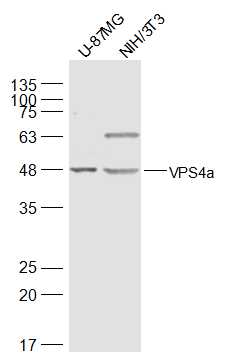 Sample:
U-87MG(Human) Cell Lysate at 30 ug
NIH/3T3(Mouse) Cell Lysate at 30 ug
Primary: Anti-VPS4a (SL7777R) at 1/500 dilution
Secondary: IRDye800CW Goat Anti-Rabbit IgG at 1/20000 dilution
Predicted band size: 48 kD
Observed band size: 48 kD
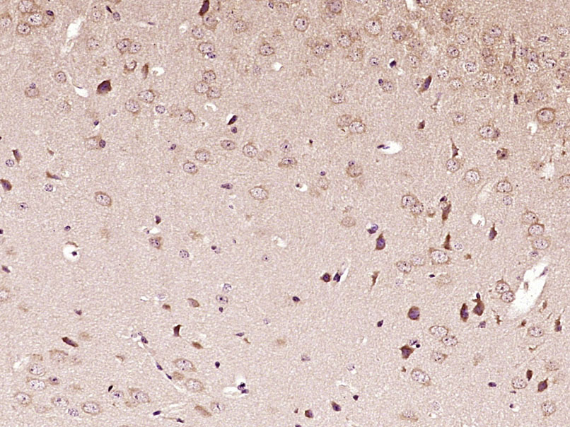 Paraformaldehyde-fixed, paraffin embedded (Mouse brain); Antigen retrieval by boiling in sodium citrate buffer (pH6.0) for 15min; Block endogenous peroxidase by 3% hydrogen peroxide for 20 minutes; Blocking buffer (normal goat serum) at 37°C for 30min; Antibody incubation with (VPS4a) Polyclonal Antibody, Unconjugated (SL7777R) at 1:400 overnight at 4°C, followed by operating according to SP Kit(Rabbit) (sp-0023) instructionsand DAB staining.
Cartpieces
Totalgoods,subtotals:¥Checkout
References (0)
No References
Bought notes(bought amounts latest0)
No one bought this product
User Comment(Total0User Comment Num)
- No comment


 +86 571 56623320
 +86 18668110335

