
Rabbit Anti-ADGRE1 antibody
F4/80; Adhesion G protein-coupled receptor E1; Cell surface glycoprotein EMR1; Cell surface glycoprotein F4/80; DD7A5 7; Egf like module containing mucin like hormone receptor like 1; Egf like module containing mucin like hormone receptor like sequence 1;
View History [Clear]
Details
Product Name ADGRE1 Chinese Name 表皮生长因子样激素受体1(EMR1)抗体 Alias F4/80; Adhesion G protein-coupled receptor E1; Cell surface glycoprotein EMR1; Cell surface glycoprotein F4/80; DD7A5 7; Egf like module containing mucin like hormone receptor like 1; Egf like module containing mucin like hormone receptor like sequence 1; EGF like module receptor 1; EGF TM7; EGF-like module receptor 1; EGF-like module-containing mucin-like hormone receptor-like 1; EGFTM7; EMR 1; EMR1; EMR-1; EMR1 hormone receptor; EMR1_HUMAN; AGRE1_HUMAN; Gpf480; Ly71; Lymphocyte antigen 71; TM7LN3. literatures Research Area immunology Growth factors and hormones G protein-coupled receptor glycoprotein G protein signal Immunogen Species Rabbit Clonality Polyclonal React Species Human, Mouse, (predicted: Rat, Pig, Guinea Pig, ) Applications WB=1:500-2000 ELISA=1:5000-10000 Flow-Cyt=2μg/Test
not yet tested in other applications.
optimal dilutions/concentrations should be determined by the end user.Theoretical molecular weight 95kDa Cellular localization The cell membrane Form Liquid Concentration 1mg/ml immunogen KLH conjugated synthetic peptide derived from human ADGRE1: 701-800/886 <Extracellular> Lsotype IgG Purification affinity purified by Protein A Buffer Solution 0.01M TBS(pH7.4) with 1% BSA, 0.03% Proclin300 and 50% Glycerol. Storage Shipped at 4℃. Store at -20 °C for one year. Avoid repeated freeze/thaw cycles. Attention This product as supplied is intended for research use only, not for use in human, therapeutic or diagnostic applications. PubMed PubMed Product Detail This gene encodes a protein that has a domain resembling seven transmembrane G protein-coupled hormone receptors (7TM receptors) at its C-terminus. The N-terminus of the encoded protein has six EGF-like modules, separated from the transmembrane segments by a serine/threonine-rich domain, a feature reminiscent of mucin-like, single-span, integral membrane glycoproteins with adhesive properties. Multiple alternatively spliced transcript variants encoding different isoforms have been found for this gene. [provided by RefSeq, Jan 2012]
Function:
Could be involved in cell-cell interactions.
Subunit:
Belongs to the G-protein coupled receptor 2 family. LN-TM7 subfamily. Contains 6 EGF-like domains. Contains 1 GPS domain.
Subcellular Location:
Cell membrane.
Tissue Specificity:
Wide expression; increased levels in peripheral blood mononuclear cells.
Similarity:
Belongs to the G-protein coupled receptor 2 family. LN-TM7 subfamily. Contains 6 EGF-like domains.
Contains 1 GPS domain.
SWISS:
Q14246
Gene ID:
2015
Database links:Entrez Gene: 2015 Human
Entrez Gene: 13733 Mouse
Omim: 600493 Human
SwissProt: Q14246 Human
SwissProt: Q61549 Mouse
Unigene: 2375 Human
Unigene: 2254 Mouse
Product Picture 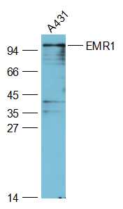 Sample:
A431(Human) Cell Lysate at 30 ug
Primary: Anti-EMR1 (SL7058R) at 1/2000 dilution
Secondary: IRDye800CW Goat Anti-Rabbit IgG at 1/20000 dilution
Predicted band size: 95 kD
Observed band size: 95 kD
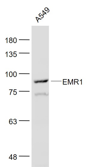 Sample:
A549(Human) Cell Lysate at 30 ug
Primary: Anti- EMR1 (SL7058R) at 1/1000 dilution
Secondary: IRDye800CW Goat Anti-Rabbit IgG at 1/20000 dilution
Predicted band size: 95 kD
Observed band size: 95 kD
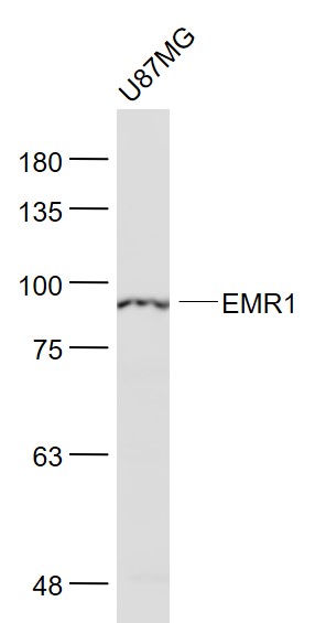 Sample:
U87MG(Human) Cell Lysate at 30 ug
Primary: Anti- EMR1 (SL7058R) at 1/1000 dilution
Secondary: IRDye800CW Goat Anti-Rabbit IgG at 1/20000 dilution
Predicted band size: 95 kD
Observed band size: 95 kD
 Blank control: Mouse kidney.
Primary Antibody (green line): Rabbit Anti-EMR1 antibody (SL7058R)
Dilution: 1μg /10^6 cells;
Isotype Control Antibody (orange line): Rabbit IgG .
Secondary Antibody : Goat anti-rabbit IgG-PE
Dilution: 1μg /test.
Protocol
The cells were incubated in 5%BSA to block non-specific protein-protein interactions for 30 min at at room temperature .Cells stained with Primary Antibody for 30 min at room temperature. The secondary antibody used for 40 min at room temperature. Acquisition of 20,000 events was performed.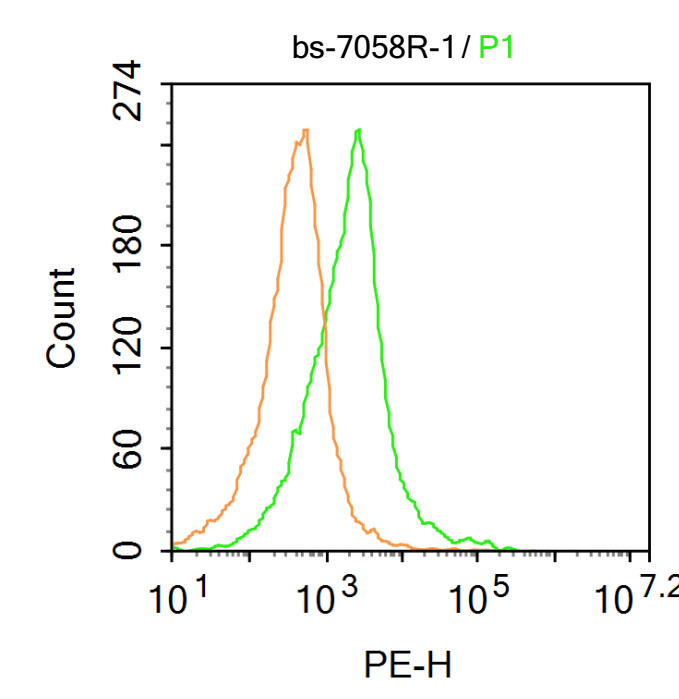 Blank control: Mouse brain.
Primary Antibody (green line): Rabbit Anti-EMR1 antibody (SL7058R)
Dilution: 1μg /10^6 cells;
Isotype Control Antibody (orange line): Rabbit IgG .
Secondary Antibody : Goat anti-rabbit IgG-PE
Dilution: 1μg /test.
Protocol
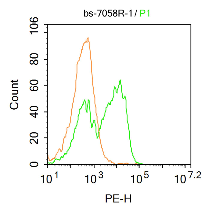
The cells were fixed with 4% PFA (10min at room temperature)and then permeabilized with 90% ice-cold methanol for 20 min at-20℃. The cells were then incubated in 5%BSA to block non-specific protein-protein interactions for 30 min at at room temperature .Cells stained with Primary Antibody for 30 min at room temperature. The secondary antibody used for 40 min at room temperature. Acquisition of 20,000 events was performed.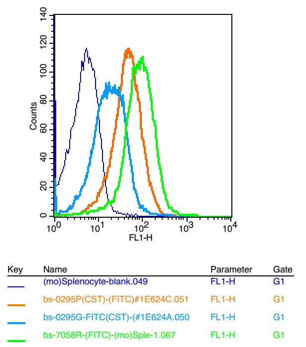 Positive control: mouse Splenocytes(2% Paraformaldehyde-fixed )
Isotype Control Antibody: Rabbit IgG Dilution: 1μg in 100 μl 1X PBS containing 0.5% BSA; Secondary Antibody: Goat anti-rabbit IgG-FITC; Dilution: 1:200 in 1 X PBS containing 0.5% BSA; Primary Antibody : rabbit Anti-EMR1 SL7058R; Dilution: 1μg in 100 μl 1X PBS containing 0.5% BSA.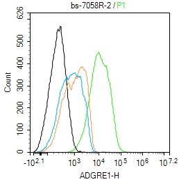 Blank control:A431.
Primary Antibody (green line): Rabbit Anti-ADGRE1 antibody (SL7058R)
Dilution: 2ug/Test;
Secondary Antibody : Goat anti-rabbit IgG-AF488
Dilution: 0.5ug/Test.
Protocol
The cells were incubated in 5%BSA to block non-specific protein-protein interactions for 30 min at room temperature .Cells stained with Primary Antibody for 30 min at room temperature. The secondary antibody used for 40 min at room temperature. Acquisition of 20,000 events was performed.
Cartpieces
Totalgoods,subtotals:¥Checkout
Bought notes(bought amounts latest0)
No one bought this product
User Comment(Total0User Comment Num)
- No comment


 +86 571 56623320


