
Rabbit Anti-CD81 antibody
26 kDa cell surface protein TAPA 1; 26 kDa cell surface protein TAPA-1; 26 kDa cell surface protein TAPA1; CD 81; CD81 antigen; CD81 molecule; CD81_HUMAN; CVID6; S5.7; TAPA 1; Target of the antiproliferative antibody 1; Tetraspanin 28; Tetraspanin-28; Tet
View History [Clear]
Details
Product Name CD81 Chinese Name CD81抗体 Alias 26 kDa cell surface protein TAPA 1; 26 kDa cell surface protein TAPA-1; 26 kDa cell surface protein TAPA1; CD 81; CD81 antigen; CD81 molecule; CD81_HUMAN; CVID6; S5.7; TAPA 1; Target of the antiproliferative antibody 1; Tetraspanin 28; Tetraspanin-28; Tetraspanin28; Tspan 28; Tspan-28; Tspan28. literatures Research Area Cell biology immunology Signal transduction Bacteria and viruses lymphocyte b-lymphocyte Immunogen Species Rabbit Clonality Polyclonal React Species Human, Mouse, (predicted: Dog, Pig, Cow, Horse, Sheep, ) Applications WB=1:500-2000 ELISA=1:5000-10000 Flow-Cyt=1μg/Test IF=1:50-200
not yet tested in other applications.
optimal dilutions/concentrations should be determined by the end user.Theoretical molecular weight 26kDa Detection molecular weight 26 kDa Cellular localization The cell membrane Form Liquid Concentration 1mg/ml immunogen KLH conjugated synthetic peptide derived from human TAPA1/CD81: 101-210/236 <Extracellular> Lsotype IgG Purification affinity purified by Protein A Buffer Solution 0.01M TBS(pH7.4) with 1% BSA, 0.03% Proclin300 and 50% Glycerol. Storage Shipped at 4℃. Store at -20 °C for one year. Avoid repeated freeze/thaw cycles. Attention This product as supplied is intended for research use only, not for use in human, therapeutic or diagnostic applications. PubMed PubMed Product Detail The protein encoded by this gene is a member of the transmembrane 4 superfamily, also known as the tetraspanin family. Most of these members are cell-surface proteins that are characterized by the presence of four hydrophobic domains. The proteins mediate signal transduction events that play a role in the regulation of cell development, activation, growth and motility. This encoded protein is a cell surface glycoprotein that is known to complex with integrins. This protein appears to promote muscle cell fusion and support myotube maintenance. Also it may be involved in signal transduction. This gene is localized in the tumor-suppressor gene region and thus it is a candidate gene for malignancies. [provided by RefSeq, Jul 2008].
Function:
May play an important role in the regulation of lymphoma cell growth. Interacts with a 16-kDa Leu-13 protein to form a complex possibly involved in signal transduction. May acts a the viral receptor for HCV.
Subunit:
Plays a critical role in HCV attachment and/or cell entry by interacting with HCV E1/E2 glycoproteins heterodimer. Interacts directly with IGSF8. Interacts with CD53 and SCIMP.
Subcellular Location:
Membrane.
Tissue Specificity:
Hematolymphoid, neuroectodermal and mesenchymal tumor cell lines.
DISEASE:
Defects in CD81 are the cause of immunodeficiency common variable type 6 (CVID6); also called antibody deficiency due to CD81 defect. CVID6 is a primary immunodeficiency characterized by antibody deficiency, hypogammaglobulinemia, recurrent bacterial infections and an inability to mount an antibody response to antigen. The defect results from a failure of B-cell differentiation and impaired secretion of immunoglobulins; the numbers of circulating B cells is usually in the normal range, but can be low.
Similarity:
Belongs to the tetraspanin (TM4SF) family.
SWISS:
P60033
Gene ID:
975
Database links:Entrez Gene: 975 Human
Omim: 186845 Human
SwissProt: P60033 Human
Unigene: 54457 Human
Product Picture 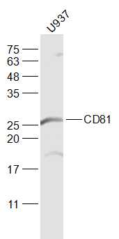 Sample:
U937(Human) Cell Lysate at 30 ug
Primary: Anti-CD81 (SL6934R) at 1/1000 dilution
Secondary: IRDye800CW Goat Anti-Rabbit IgG at 1/20000 dilution
Predicted band size: 26 kD
Observed band size: 26 kD
 Sample:
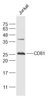
Jurkat(Human) Cell Lysate at 30 ug
Primary: Anti-CD81 (SL6934R) at 1/1000 dilution
Secondary: IRDye800CW Goat Anti-Rabbit IgG at 1/20000 dilution
Predicted band size: 26 kD
Observed band size: 26 kD
 Sample:
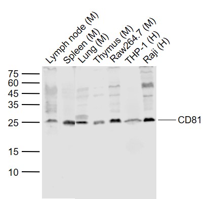
Lane 1: Lymph node (Mouse) Lysate at 40 ug
Lane 2: Spleen (Mouse) Lysate at 40 ug
Lane 3: Lung (Mouse) Lysate at 40 ug
Lane 4: Thymus (Mouse) Lysate at 40 ug
Lane 5: Raw264.7 (Mouse) Cell Lysate at 30 ug
Lane 6: THP-1 (Human) Cell Lysate at 30 ug
Lane 7: Raji (Human) Cell Lysate at 30 ug
Primary: Anti-CD81 (SL6934R) at 1/1000 dilution
Secondary: IRDye800CW Goat Anti-Rabbit IgG at 1/20000 dilution
Predicted band size: 26 kD
Observed band size: 25 kD
 Sample:
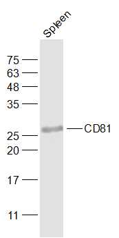
Spleen (Mouse) Lysate at 40 ug
Primary: Anti-CD81 (SL6934R) at 1/1000 dilution
Secondary: IRDye800CW Goat Anti-Rabbit IgG at 1/20000 dilution
Predicted band size: 26 kD
Observed band size: 26 kD
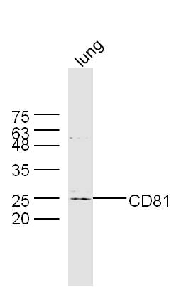 Sample: Lung (Mouse) Lysate at 40 ug
Primary: Anti-CD81 (SL6934R) at 1/300 dilution
Secondary: IRDye800CW Goat Anti-Rabbit IgG at 1/20000 dilution
Predicted band size: 26 kD
Observed band size: 25 kD
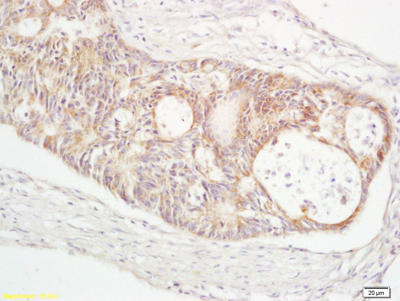 Tissue/cell: human rectal carcinoma; 4% Paraformaldehyde-fixed and paraffin-embedded;
Antigen retrieval: citrate buffer ( 0.01M, pH 6.0 ), Boiling bathing for 15min; Block endogenous peroxidase by 3% Hydrogen peroxide for 30min; Blocking buffer (normal goat serum,C-0005) at 37℃ for 20 min;
Incubation: Anti-TAPA1/CD81 Polyclonal Antibody, Unconjugated(SL6934R) 1:200, overnight at 4°C, followed by conjugation to the secondary antibody(SP-0023) and DAB(C-0010) staining
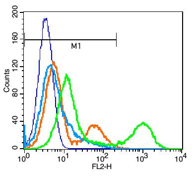 Blank control(blue): Jurkat cells(fixed with 2% paraformaldehyde (10 min)).
Primary Antibody:Rabbit Anti-CD81 antibody(SL6934R), Dilution: 1μg in 100 μL 1X PBS containing 0.5% BSA;
Isotype Control Antibody: Rabbit IgG(orange) ,used under the same conditions );
Secondary Antibody: Goat anti-rabbit IgG-PE(white blue), Dilution: 1:200 in 1 X PBS containing 0.5% BSA.
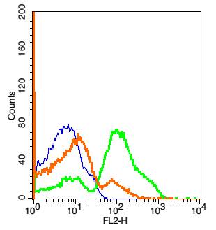 Blank control: Mouse Brain cells(blue).
Primary Antibody: Rabbit Anti- CD81 /PEConjugated antibody (SL6934R-PE), Dilution: 5μg in 100 μL 1X PBS containing 0.5% BSA;
Isotype Control Antibody: Rabbit IgG/PE(orange) ,used under the same conditions.
Cartpieces
Totalgoods,subtotals:¥Checkout
References (0)
No References
Bought notes(bought amounts latest0)
No one bought this product
User Comment(Total0User Comment Num)
- No comment


 +86 571 56623320
 +86 18668110335

