
Rabbit Anti-ASC/TMS1 antibody
TMS1; Apoptosis associated speck like protein containing a CARD; Apoptosis-associated speck-like protein containing a CARD; ASC_HUMAN; CARD 5; CARD5; Caspase recruitment domain containing protein 5; Caspase recruitment domain protein 5; Caspase recruitmen
View History [Clear]
Details
Product Name ASC/TMS1 Chinese Name 凋亡相关斑点样蛋白ASC抗体 Alias TMS1; Apoptosis associated speck like protein containing a CARD; Apoptosis-associated speck-like protein containing a CARD; ASC_HUMAN; CARD 5; CARD5; Caspase recruitment domain containing protein 5; Caspase recruitment domain protein 5; Caspase recruitment domain-containing protein 5; hASC; MGC10332; PYCARD; TMS-1; PYD and CARD domain containing; PYD and CARD domain containing protein; PYD and CARD domain-containing protein; Target of methylation induced silencing 1; Target of methylation-induced silencing 1; TMS 1. literatures Research Area Tumour Cell biology immunology Signal transduction Apoptosis transcriptional regulatory factor TumourCell biologyMaker Immunogen Species Rabbit Clonality Polyclonal React Species Human, Mouse, Rat, (predicted: Dog, Pig, Cow, Horse, Rabbit, ) Applications WB=1:500-2000 ELISA=1:5000-10000 IHC-P=1:100-500 IHC-F=1:100-500 Flow-Cyt=1ug/test ICC=1:100 IF=1:100-500 (Paraffin sections need antigen repair)
not yet tested in other applications.
optimal dilutions/concentrations should be determined by the end user.Theoretical molecular weight 22kDa Cellular localization The nucleus cytoplasmic Form Liquid Concentration 1mg/ml immunogen KLH conjugated synthetic peptide derived from human ASC: 31-130/195 Lsotype IgG Purification affinity purified by Protein A Buffer Solution 0.01M TBS(pH7.4) with 1% BSA, 0.03% Proclin300 and 50% Glycerol. Storage Shipped at 4℃. Store at -20 °C for one year. Avoid repeated freeze/thaw cycles. Attention This product as supplied is intended for research use only, not for use in human, therapeutic or diagnostic applications. PubMed PubMed Product Detail Promotes caspase-mediated apoptosis. This proapoptotic activity is mediated predominantly through the activation of caspase-9. May be a component of the inflammasome, a protein complex which also includes NALP2, CARD8 and CASP1 and whose function would be the activation of proinflammatory caspases. Tissue specificity: Widely expressed at low levels. Detected in peripheral blood leukocytes, lung, small intestine, spleen, thymus, colon and at lower levels in placenta, liver and kidney. Very low expression in skeletal muscle, heart and brain. Detected in the leukemia cell lines HL-60 and U937, but not in Jurkat T-cell lymphoma and Daudi Burkitt's lymphoma.
Function:
Promotes caspase-mediated apoptosis. This proapoptotic activity is mediated predominantly through the activation of caspase-9. May be a component of the inflammasome, a protein complex which also includes NALP2, CARD8 and CASP1 and whose function would be the activation of proinflammatory caspases.
Subunit:
Forms complexes with other DAPIN domain-containing proteins. Interacts with CIAS1/PYPAF1 and PYDC1.
Subcellular Location:
Cytoplasm. Note=Upstream of caspase activation, a redistribution from the cytoplasm to the aggregates occurs. These appear as hollow, perinuclear spherical, ball-like structures.
Tissue Specificity:
Widely expressed at low levels. Detected in peripheral blood leukocytes, lung, small intestine, spleen, thymus, colon and at lower levels in placenta, liver and kidney. Very low expression in skeletal muscle, heart and brain. Detected in the leukemia cell lines HL-60 and U937, but not in Jurkat T-cell lymphoma and Daudi Burkitt's lymphoma. Detected in the melanoma cell line WM35, but not in WM793. Not detected in HeLa cervical carcinoma cells and Molt 4 lymphocytic leukemia cells.
Post-translational modifications:
Phosphorylated.
Similarity:
Contains 1 CARD domain.
Contains 1 DAPIN domain.
SWISS:
Q9ULZ3
Gene ID:
29108
Database links:Entrez Gene: 29108 Human
Omim: 606838 Human
SwissProt: Q9ULZ3 Human
Unigene: 499094 Human
Product Picture 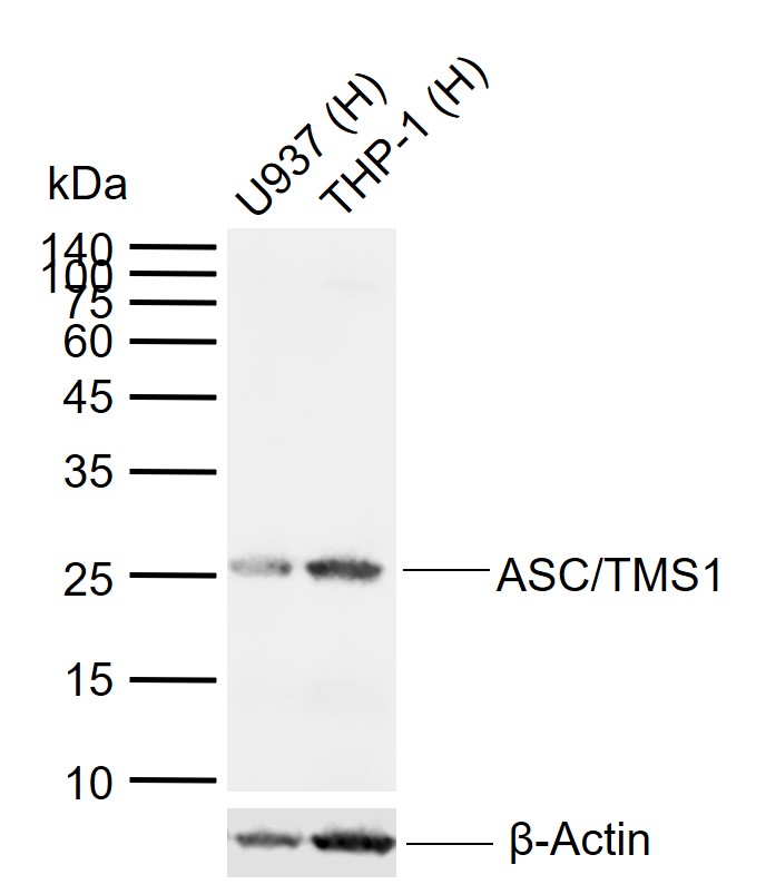 Sample:
Lane 1: Human U937 cell lysates
Lane 2: Human THP-1 cell lysates
Primary: Anti-ASC/TMS1 (SL6741R) at 1/1000 dilution
Secondary: IRDye800CW Goat Anti-Rabbit IgG at 1/20000 dilution
Predicted band size: 22 kDa
Observed band size: 25 kDa
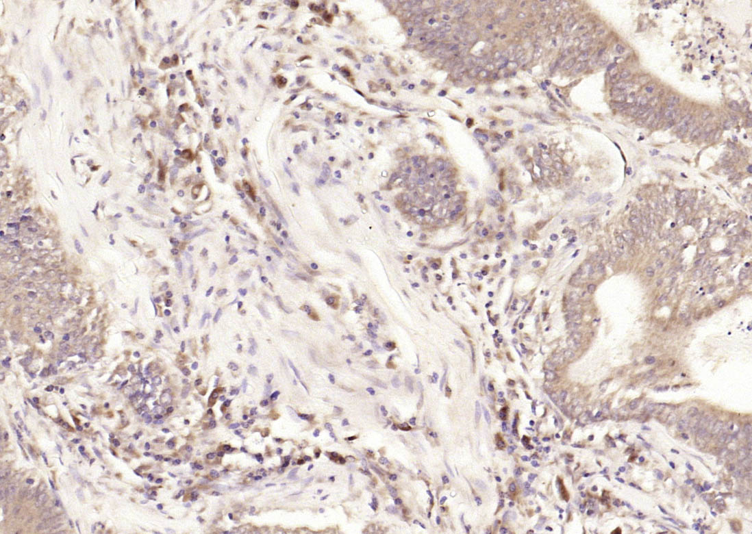 Paraformaldehyde-fixed, paraffin embedded (human colon carcinoma); Antigen retrieval by boiling in sodium citrate buffer (pH6.0) for 15min; Block endogenous peroxidase by 3% hydrogen peroxide for 20 minutes; Blocking buffer (normal goat serum) at 37°C for 30min; Antibody incubation with (ASC) Polyclonal Antibody, Unconjugated (SL6741R) at 1:2000 overnight at 4°C, followed by operating according to SP Kit(Rabbit) (sp-0023) instructionsand DAB staining.
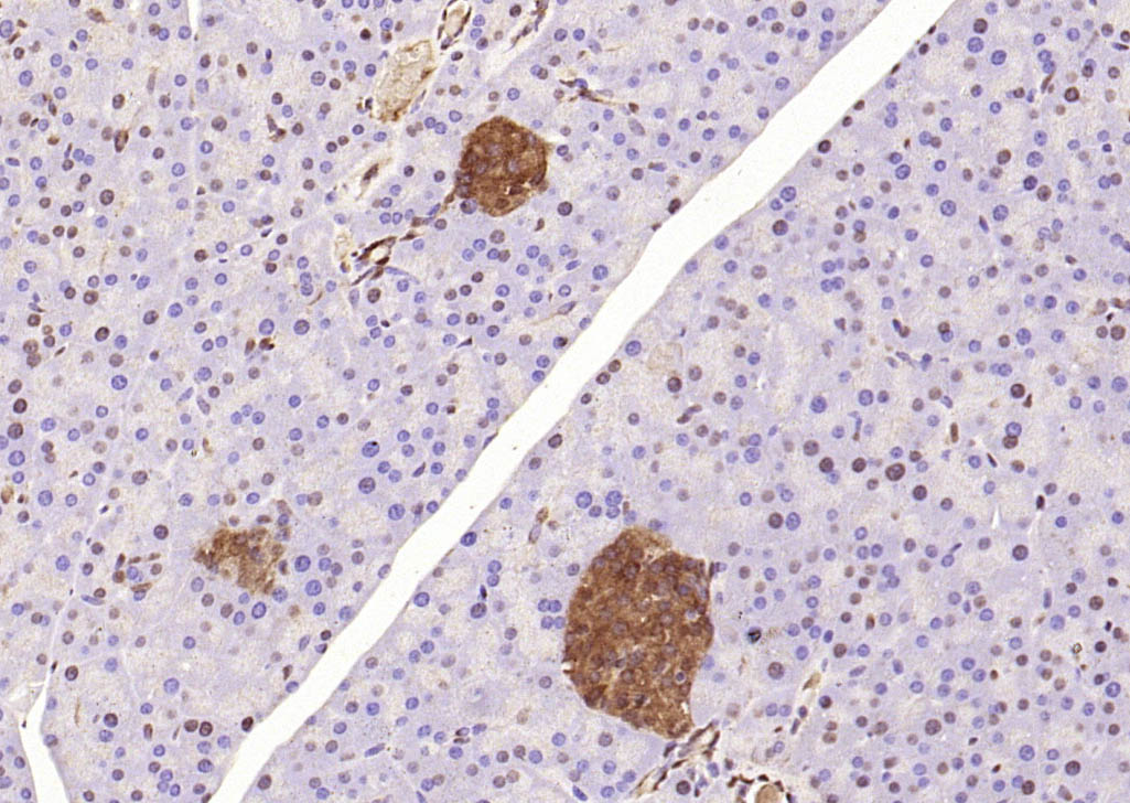 Paraformaldehyde-fixed, paraffin embedded (mouse pancreas); Antigen retrieval by boiling in sodium citrate buffer (pH6.0) for 15min; Block endogenous peroxidase by 3% hydrogen peroxide for 20 minutes; Blocking buffer (normal goat serum) at 37°C for 30min; Antibody incubation with (ASC) Polyclonal Antibody, Unconjugated (SL6741R) at 1:2000 overnight at 4°C, followed by operating according to SP Kit(Rabbit) (sp-0023) instructionsand DAB staining.
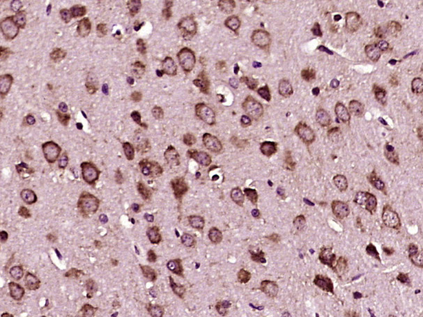 Paraformaldehyde-fixed, paraffin embedded (mouse brain tissue); Antigen retrieval by boiling in sodium citrate buffer (pH6.0) for 15min; Block endogenous peroxidase by 3% hydrogen peroxide for 20 minutes; Blocking buffer (normal goat serum) at 37°C for 30min; Antibody incubation with (ASC) Polyclonal Antibody, Unconjugated (SL6741R) at 1:400 overnight at 4°C, followed by operating according to SP Kit(Rabbit) (sp-0023) instructionsand DAB staining.
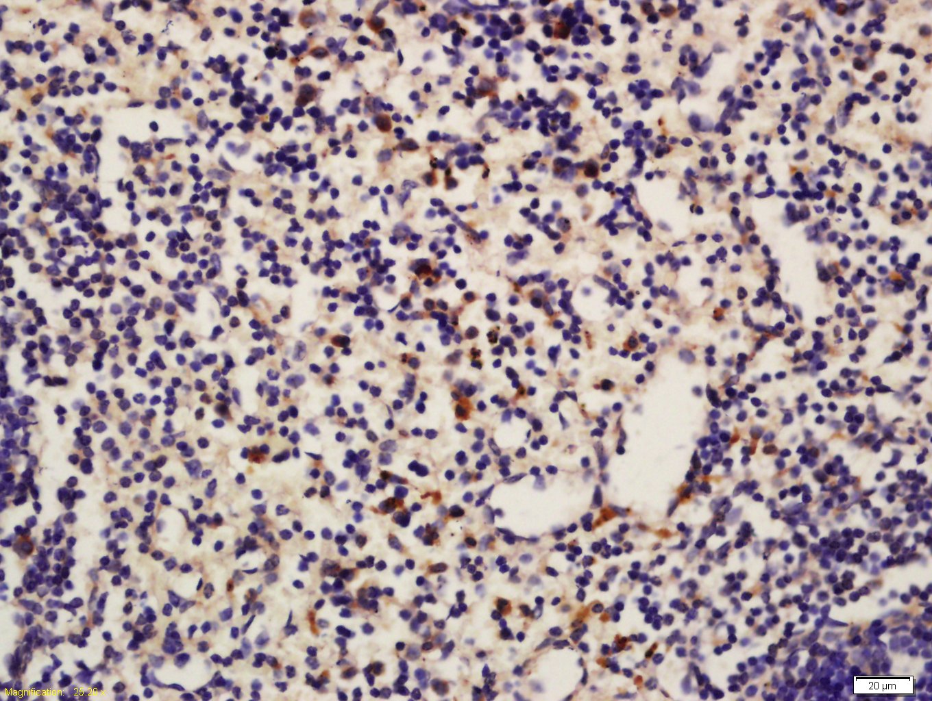 Tissue/cell: Mouse spleen tissue; 4% Paraformaldehyde-fixed and paraffin-embedded;
Antigen retrieval: citrate buffer ( 0.01M, pH 6.0 ), Boiling bathing for 15min; Block endogenous peroxidase by 3% Hydrogen peroxide for 30min; Blocking buffer (normal goat serum,C-0005) at 37℃ for 20 min;
Incubation: Anti-ASC Polyclonal Antibody, Unconjugated(SL6741R) 1:200, overnight at 4°C, followed by conjugation to the secondary antibody(SP-0023) and DAB(C-0010) staining
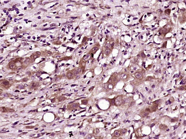 Paraformaldehyde-fixed, paraffin embedded (human gastric carcinoma); Antigen retrieval by boiling in sodium citrate buffer (pH6.0) for 15min; Block endogenous peroxidase by 3% hydrogen peroxide for 20 minutes; Blocking buffer (normal goat serum) at 37°C for 30min; Antibody incubation with (ASC) Polyclonal Antibody, Unconjugated (SL6741R) at 1:400 overnight at 4°C, followed by operating according to SP Kit(Rabbit) (sp-0023) instructionsand DAB staining.
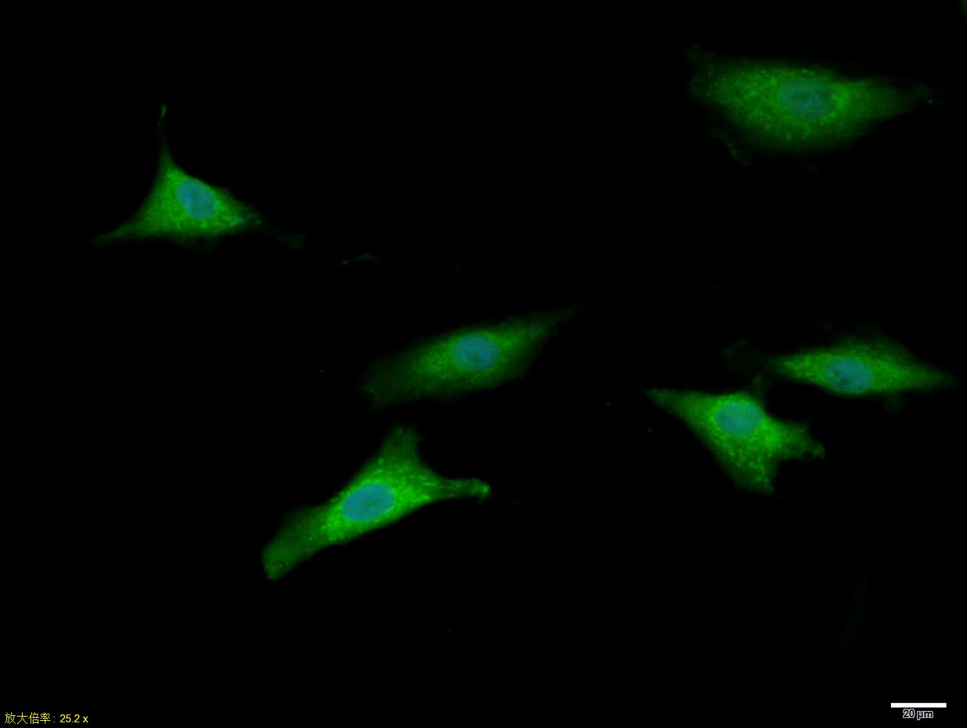 A549 cell; 4% Paraformaldehyde-fixed; Triton X-100 at room temperature for 20 min; Blocking buffer (normal goat serum, C-0005) at 37°C for 20 min; Antibody incubation with (ASC) polyclonal Antibody, Unconjugated (SL6741R) 1:100, 90 minutes at 37°C; followed by a conjugated Goat Anti-Rabbit IgG antibody at 37°C for 90 minutes, DAPI (blue, C02-04002) was used to stain the cell nuclei.
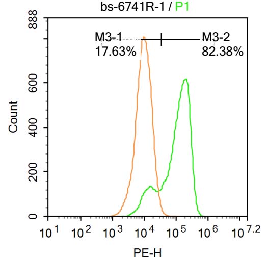 Blank control:A549.
Primary Antibody (green line): Rabbit Anti-ASC antibody (SL6741R)
Dilution: 1μg /10^6 cells;
Isotype Control Antibody (orange line): Rabbit IgG .
Secondary Antibody : Goat anti-rabbit IgG-PE
Dilution: 1μg /test.
Protocol
The cells were fixed with 4% PFA (10min at room temperature)and then permeabilized with 20% PBST for 20 min at-20℃. The cells were then incubated in 5%BSA to block non-specific protein-protein interactions for 30 min at at room temperature .Cells stained with Primary Antibody for 30 min at room temperature. The secondary antibody used for 40 min at room temperature. Acquisition of 20,000 events was performed.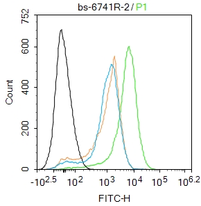 Blank control:Mosue spleen.
Primary Antibody (green line): Rabbit Anti-ASC antibody (SL6741R)
Dilution: 2μg /10^6 cells;
Isotype Control Antibody (orange line): Rabbit IgG .
Secondary Antibody : Goat anti-rabbit IgG-FITC
Dilution: 1μg /test.
Protocol
The cells were fixed with 4% PFA (10min at room temperature)and then permeabilized with 90% ice-cold methanol for 20 min at -20℃. The cells were then incubated in 5%BSA to block non-specific protein-protein interactions for 30 min at room temperature .Cells stained with Primary Antibody for 30 min at room temperature. The secondary antibody used for 40 min at room temperature. Acquisition of 20,000 events was performed.
Cartpieces
Totalgoods,subtotals:¥Checkout
Bought notes(bought amounts latest0)
No one bought this product
User Comment(Total0User Comment Num)
- No comment


 +86 571 56623320
 +86 18668110335

