
Rabbit Anti-COG1 antibody
Ldlbc; CDG2Gv Component of oligomeric golgi complex 1; Conserved oligomeric Golgi complex protein 1; Low density lipoprotein receptor defect B complementing; COG1_HUMAN.
View History [Clear]
Details
Product Name COG1 Chinese Name COG1蛋白抗体 Alias Ldlbc; CDG2Gv Component of oligomeric golgi complex 1; Conserved oligomeric Golgi complex protein 1; Low density lipoprotein receptor defect B complementing; COG1_HUMAN. literatures Research Area Cell biology Signal transduction Cell type markers Immunogen Species Rabbit Clonality Polyclonal React Species Human, Rat, Applications WB=1:500-2000 ELISA=1:5000-10000 IHC-P=1:100-500 IHC-F=1:100-500 IF=1:100-500 (Paraffin sections need antigen repair)
not yet tested in other applications.
optimal dilutions/concentrations should be determined by the end user.Theoretical molecular weight 109kDa Cellular localization cytoplasmic The cell membrane Form Liquid Concentration 1mg/ml immunogen KLH conjugated synthetic peptide derived from human COG1: 501-600/980 Lsotype IgG Purification affinity purified by Protein A Buffer Solution 0.01M TBS(pH7.4) with 1% BSA, 0.03% Proclin300 and 50% Glycerol. Storage Shipped at 4℃. Store at -20 °C for one year. Avoid repeated freeze/thaw cycles. Attention This product as supplied is intended for research use only, not for use in human, therapeutic or diagnostic applications. PubMed PubMed Product Detail There are eight COG proteins (COG1-8) which form a Golgi-localized complex (COG) required for normal Golgi morphology and function. It is thought that COG1 is required for steps in the normal medial and trans Golgi-associated processing of glycoconjugates and plays a role in the organization of the Golgi-localized complex.
Function:
Required for normal Golgi function (By similarity).
Subunit:
Component of the conserved oligomeric Golgi complex which is composed of eight different subunits and is required for normal Golgi morphology and localization.
Subcellular Location:
Golgi apparatus membrane; Peripheral membrane protein; Cytoplasmic side.
DISEASE:
Defects in COG1 are the cause of congenital disorder of glycosylation type 2G (CDG2G) [MIM:611209]; also known as CDG-II caused by COG1 deficiency. CDGs are a family of severe inherited diseases caused by a defect in glycoprotein biosynthesis. They are characterized by under-glycosylated serum glycoproteins. These multisystem disorders present with a wide variety of clinical features, such as disorders of the nervous system development, psychomotor retardation, dysmorphic features, hypotonia, coagulation disorders and immunodeficiency. The broad spectrum of features reflects the critical role of N-glycoproteins during embryonic development, differentiation, and maintenance of cell functions. Clinical features of CDG2G include failure to thrive, generalized hypotonia, growth retardation and mild psychomotor retardation. CDG2G is biochemically characterized by a defect in O-glycosylation as well as N-glycosylation.
Similarity:
Belongs to the COG1 family.
SWISS:
Q8WTW3
Gene ID:
9382
Database links:
Entrez Gene: 9382 Human
Entrez Gene: 16834 Mouse
Omim: 606973 Human
SwissProt: Q8WTW3 Human
SwissProt: Q9Z160 Mouse
Unigene: 103555 Human
Unigene: 261620 Mouse
Product Picture 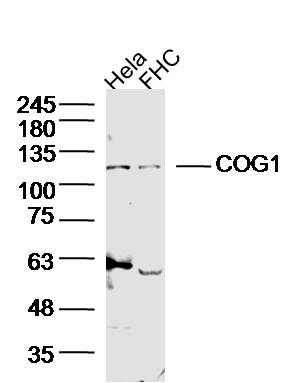 Sample:
Hela Cell (Human) Lysate at 40 ug
FHC Cell (Human) Lysate at 40 ug
Primary: Anti-COG1 (SL6647R) at 1/300 dilution
Secondary: IRDye800CW Goat Anti-Rabbit IgG at 1/20000 dilution
Predicted band size: 109 kD
Observed band size: 109 kD
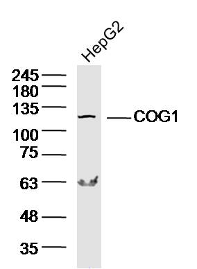 Sample: HepG2 Cell (Human) Lysate at 40 ug
Primary: Anti-COG1 (SL6647R) at 1/300 dilution
Secondary: IRDye800CW Goat Anti-Rabbit IgG at 1/20000 dilution
Predicted band size: 109 kD
Observed band size: 109 kD
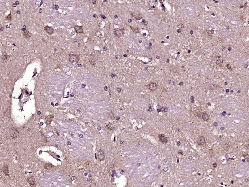 Paraformaldehyde-fixed, paraffin embedded (Rat brain); Antigen retrieval by boiling in sodium citrate buffer (pH6.0) for 15min; Block endogenous peroxidase by 3% hydrogen peroxide for 20 minutes; Blocking buffer (normal goat serum) at 37°C for 30min; Antibody incubation with (COG1) Polyclonal Antibody, Unconjugated (SL6647R) at 1:400 overnight at 4°C, followed by operating according to SP Kit(Rabbit) (sp-0023) instructionsand DAB staining.
Cartpieces
Totalgoods,subtotals:¥Checkout
References (0)
No References
Bought notes(bought amounts latest0)
No one bought this product
User Comment(Total0User Comment Num)
- No comment


 +86 571 56623320
 +86 18668110335

