
Rabbit Anti-Complement fragment 3c antibody
AHUS5; acylation-stimulating protein cleavage product; ARMD9; ASP; C3 and PZP-like alpha-2-macroglobulin domain-containing protein 1; C3; CO3_HUMAN; Complement C3; Complement C3c alpha' chain fragment 2; Complement C3c; Complement component 3; Complement
View History [Clear]
Details
Product Name Complement fragment 3c Chinese Name 补体片段C3c抗体 Alias AHUS5; acylation-stimulating protein cleavage product; ARMD9; ASP; C3 and PZP-like alpha-2-macroglobulin domain-containing protein 1; C3; CO3_HUMAN; Complement C3; Complement C3c alpha' chain fragment 2; Complement C3c; Complement component 3; Complement component C3; Complement factor 3; CPAMD1. literatures Research Area Cell biology immunology Signal transduction G protein-coupled receptor Immunogen Species Rabbit Clonality Polyclonal React Species Human, Rat, (predicted: Mouse, Dog, Pig, ) Applications WB=1:500-2000 ELISA=1:5000-10000 IHC-P=1:100-500 IHC-F=1:100-500 IF=1:100-500 (Paraffin sections need antigen repair)
not yet tested in other applications.
optimal dilutions/concentrations should be determined by the end user.Theoretical molecular weight 183kDa Cellular localization Secretory protein Form Liquid Concentration 1mg/ml immunogen KLH conjugated synthetic peptide derived from human Complement C3c alpha' chain fragment 1: 1051-1200/1663 Lsotype IgG Purification affinity purified by Protein A Buffer Solution 0.01M TBS(pH7.4) with 1% BSA, 0.03% Proclin300 and 50% Glycerol. Storage Shipped at 4℃. Store at -20 °C for one year. Avoid repeated freeze/thaw cycles. Attention This product as supplied is intended for research use only, not for use in human, therapeutic or diagnostic applications. PubMed PubMed Product Detail C3 plays a central role in the activation of the complement system. Its processing by C3 convertase is the central reaction in both classical and alternative complement pathways. After activation C3b can bind covalently, via its reactive thioester, to cell surface carbohydrates or immune aggregates. Derived from proteolytic degradation of complement C3, C3a anaphylatoxin is a mediator of local inflammatory process. It induces the contraction of smooth muscle, increases vascular permeability and causes histamine release from mast cells and basophilic leukocytes.
Function:
C3 plays a central role in the activation of the complement system. Its processing by C3 convertase is the central reaction in both classical and alternative complement pathways. After activation C3b can bind covalently, via its reactive thioester, to cell surface carbohydrates or immune aggregates.
Derived from proteolytic degradation of complement C3, C3a anaphylatoxin is a mediator of local inflammatory process. It induces the contraction of smooth muscle, increases vascular permeability and causes histamine release from mast cells and basophilic leukocytes.
Acylation stimulating protein (ASP): adipogenic hormone that stimulates triglyceride (TG) synthesis and glucose transport in adipocytes, regulating fat storage and playing a role in postprandial TG clearance. Appears to stimulate TG synthesis via activation of the PLC, MAPK and AKT signaling pathways. Ligand for GPR77. Promotes the phosphorylation, ARRB2-mediated internalization and recycling of GPR77.
Subunit:
C3 precursor is first processed by the removal of 4 Arg residues, forming two chains, beta and alpha, linked by a disulfide bond. C3 convertase activates C3 by cleaving the alpha chain, releasing C3a anaphylatoxin and generating C3b (beta chain + alpha' chain). C3dg interacts with CR2 (via the N-terminal Sushi domains 1 and 2). During pregnancy, C3dg exists as a complex (probably a 2:2:2 heterohexamer) with AGT and the proform of PRG2. Interacts with VSIG4. C3b interacts with herpes simplex virus 1 (HHV-1) and herpes simplex virus 2 (HHV-2) envelope glycoprotein C; this interaction inhibits the activation of the complement system. Interacts with S.aureus immunoglobulin-binding protein sbi, this prevents interaction between C3dg and CR2. Interacts with S.aureus fib.
Subcellular Location:
Secreted.
Tissue Specificity:
Plasma. The acylation stimulating protein (ASP) is expressed in adiopocytes and released into the plasma during both the fasting and postprandial periods.
Post-translational modifications:
C3b is rapidly split in two positions by factor I and a cofactor to form iC3b (inactivated C3b) and C3f which is released. Then iC3b is slowly cleaved (possibly by factor I) to form C3c (beta chain + alpha' chain fragment 1 + alpha' chain fragment 2), C3dg and C3f. Other proteases produce other fragments such as C3d or C3g. C3a is further processed by carboxypeptidases to release the C-terminal arginine residue generating the acylation stimulating protein (ASP). Levels of ASP are increased in adipocytes in the postprandial period and by insulin and dietary chylomicrons.
Phosphorylation sites are present in the extracellular medium.
DISEASE:
Defects in C3 are the cause of complement component 3 deficiency (C3D) [MIM:613779]. A rare defect of the complement classical pathway. Patients develop recurrent, severe, pyogenic infections because of ineffective opsonization of pathogens. Some patients may also develop autoimmune disorders, such as arthralgia and vasculitic rashes, lupus-like syndrome and membranoproliferative glomerulonephritis.
Genetic variation in C3 is associated with susceptibility to age-related macular degeneration type 9 (ARMD9) [MIM:611378]. ARMD is a multifactorial eye disease and the most common cause of irreversible vision loss in the developed world. In most patients, the disease is manifest as ophthalmoscopically visible yellowish accumulations of protein and lipid that lie beneath the retinal pigment epithelium and within an elastin-containing structure known as Bruch membrane.
Defects in C3 are a cause of susceptibility to hemolytic uremic syndrome atypical type 5 (AHUS5) [MIM:612925]. An atypical form of hemolytic uremic syndrome. It is a complex genetic disease characterized by microangiopathic hemolytic anemia, thrombocytopenia, renal failure and absence of episodes of enterocolitis and diarrhea. In contrast to typical hemolytic uremic syndrome, atypical forms have a poorer prognosis, with higher death rates and frequent progression to end-stage renal disease. Note=Susceptibility to the development of atypical hemolytic uremic syndrome can be conferred by mutations in various components of or regulatory factors in the complement cascade system. Other genes may play a role in modifying the phenotype.
Note=Increased levels of C3 and its cleavage product ASP, are associated with obesity, diabetes and coronary heart disease. Short-term endurance training reduces baseline ASP levels and subsequently fat storage.
Similarity:
Contains 1 anaphylatoxin-like domain.
Contains 1 NTR domain.
SWISS:
P01024
Gene ID:
718
Database links:Entrez Gene: 718 Human
Omim: 120700 Human
SwissProt: P01024 Human
Unigene: 529053 Human
Product Picture 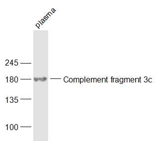 Sample:
Plasma (Rat) Lysate at 40 ug
Primary: Anti-Complement fragment 3c (SL6416R) at 1/1000 dilution
Secondary: IRDye800CW Goat Anti-Rabbit IgG at 1/20000 dilution
Predicted band size: 183 kD
Observed band size: 183 kD
 Sample:
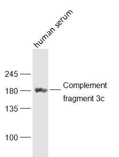
human serum Lysate at 40 ug
Primary: Anti-Complement fragment 3c (SL6416R) at 1/1000 dilution
Secondary: IRDye800CW Goat Anti-Rabbit IgG at 1/20000 dilution
Predicted band size: 183 kD
Observed band size: 183 kD
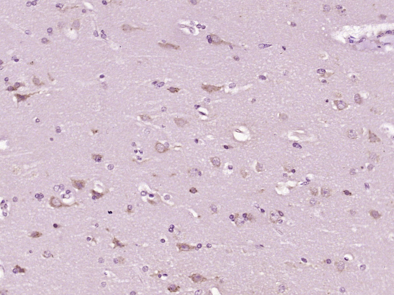 Paraformaldehyde-fixed, paraffin embedded (Human brain glioma); Antigen retrieval by boiling in sodium citrate buffer (pH6.0) for 15min; Block endogenous peroxidase by 3% hydrogen peroxide for 20 minutes; Blocking buffer (normal goat serum) at 37°C for 30min; Antibody incubation with (Complement fragment 3c) Polyclonal Antibody, Unconjugated (SL6416R) at 1:400 overnight at 4°C, followed by operating according to SP Kit(Rabbit) (sp-0023) instructionsand DAB staining.
Cartpieces
Totalgoods,subtotals:¥Checkout
References (0)
No References
Bought notes(bought amounts latest0)
No one bought this product
User Comment(Total0User Comment Num)
- No comment


 +86 571 56623320
 +86 18668110335

