
Rabbit Anti-SHC3 antibody
N SHC; N-Shc; Neuronal Shc; NSHC; Protein Rai; Rai; SH2 domain protein C3; SHC (Src homology 2 domain containing) transforming protein 3; SHC protein C; SHC-like protein, neuronal; SHC-transforming protein 3; SHC-transforming protein C; Shc3; SHC3_HUMAN;
View History [Clear]
Details
Product Name SHC3 Chinese Name SH2结构域蛋白C3抗体 Alias N SHC; N-Shc; Neuronal Shc; NSHC; Protein Rai; Rai; SH2 domain protein C3; SHC (Src homology 2 domain containing) transforming protein 3; SHC protein C; SHC-like protein, neuronal; SHC-transforming protein 3; SHC-transforming protein C; Shc3; SHC3_HUMAN; SHCC; Src homology 2 domain-containing transforming protein; Src homology 2 domain-containing-transforming protein C3. Research Area Tumour Neurobiology Signal transduction G protein-coupled receptor G protein signal Immunogen Species Rabbit Clonality Polyclonal React Species Mouse, (predicted: Human, Rat, Dog, Horse, ) Applications WB=1:500-2000 ELISA=1:5000-10000 IHC-P=1:100-500 IHC-F=1:100-500 IF=1:100-500 (Paraffin sections need antigen repair)
not yet tested in other applications.
optimal dilutions/concentrations should be determined by the end user.Theoretical molecular weight 64kDa Cellular localization cytoplasmic The cell membrane Form Liquid Concentration 1mg/ml immunogen KLH conjugated synthetic peptide derived from human SHC3: 501-594/594 Lsotype IgG Purification affinity purified by Protein A Buffer Solution 0.01M TBS(pH7.4) with 1% BSA, 0.03% Proclin300 and 50% Glycerol. Storage Shipped at 4℃. Store at -20 °C for one year. Avoid repeated freeze/thaw cycles. Attention This product as supplied is intended for research use only, not for use in human, therapeutic or diagnostic applications. PubMed PubMed Product Detail Signaling adapter that couples activated growth factor receptors to signaling pathway in neurons. Involved in the signal transduction pathways of neurotrophin-activated Trk receptors in cortical neurons.
Function:
Signaling adapter that couples activated growth factor receptors to signaling pathway in neurons. Involved in the signal transduction pathways of neurotrophin-activated Trk receptors in cortical neurons.
Subunit:
Interacts with the Trk receptors in a phosphotyrosine-dependent manner. Once activated, binds to GRB2. Interacts with activated EGF receptors.
Tissue Specificity:
Mainly expressed in brain. Hardly detectable in other tissues, except in pancreas. Highly expressed in the cerebral cortex, frontal and temporal lobes, occipital pole, hippocampus, caudate nucleus and amygdala. Expressed at low level in the cerebellum, medulla and spinal cord.
Post-translational modifications:
Tyrosine phosphorylated.
Similarity:
Contains 1 PID domain.
Contains 1 SH2 domain.
SWISS:
Q92529
Gene ID:
53358
Database links:Entrez Gene: 53358 Human
Entrez Gene: 20418 Mouse
Omim: 605263 Human
SwissProt: Q92529 Human
SwissProt: Q61120 Mouse
Unigene: 292737 Human
Unigene: 131870 Mouse
Unigene: 210551 Rat
Product Picture 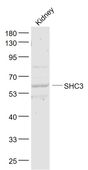 Sample:
Kidney (Mouse) Lysate at 40 ug
Primary: Anti- SHC3 (SL6199R) at 1/1000 dilution
Secondary: IRDye800CW Goat Anti-Rabbit IgG at 1/20000 dilution
Predicted band size: 64 kD
Observed band size: 64 kD
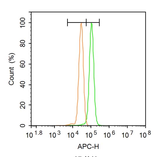 Blank control (Black line): A431(Black).
Primary Antibody (green line): Rabbit Anti-ZNF185 antibody (SL6119R)
Dilution: 1μg /10^6 cells;
Isotype Control Antibody (orange line): Rabbit IgG .
Secondary Antibody (white blue line): Goat anti-rabbit IgG-AF647
Dilution: 1μg /test.
Protocol
The cells were fixed with 4% PFA (10min at room temperature)and then permeabilized with 20% PBST for 20 min at room temperature. The cells were then incubated in 5%BSA to block non-specific protein-protein interactions for 30 min at -20℃ .Cells stained with Primary Antibody for 30 min at room temperature. The secondary antibody used for 40 min at room temperature. Acquisition of 20,000 events was performed.
Cartpieces
Totalgoods,subtotals:¥Checkout
Bought notes(bought amounts latest0)
No one bought this product
User Comment(Total0User Comment Num)
- No comment


 +86 571 56623320


