
Rabbit Anti-phospho-NFKB p65 (Thr435)antibody
NF-kB p65 (phospho T435); p-NF-kB p65 (phospho T435); NFkB-p65(Phospho-Thr435); RELA(phospho T435); NF kB P65; NF-kB p65; NFKBp65; NF-κBp65; p65 NF kappaB; p65 NFkB; NFKBp65; RELA; Transcription Factor p65; v rel avian reticuloendotheliosis viral oncogene
View History [Clear]
Details
Product Name phospho-NFKB p65 (Thr435) Chinese Name 磷酸化The nucleus因子抗体 Alias NF-kB p65 (phospho T435); p-NF-kB p65 (phospho T435); NFkB-p65(Phospho-Thr435); RELA(phospho T435); NF kB P65; NF-kB p65; NFKBp65; NF-κBp65; p65 NF kappaB; p65 NFkB; NFKBp65; RELA; Transcription Factor p65; v rel avian reticuloendotheliosis viral oncogene homolog A (nuclear factor of kappa light polypeptide gene enhancer in B cells 3 (p65)); V Rel Avian Reticuloendotheliosis Viral Oncogene Homolog A; v rel reticuloendotheliosis viral oncogene homolog A (avian); v-rel reticuloendotheliosis viral oncogene homolog A; p65NFKB; Avian reticuloendotheliosis viral (v rel) oncogene homolog A; MGC131774; NFKB 3; NFKB3; Nuclear Factor NF Kappa B p65 Subunit; Nuclear factor of kappa light polypeptide gene enhancer in B cells 3; Nuclear Factor Of Kappa Light Polypeptide Gene Enhancer In B Cells. NFκB-p65; NFκB p65; NF κB-p65; NFκBp65; literatures Product Type Phosphorylated anti Research Area Tumour immunology Chromatin and nuclear signals Signal transduction Apoptosis transcriptional regulatory factor Kinases and Phosphatases Immunogen Species Rabbit Clonality Polyclonal React Species Human, Mouse, (predicted: Rat, ) Applications WB=1:500-2000 IHC-P=1:100-500 IHC-F=1:100-500 Flow-Cyt=2ug/Test ICC=1:100 IF=1:100-500 (Paraffin sections need antigen repair)
not yet tested in other applications.
optimal dilutions/concentrations should be determined by the end user.Theoretical molecular weight 61kDa Cellular localization The nucleus cytoplasmic Form Liquid Concentration 1mg/ml immunogen KLH conjugated Synthesised phosphopeptide derived from human NFKBp65 around the phosphorylation site of Thr435: EG(p-T)LS Lsotype IgG Purification affinity purified by Protein A Buffer Solution 0.01M TBS(pH7.4) with 1% BSA, 0.03% Proclin300 and 50% Glycerol. Storage Shipped at 4℃. Store at -20 °C for one year. Avoid repeated freeze/thaw cycles. Attention This product as supplied is intended for research use only, not for use in human, therapeutic or diagnostic applications. PubMed PubMed Product Detail NF-kappa-B is a ubiquitous transcription factor involved in several biological processes. It is held in the cytoplasm in an inactive state by specific inhibitors. Upon degradation of the inhibitor, NF-kappa-B moves to the nucleus and activates transcription of specific genes. NF-kappa-B is composed of NFKB1 or NFKB2 bound to either REL, RELA, or RELB. The most abundant form of NF-kappa-B is NFKB1 complexed with the product of this gene, RELA. Four transcript variants encoding different isoforms have been found for this gene. [provided by RefSeq, Sep 2011].
Function:
NF-kappa-B is a pleiotropic transcription factor present in almost all cell types and is the endpoint of a series of signal transduction events that are initiated by a vast array of stimuli related to many biological processes such as inflammation, immunity, differentiation, cell growth, tumorigenesis and apoptosis. NF-kappa-B is a homo- or heterodimeric complex formed by the Rel-like domain-containing proteins RELA/p65, RELB, NFKB1/p105, NFKB1/p50, REL and NFKB2/p52 and the heterodimeric p65-p50 complex appears to be most abundant one. The dimers bind at kappa-B sites in the DNA of their target genes and the individual dimers have distinct preferences for different kappa-B sites that they can bind with distinguishable affinity and specificity. Different dimer combinations act as transcriptional activators or repressors, respectively. NF-kappa-B is controlled by various mechanisms of post-translational modification and subcellular compartmentalization as well as by interactions with other cofactors or corepressors. NF-kappa-B complexes are held in the cytoplasm in an inactive state complexed with members of the NF-kappa-B inhibitor (I-kappa-B) family. In a conventional activation pathway, I-kappa-B is phosphorylated by I-kappa-B kinases (IKKs) in response to different activators, subsequently degraded thus liberating the active NF-kappa-B complex which translocates to the nucleus. NF-kappa-B heterodimeric p65-p50 and p65-c-Rel complexes are transcriptional activators. The NF-kappa-B p65-p65 complex appears to be involved in invasin-mediated activation of IL-8 expression. The inhibitory effect of I-kappa-B upon NF-kappa-B the cytoplasm is exerted primarily through the interaction with p65. p65 shows a weak DNA-binding site which could contribute directly to DNA binding in the NF-kappa-B complex. Associates with chromatin at the NF-kappa-B promoter region via association with DDX1.
Subunit:
Component of the NF-kappa-B p65-p50 complex. Component of the NF-kappa-B p65-c-Rel complex. Homodimer; component of the NF-kappa-B p65-p65 complex. Component of the NF-kappa-B p65-p52 complex. May interact with ETHE1. Binds AES and TLE1. Interacts with TP53BP2. Binds to and is phosphorylated by the activated form of either RPS6KA4 or RPS6KA5. Interacts with ING4 and this interaction may be indirect. Interacts with CARM1, USP48 and UNC5CL. Interacts with IRAK1BP1 (By similarity). Interacts with NFKBID (By similarity). Interacts with NFKBIA. Interacts with GSK3B. Interacts with NFKBIB (By similarity). Interacts with NFKBIE. Interacts with NFKBIZ. Interacts with EHMT1 (via ANK repeats) (By similarity). Part of a 70-90 kDa complex at least consisting of CHUK, IKBKB, NFKBIA, RELA, IKBKAP and MAP3K14. Interacts with HDAC3; HDAC3 mediates the deacetylation of RELA. Interacts with HDAC1; the interaction requires non-phosphorylated RELA. Interacts with CBP; the interaction requires phosphorylated RELA. Interacts (phosphorylated at 'Thr-254') with PIN1; the interaction inhibits p65 binding to NFKBIA. Interacts with SOCS1. Interacts with UXT. Interacts with MTDH and PHF11. Interacts with ARRB2. Interacts with human respiratory syncytial virus (HRSV) protein M2-1. Interacts with NFKBIA (when phosphorylated), the interaction is direct; phosphorylated NFKBIA is part of a SCF(BTRC)-like complex lacking CUL1. Interacts with RNF25. Interacts (via C-terminus) with DDX1. Interacts with UFL1 and COMMD1. Interacts with BRMS1; this promotes deacetylation of 'Lys-310'. Interacts with NOTCH2 (By similarity). Directly interacts with MEN1; this interaction represses NFKB-mediated transactivation. Interacts with AKIP1, which promotes the phosphorylation and nuclear retention of RELA. Interacts (via the RHD) with GFI1; the interaction, after bacterial lipopolysaccharide (LPS) stimulation, inhibits the transcriptional activity by interfering with the DNA-binding activity to target gene promoter DNA.
Subcellular Location:
Nucleus. Cytoplasm. Note=Colocalized with DDX1 in the nucleus upon TNF-alpha induction. Nuclear, but also found in the cytoplasm in an inactive form complexed to an inhibitor (I-kappa-B). Colocalizes with GFI1 in the nucleus after LPS stimulation.
Post-translational modifications:
Ubiquitinated, leading to its proteasomal degradation. Degradation is required for termination of NF-kappa-B response.
Monomethylated at Lys-310 by SETD6. Monomethylation at Lys-310 is recognized by the ANK repeats of EHMT1 and promotes the formation of repressed chromatin at target genes, leading to down-regulation of NF-kappa-B transcription factor activity. Phosphorylation at Ser-311 disrupts the interaction with EHMT1 without preventing monomethylation at Lys-310 and relieves the repression of target genes.
Phosphorylation at Ser-311 disrupts the interaction with EHMT1 and promotes transcription factor activity. Phosphorylation on Ser-536 stimulates acetylation on Lys-310 and interaction with CBP; the phosphorylated and acetylated forms show enhanced transcriptional activity. Phosphorylation at Ser-276 by RPS6KA4 and RPS6KA5 promotes its transactivation and transcriptional activities.
Reversibly acetylated; the acetylation seems to be mediated by CBP, the deacetylation by HDAC3 and SIRT2. Acetylation at Lys-122 enhances DNA binding and impairs association with NFKBIA. Acetylation at Lys-310 is required for full transcriptional activity in the absence of effects on DNA binding and NFKBIA association. Acetylation can also lower DNA-binding and results in nuclear export. Interaction with BRMS1 promotes deacetylation of Lys-310. Lys-310 is deacetylated by SIRT2.
S-nitrosylation of Cys-38 inactivates the enzyme activity.
Sulfhydration at Cys-38 mediates the anti-apoptotic activity by promoting the interaction with RPS3 and activating the transcription factor activity.
Sumoylation by PIAS3 negatively regulates DNA-bound activated NF-kappa-B.
Similarity:
Contains 1 RHD (Rel-like) domain.
SWISS:
Q04206
Gene ID:
5970
Database links:Entrez Gene: 5970 Human
Omim: 164014 Human
SwissProt: Q04206 Human
Unigene: 502875 Human
NF-κBp65是一种重要的转录因子,NF-kBp65可激活参与炎症、细胞增殖、Apoptosis等基因的调节,影响着细胞的凋亡,同时影响着Tumour细胞对细胞毒性药物及离子辐射的敏感性。ras基因诱导的致癌突变作用需NFkB的活化,提示NFkB在致癌发生方面可能起一定作用;另有文献报道,在乳腺癌、非小细胞性肺癌、甲状腺癌、T或Blymphocyte白血病及病毒诱变导致的Tumour等人类Tumour中,NFkB活化或表达。 NF-кB可以保护细胞免受Tumour坏死因子以及电离辐射等引起的凋亡作用,而抑制NFkB的表达可以增加TNF等引起的Apoptosis,以及增加化疗及放疗对Tumour细胞的敏感性。Product Picture 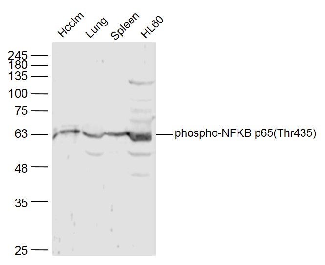 Sample:
Hcclm3 Cell(Human) Lysate at 40 ug
Lung (Mouse) Lysate at 40 ug
Spleen (Mouse) Lysate at 40 ug
HL60 Cell (Human) Lysate at 40 ug
Primary: Anti-phospho-NFKB p65(Thr435) (SL5661R) at 1/300 dilution
Secondary: IRDye800CW Goat Anti-Rabbit IgG at 1/20000 dilution
Predicted band size: 61 kD
Observed band size: 61 kD
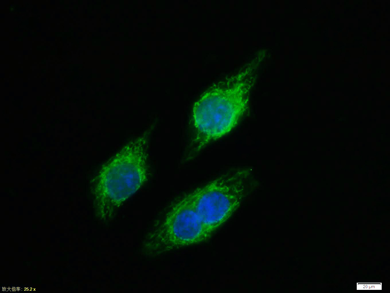 Tissue/cell:MCF7 cell; 4% Paraformaldehyde-fixed; Triton X-100 at room temperature for 20 min; Blocking buffer (normal goat serum,C-0005) at 37°C for 20 min; Antibody incubation with (phospho-NFKB p65 (Thr435)) polyclonal Antibody, Unconjugated (SL5661R) 1:100, 90 minutes at 37°C; followed by a FITC conjugated Goat Anti-Rabbit IgG antibody at 37°C for 90 minutes, DAPI (blue, C02-04002) was used to stain the cell nuclei.
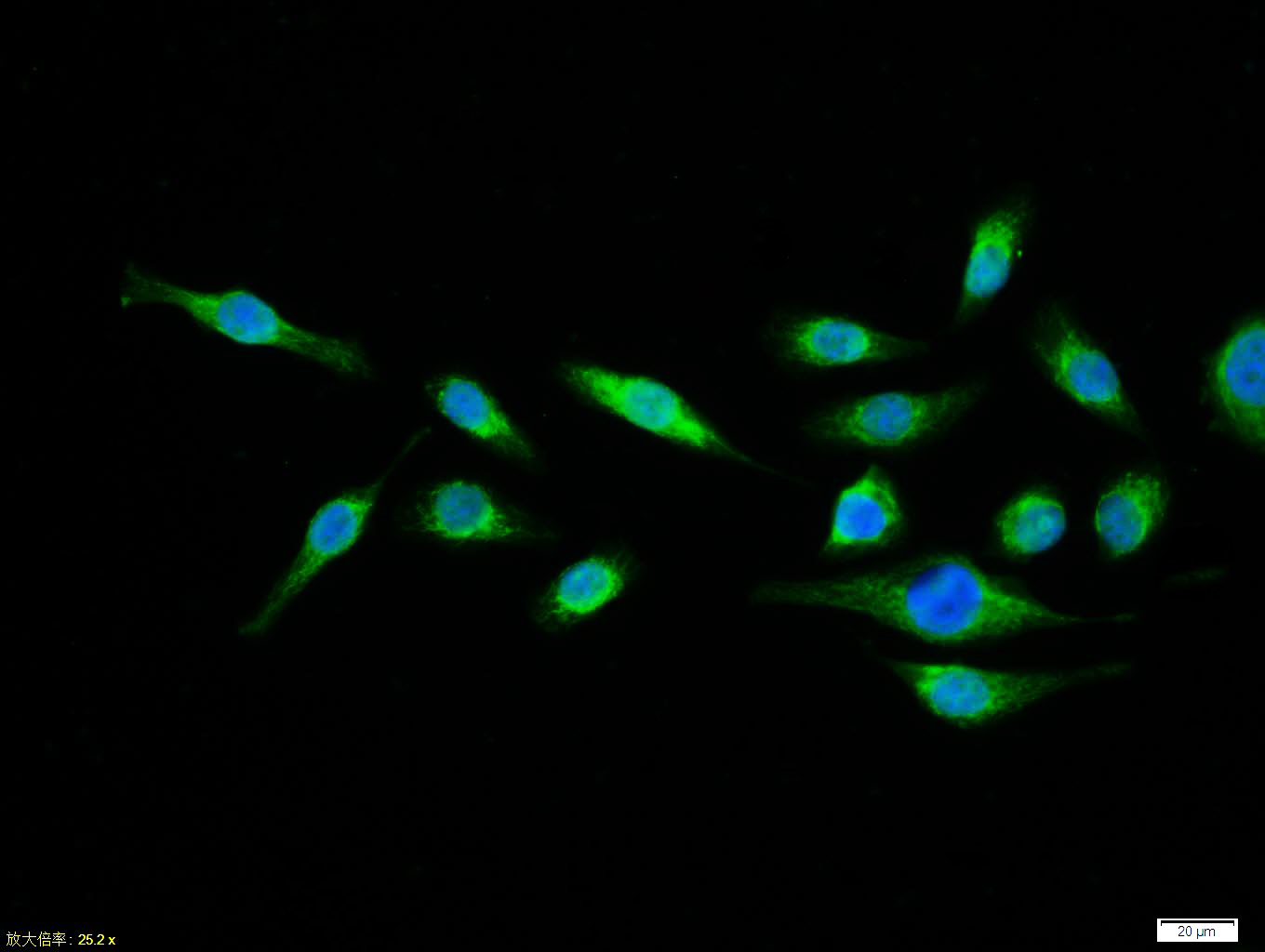 Tissue/cell:MCF7 cell; 4% Paraformaldehyde-fixed; Triton X-100 at room temperature for 20 min; Blocking buffer (normal goat serum,C-0005) at 37°C for 20 min; Antibody incubation with (phospho-NFKB p65 (Thr435)) polyclonal Antibody, Unconjugated (SL5661R) 1:100, 90 minutes at 37°C; followed by a FITC conjugated Goat Anti-Rabbit IgG antibody at 37°C for 90 minutes, DAPI (blue, C02-04002) was used to stain the cell nuclei.
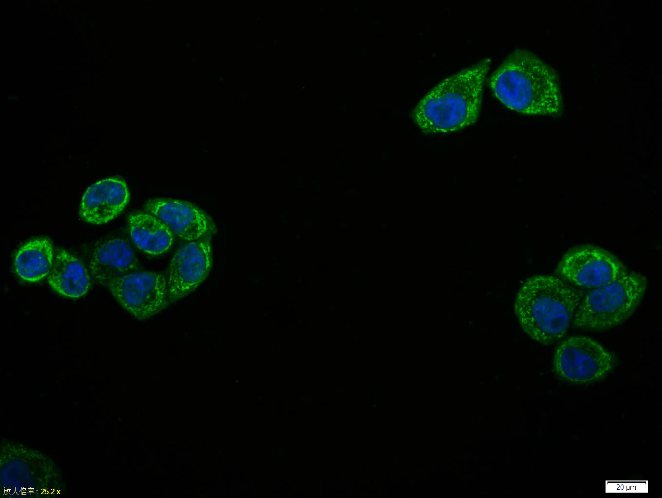 Tissue/cell:Hela cell; 4% Paraformaldehyde-fixed; Triton X-100 at room temperature for 20 min; Blocking buffer (normal goat serum,C-0005) at 37°C for 20 min; Antibody incubation with (phospho-NFKB p65 (Thr435)) polyclonal Antibody, Unconjugated (SL5661R) 1:100, 90 minutes at 37°C; followed by a FITC conjugated Goat Anti-Rabbit IgG antibody at 37°C for 90 minutes, DAPI (blue, C02-04002) was used to stain the cell nuclei.
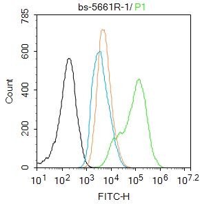 Blank control:A431.
Primary Antibody (green line): Rabbit Anti-phospho-NFKB p65 (Thr435) antibody (SL5661R)
Dilution: 1μg /10^6 cells;
Isotype Control Antibody (orange line): Rabbit IgG .
Secondary Antibody : Goat anti-rabbit IgG-FITC
Dilution: 1μg /test.
Protocol
The cells were fixed with 4% PFA (10min at room temperature)and then permeabilized with 90% ice-cold methanol for 20 min at-20℃. The cells were then incubated in 5%BSA to block non-specific protein-protein interactions for 30 min at room temperature .Cells stained with Primary Antibody for 30 min at room temperature. The secondary antibody used for 40 min at room temperature. Acquisition of 20,000 events was performed.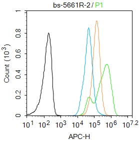 Blank control:MCF7.
Primary Antibody (green line): Rabbit Anti-phospho-NFKB p65 (Thr435) antibody (SL5661R)
Dilution: 2μg /10^6 cells;
Isotype Control Antibody (orange line): Rabbit IgG .
Secondary Antibody : Goat anti-rabbit IgG-AF647
Dilution: 1μg /test.
Protocol
The cells were fixed with 4% PFA (10min at room temperature)and then permeabilized with 90% ice-cold methanol for 20 min at -20℃. The cells were then incubated in 5%BSA to block non-specific protein-protein interactions for 30 min at room temperature .Cells stained with Primary Antibody for 30 min at room temperature. The secondary antibody used for 40 min at room temperature. Acquisition of 20,000 events was performed.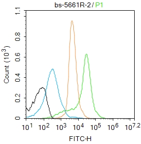 Blank control:Mouse spleen.
Primary Antibody (green line): Rabbit Anti-phospho-NFKB p65 (Thr435) antibody (SL5661R)
Dilution: 2μg /10^6 cells;
Isotype Control Antibody (orange line): Rabbit IgG .
Secondary Antibody : Goat anti-rabbit IgG-AF488
Dilution: 1μg /test.
Protocol
The cells were fixed with 4% PFA (10min at room temperature)and then permeabilized with 90% ice-cold methanol for 20 min at-20℃. The cells were then incubated in 5%BSA to block non-specific protein-protein interactions for 30 min at room temperature.Cells stained with Primary Antibody for 30 min at room temperature. The secondary antibody used for 40 min at room temperature. Acquisition of 20,000 events was performed.
Cartpieces
Totalgoods,subtotals:¥Checkout
Bought notes(bought amounts latest0)
No one bought this product
User Comment(Total0User Comment Num)
- No comment


 +86 571 56623320
 +86 18668110335

