
Rabbit Anti-phospho-PTPN6 (Tyr536)antibody
SHP-1(Phospho-Tyr536); PTPN6(phospho Y536); SHP1(phospho Y536); SHP1; 70 kda SHP 1L protein; 70 kda SHP1L protein; 70Z-SHP; EC 3.1.3.48; HCP; HCPH; Hematopoietic cell phosphatase; Hematopoietic cell protein tyrosine phosphatase; Hematopoietic cell protein
View History [Clear]
Details
Product Name phospho-PTPN6 (Tyr536) Chinese Name 磷酸化蛋白酪氨酸磷酸酶1C抗体 Alias SHP-1(Phospho-Tyr536); PTPN6(phospho Y536); SHP1(phospho Y536); SHP1; 70 kda SHP 1L protein; 70 kda SHP1L protein; 70Z-SHP; EC 3.1.3.48; HCP; HCPH; Hematopoietic cell phosphatase; Hematopoietic cell protein tyrosine phosphatase; Hematopoietic cell protein-tyrosine phosphatase; HPTP 1C; HPTP1C; MGC124580; Protein tyrosine phosphatase 1C; Protein tyrosine phosphatase non receptor type 6; Protein tyrosine phosphatase SHP 1; Protein tyrosine phosphatase SHP1; Protein-tyrosine phosphatase 1C; Protein-tyrosine phosphatase SHP-1; PTN6_HUMAN; PTP 1C; PTP-1C; PTP1C; PTPN 6; PTPN6; SH PTP 1; SH PTP1; SH-PTP1; SHP 1; SHP 1L; SHP1L; SHPTP 1; SHPTP1; Tyrosine protein phosphatase non receptor type 6; Tyrosine-protein phosphatase non-receptor type 6. Product Type Phosphorylated anti Research Area Tumour immunology Developmental biology Signal transduction transcriptional regulatory factor Kinases and Phosphatases Immunogen Species Rabbit Clonality Polyclonal React Species Human, (predicted: Dog, Horse, ) Applications WB=1:500-2000 IHC-P=1:100-500 IHC-F=1:100-500 Flow-Cyt=2ug/test (Paraffin sections need antigen repair)
not yet tested in other applications.
optimal dilutions/concentrations should be determined by the end user.Theoretical molecular weight 65kDa Cellular localization The nucleus cytoplasmic Form Liquid Concentration 1mg/ml immunogen KLH conjugated Synthesised phosphopeptide derived from human PTPN6 around the phosphorylation site of Tyr536: SE(p-Y)GN Lsotype IgG Purification affinity purified by Protein A Buffer Solution 0.01M TBS(pH7.4) with 1% BSA, 0.03% Proclin300 and 50% Glycerol. Storage Shipped at 4℃. Store at -20 °C for one year. Avoid repeated freeze/thaw cycles. Attention This product as supplied is intended for research use only, not for use in human, therapeutic or diagnostic applications. PubMed PubMed Product Detail The protein encoded by this gene is a member of the protein tyrosine phosphatase (PTP) family. PTPs are known to be signaling molecules that regulate a variety of cellular processes including cell growth, differentiation, mitotic cycle, and oncogenic transformation. N-terminal part of this PTP contains two tandem Src homolog (SH2) domains, which act as protein phospho-tyrosine binding domains, and mediate the interaction of this PTP with its substrates. This PTP is expressed primarily in hematopoietic cells, and functions as an important regulator of multiple signaling pathways in hematopoietic cells. This PTP has been shown to interact with, and dephosphorylate a wide spectrum of phospho-proteins involved in hematopoietic cell signaling. Multiple alternatively spliced variants of this gene, which encode distinct isoforms, have been reported. [provided by RefSeq, Jul 2008]
Function:
Modulates signaling by tyrosine phosphorylated cell surface receptors such as KIT and the EGF receptor/EGFR. The SH2 regions may interact with other cellular components to modulate its own phosphatase activity against interacting substrates. Together with MTUS1, induces UBE2V2 expression upon angiotensin II stimulation. Plays a key role in hematopoiesis.
Subunit:
Monomer. Interacts with MTUS1. Interacts with MILR1 (tyrosine-phosphorylated). Interacts with KIT. Binds PTPNS1, LILRB1 and LILRB2. Interacts with FCRL2, FCRL3, FCRL4, CD300LF, CDK2 and CD84. Interacts with KIR2DL1; the interaction is enhanced by ARRB2. Interacts (via SH2 1 domain) with ROS1; the interaction is direct and promotes ROS1 dephosphorylation. Interacts with EGFR; inhibits EGFR-dependent activation of MAPK/ERK. Interacts with LYN. Interacts with the tyrosine phosphorylated form of PDPK1.
Subcellular Location:
Cytoplasm. Nucleus. Note=In neurons, translocates into the nucleus after treatment with angiotensin II. Shuttles between the cytoplasm and nucleus via its association with PDPK1.
Tissue Specificity:
Isoform 1 is expressed in hematopoietic cells. Isoform 2 is expressed in non-hematopoietic cells.
Post-translational modifications:
Phosphorylated on tyrosine residues. Binding of KITLG/SCF to KIT increases tyrosine phosphorylation. Phosphorylation at Tyr-564 enhances phosphatase activity.
Similarity:
Belongs to the protein-tyrosine phosphatase family. Non-receptor class 2 subfamily.
Contains 2 SH2 domains.
Contains 1 tyrosine-protein phosphatase domain.
SWISS:
P29350
Gene ID:
5777
Database links:Entrez Gene: 5777 Human
Entrez Gene: 15170 Mouse
Omim: 176883 Human
SwissProt: P29350 Human
SwissProt: P29351 Mouse
Unigene: 63489 Human
Unigene: 271799 Mouse
Unigene: 18985 Rat
Product Picture 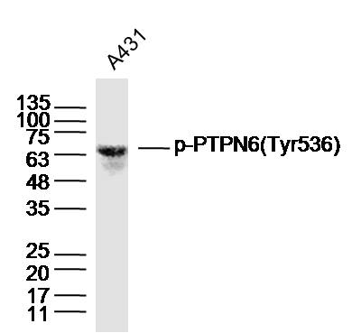 Sample: A431 Cell (Human) Lysate at 40 ug
Primary: Anti-phospho-PTPN6(Tyr564)(SL5577R)at 1/300 dilution
Secondary: IRDye800CW Goat Anti-Rabbit IgG at 1/20000 dilution
Predicted band size: 65kD
Observed band size: 65kD
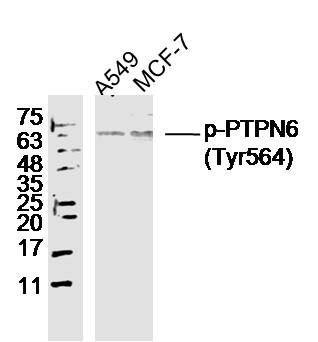 Sample:
A549 Cell (Human) Lysate at 40 ug
MCF-7 Cell (Human) Lysate at 40 ug
Primary: Anti- phospho-PTPN6(Tyr564) (SL5577R) at 1/300 dilution
Secondary: IRDye800CW Goat Anti-Rabbit IgG at 1/20000 dilution
Predicted band size: 65 kD
Observed band size: 65 kD
 Paraformaldehyde-fixed, paraffin embedded (Human glioma); Antigen retrieval by boiling in sodium citrate buffer (pH6.0) for 15min; Block endogenous peroxidase by 3% hydrogen peroxide for 20 minutes; Blocking buffer (normal goat serum) at 37°C for 30min; Antibody incubation with (phospho-PTPN6(Tyr564)) Polyclonal Antibody, Unconjugated (SL5577R) at 1:400 overnight at 4°C, followed by operating according to SP Kit(Rabbit) (sp-0023) instructionsand DAB staining.
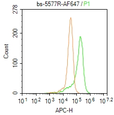 Blank control: Molt4.
Primary Antibody (green line): Rabbit Anti- phospho-PTPN6(Tyr564) / AF647 Conjugated antibody (SL5577R-AF647)
Dilution: 2μg /10^6 cells;
Isotype Control Antibody (orange line): Rabbit IgG- AF647 .
Protocol
The cells were fixed with 4% PFA (10min at room temperature)and then permeabilized with 0.1% PBST for 20 min at-20℃. The cells were then incubated in 5% BSA to block non-specific protein-protein interactions for 30 min at room temperature. The cells were stained with Primary Antibody for 30 min at room temperature. Acquisition of 20,000 events was performed.
Cartpieces
Totalgoods,subtotals:¥Checkout
Bought notes(bought amounts latest0)
No one bought this product
User Comment(Total0User Comment Num)
- No comment


 +86 571 56623320
 +86 18668110335

