
Rabbit Anti-phospho-NFATc2 (Ser330)antibody
NFATC2(phospho Ser330); NFATC2(phospho S330); AI607462; KIAA0611; NF ATp; NF-ATc2; NFAT 1; NFAT pre existing subunit; NFAT transcription complex, preexisting component; NFAT1; NFAT1-D; NFATc2; NFATp; Nuclear factor of activated T cells cytoplasmic 2; Nucl
View History [Clear]
Details
Product Name phospho-NFATc2 (Ser330) Chinese Name 磷酸化核因子活化T细胞胞浆蛋白2抗体 Alias NFATC2(phospho Ser330); NFATC2(phospho S330); AI607462; KIAA0611; NF ATp; NF-ATc2; NFAT 1; NFAT pre existing subunit; NFAT transcription complex, preexisting component; NFAT1; NFAT1-D; NFATc2; NFATp; Nuclear factor of activated T cells cytoplasmic 2; Nuclear factor of activated T cells cytoplasmic calcineurin dependent 2; Nuclear factor of activated T cells pre-existing component; T cell transcription factor NFAT; NF2IP_HUMAN; Nuclear factor of activated T-cells, cytoplasmic 2; NFAT pre-existing subunit; NF-ATp; T-cell transcription factor NFAT1. Product Type Phosphorylated anti Research Area immunology Neurobiology Signal transduction transcriptional regulatory factor Kinases and Phosphatases t-lymphocyte Immunogen Species Rabbit Clonality Polyclonal React Species Human, Mouse, Rat, (predicted: Dog, Pig, Cow, Horse, ) Applications WB=1:500-2000 ELISA=1:5000-10000 IHC-P=1:100-500 Flow-Cyt=1ug/Test ICC=1:100 (Paraffin sections need antigen repair)
not yet tested in other applications.
optimal dilutions/concentrations should be determined by the end user.Theoretical molecular weight 100kDa Cellular localization The nucleus cytoplasmic Form Liquid Concentration 1mg/ml immunogen KLH conjugated Synthesised phosphopeptide derived from human NFATc2 around the phosphorylation site of Ser330: DP(p-S)PV Lsotype IgG Purification affinity purified by Protein A Buffer Solution 0.01M TBS(pH7.4) with 1% BSA, 0.03% Proclin300 and 50% Glycerol. Storage Shipped at 4℃. Store at -20 °C for one year. Avoid repeated freeze/thaw cycles. Attention This product as supplied is intended for research use only, not for use in human, therapeutic or diagnostic applications. PubMed PubMed Product Detail This gene is a member of the nuclear factor of activated T cells (NFAT) family. The product of this gene is a DNA-binding protein with a REL-homology region (RHR) and an NFAT-homology region (NHR). This protein is present in the cytosol and only translocates to the nucleus upon T cell receptor (TCR) stimulation, where it becomes a member of the nuclear factors of activated T cells transcription complex. This complex plays a central role in inducing gene transcription during the immune response. Alternate transcriptional splice variants encoding different isoforms have been characterized. [provided by RefSeq, Apr 2012]
Function:
Plays a role in the inducible expression of cytokine genes in T-cells, especially in the induction of the IL-2, IL-3, IL-4, TNF-alpha or GM-CSF. Promotes invasive migration through the activation of GPC6 expression and WNT5A signaling pathway.
Subunit:
Member of the multicomponent NFATC transcription complex that consists of at least two components, a pre-existing cytoplasmic component NFATC2 and an inducible nuclear component NFATC1. Other members such as NFATC4, NFATC3 or members of the activating protein-1 family, MAF, GATA4 and Cbp/p300 can also bind the complex. The phosphorylated form specifically interacts with XPO1; which mediates nuclear export. NFATC proteins bind to DNA as monomers. Interacts with NFATC2IP (By similarity).
Subcellular Location:
Cytoplasm. Nucleus. Note=Cytoplasmic for the phosphorylated form and nuclear after activation that is controlled by calcineurin-mediated dephosphorylation. Rapid nuclear exit of NFATC is thought to be one mechanism by which cells distinguish between sustained and transient calcium signals. The subcellular localization of NFATC plays a key role in the regulation of gene transcription.
Tissue Specificity:
Expressed in thymus, spleen, heart, testis, brain, placenta, muscle and pancreas.
Post-translational modifications:
In resting cells, phosphorylated by NFATC-kinase on at least 18 sites in the 99-365 region. Upon cell stimulation, all these sites except Ser-245 are dephosphorylated by calcineurin. Dephosphorylation induces a conformational change that simultaneously exposes an NLS and masks an NES, which results in nuclear localization. Simultaneously, one site among Ser-53; Ser-54 and Ser-56 is phosphorylated; which is required for full transcriptional activity.
Similarity:
Contains 1 RHD (Rel-like) domain.
SWISS:
Q13469
Gene ID:
4773
Database links:Entrez Gene: 4773 Human
Entrez Gene: 18019 Mouse
Omim: 600490 Human
SwissProt: Q13469 Human
SwissProt: Q60591 Mouse
Unigene: 713650 Human
Unigene: 744148 Human
Unigene: 116802 Mouse
Unigene: 33679 Rat
Product Picture 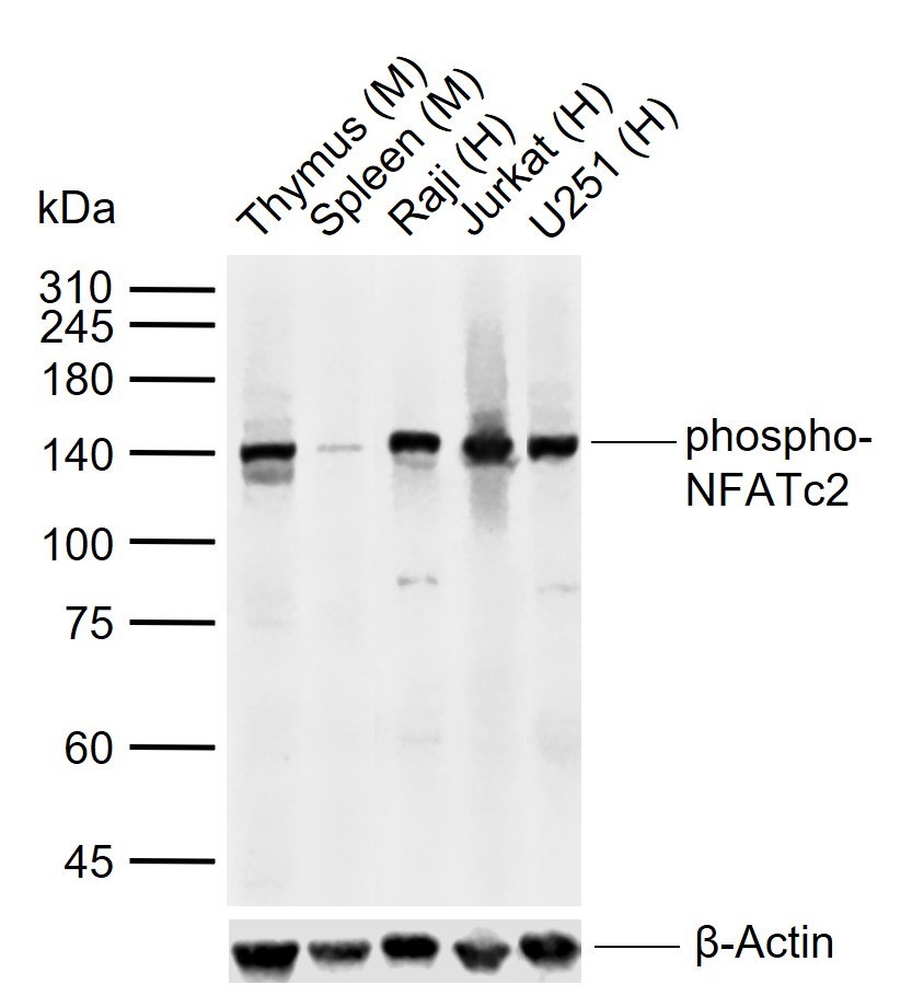 Sample:
Lane 1: Mouse Thymus tissue lysates
Lane 2: Mouse Spleen tissue lysates
Lane 3: Human Raji cell lysates
Lane 4: Human Jurkat cell lysates
Lane 5: Human U251 cell lysates
Primary: Anti-phospho-NFATc2 (Ser330) (SL5516R) at 1/1000 dilution
Secondary: IRDye800CW Goat Anti-Rabbit IgG at 1/20000 dilution
Predicted band size: 100 kDa
Observed band size: 140 kDa
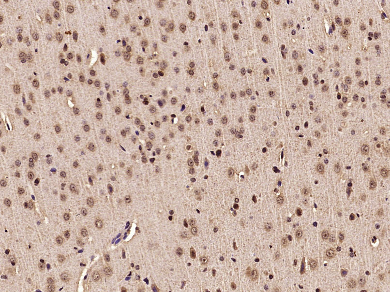 Paraformaldehyde-fixed, paraffin embedded (Rat brain); Antigen retrieval by microwave in sodium citrate buffer (pH6.0) ; Block endogenous peroxidase by 3% hydrogen peroxide for 30 minutes; Blocking buffer (3% BSA) at RT for 30min; Antibody incubation with (phospho-NFATc2(Ser330)) Polyclonal Antibody, Unconjugated (SL5516R) at 1:400 overnight at 4°C, followed by conjugation to the secondary antibody (labeled with HRP)and DAB staining.
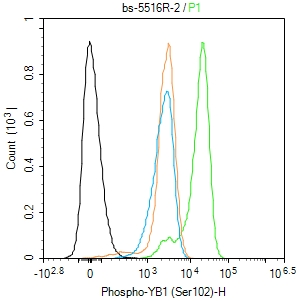 Blank control(black line):Molt4.
Primary Antibody (green line): Rabbit Anti-phospho-NFATc2 (Ser330) antibody (SL5516R)
Dilution:1ug/Test;
Secondary Antibody(white blue line): Goat anti-rabbit IgG-AF488
Dilution: 0.5ug/Test.
Isotype control(orange line): Normal Rabbit IgG
Protocol
The cells were fixed with 4% PFA (10min at room temperature)and then permeabilized with 90% ice-cold methanol for 20 min at -20℃, The cells were then incubated in 5%BSA to block non-specific protein-protein interactions for 30 min at room temperature .Cells stained with Primary Antibody for 30 min at room temperature. The secondary antibody used for 40 min at room temperature. Acquisition of 20,000 events was performed.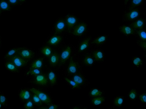 HUVEC cell; 4% Paraformaldehyde-fixed; Triton X-100 at room temperature for 20 min; Blocking buffer (normal goat serum, C-0005) at 37°C for 20 min; Antibody incubation with (phospho-NFATc2 (Ser330)) polyclonal Antibody, Unconjugated (SL5516R) 1:100, 90 minutes at 37°C; followed by a conjugated Goat Anti-Rabbit IgG antibody at 37°C for 90 minutes, DAPI (blue, C02-04002) was used to stain the cell nuclei.
Cartpieces
Totalgoods,subtotals:¥Checkout
Bought notes(bought amounts latest0)
No one bought this product
User Comment(Total0User Comment Num)
- No comment


 +86 571 56623320
 +86 18668110335

