
Rabbit Anti-phospho-AR/Androgen receptor (Ser213)antibody
Androgen Receptor (phospho S213); Androgen Receptor (Phospho-Ser213); Androgen Receptor (phospho Ser213); p-Androgen Receptor (Ser213); ANDR_HUMAN; HYSP1; AIS; Androgen receptor (dihydrotestosterone receptor; testicular feminization; spinal and bulbar mus
View History [Clear]
Details
Product Name phospho-AR/Androgen receptor (Ser213) Chinese Name 磷酸化雄激素受体(AR)抗体 Alias Androgen Receptor (phospho S213); Androgen Receptor (Phospho-Ser213); Androgen Receptor (phospho Ser213); p-Androgen Receptor (Ser213); ANDR_HUMAN; HYSP1; AIS; Androgen receptor (dihydrotestosterone receptor; testicular feminization; spinal and bulbar muscular atrophy; Kennedy disease); AR; DHTR; Dihydro Testosterone Receptor; Dihydrotestosterone receptor; HUMARA; Nuclear receptor subfamily 3 group C member 4; SBMA; SMAX1; Spinal and bulbar muscular atrophy; TFM. Product Type Phosphorylated anti Research Area Tumour immunology Immunogen Species Rabbit Clonality Polyclonal React Species Human, Applications WB=1:500-2000 ELISA=1:5000-10000 IHC-P=1:100-500 IHC-F=1:100-500 ICC=1:100 IF=1:100-500 (Paraffin sections need antigen repair)
not yet tested in other applications.
optimal dilutions/concentrations should be determined by the end user.Theoretical molecular weight 101kDa Cellular localization The nucleus cytoplasmic Form Liquid Concentration 1mg/ml immunogen KLH conjugated Synthesised phosphopeptide derived from human Androgen Receptor around the phosphorylation site of Ser213: ER(p-S)GA Lsotype IgG Purification affinity purified by Protein A Buffer Solution 0.01M TBS(pH7.4) with 1% BSA, 0.03% Proclin300 and 50% Glycerol. Storage Shipped at 4℃. Store at -20 °C for one year. Avoid repeated freeze/thaw cycles. Attention This product as supplied is intended for research use only, not for use in human, therapeutic or diagnostic applications. PubMed PubMed Product Detail The androgen receptor gene is more than 90 kb long and codes for a protein that has 3 major functional domains: the N-terminal domain, DNA-binding domain, and androgen-binding domain. The protein functions as a steroid-hormone activated transcription factor. Upon binding the hormone ligand, the receptor dissociates from accessory proteins, translocates into the nucleus, dimerizes, and then stimulates transcription of androgen responsive genes. This gene contains 2 polymorphic trinucleotide repeat segments that encode polyglutamine and polyglycine tracts in the N-terminal transactivation domain of its protein. Expansion of the polyglutamine tract causes spinal bulbar muscular atrophy (Kennedy disease). Mutations in this gene are also associated with complete androgen insensitivity (CAIS). Two alternatively spliced variants encoding distinct isoforms have been described. [provided by RefSeq, Jul 2008]
Function:
Steroid hormone receptors are ligand-activated transcription factors that regulate eukaryotic gene expression and affect cellular proliferation and differentiation in target tissues. Transcription factor activity is modulated by bound coactivator and corepressor proteins. Transcription activation is down-regulated by NR0B2. Activated, but not phosphorylated, by HIPK3 and ZIPK/DAPK3. [ENZYME REGULATION] AIM-100 (4-amino-5,6-biaryl-furo[2,3-d]pyrimidine) suppresses TNK2-mediated phosphorylation at Tyr-267. Inhibits the binding of the Tyr-267 phosphorylated form to androgen-responsive enhancers (AREs) and its transcriptional activity.
Subunit:
Binds DNA as a homodimer. Part of a ternary complex containing AR, EFCAB6/DJBP and PARK7. Interacts with HIPK3 and NR0B2 in the presence of androgen. The ligand binding domain interacts with KAT7/HBO1 in the presence of dihydrotestosterone. Interacts with EFCAB6/DJBP, PELP1, PQBP1, RANBP9, RBAK, SPDEF, SRA1, TGFB1I1, ZNF318 and RREB1. Interacts with ZMIZ1/ZIMP10 and ZMIZ2/ZMIP7 which both enhance its transactivation activity. Interacts with SLC30A9 and RAD54L2/ARIP4. Interacts via the ligand-binding domain with LXXLL and FXXLF motifs from NCOA1, NCOA2, NCOA3, NCOA4 and MAGEA11. The AR N-terminal poly-Gln region binds Ran resulting in enhancement of AR-mediated transactivation. Ran-binding decreases as the poly-Gln length increases. Interacts with HIP1 (via coiled coil domain). Interacts (via ligand-binding domain) with TRIM68. Interacts with TNK2. Interacts with USP26. Interacts with RNF6. Interacts (regulated by RNF6 probably through polyubiquitination) with RNF14; regulates AR transcriptional activity. Interacts with PRMT2 and TRIM24. Interacts with GNB2L1/RACK1. Interacts with RANBP10; this interaction enhances dihydrotestosterone-induced AR transcriptional activity. Interacts with PRPF6 in a hormone-independent way; this interaction enhances dihydrotestosterone-induced AR transcriptional activity. Interacts with STK4/MST1. Interacts with ZIPK/DAPK3. Interacts with LPXN. Interacts with MAK. Part of a complex containing AR, MAK and NCOA3.
Subcellular Location:
Nucleus. Cytoplasm. Note=Predominantly cytoplasmic in unligated form but translocates to the nucleus upon ligand-binding. Can also translocate to the nucleus in unligated form in the presence of GNB2L1.
Tissue Specificity:
Isoform 2 is mainly expressed in heart and skeletal muscle.
Post-translational modifications:
Sumoylated on Lys-386 (major) and Lys-520. Ubiquitinated. Deubiquitinated by USP26. 'Lys-6' and 'Lys-27'-linked polyubiquitination by RNF6 modulates AR transcriptional activity and specificity.
Phosphorylated in prostate cancer cells in response to several growth factors including EGF. Phosphorylation is induced by c-Src kinase (CSK). Tyr-534 is one of the major phosphorylation sites and an increase in phosphorylation and Src kinase activity is associated with prostate cancer progression. Phosphorylation by TNK2 enhances the DNA-binding and transcriptional activity and may be responsible for androgen-independent progression of prostate cancer. Phosphorylation at Ser-81 by CDK9 regulates AR promoter selectivity and cell growth. Phosphorylation by PAK6 leads to AR-mediated transcription inhibition.
Palmitoylated by ZDHHC7 and ZDHHC21. Palmitoylation is required for plasma membrane targeting and for rapid intracellular signaling via ERK and AKT kinases and cAMP generation.
DISEASE:
Defects in AR are the cause of androgen insensitivity syndrome (AIS) [MIM:300068]; previously known as testicular feminization syndrome (TFM). AIS is an X-linked recessive form of pseudohermaphroditism due end-organ resistance to androgen. Affected males have female external genitalia, female breast development, blind vagina, absent uterus and female adnexa, and abdominal or inguinal testes, despite a normal 46,XY karyotype.
Defects in AR are the cause of spinal and bulbar muscular atrophy X-linked type 1 (SMAX1) [MIM:313200]; also known as Kennedy disease. SMAX1 is an X-linked recessive form of spinal muscular atrophy. Spinal muscular atrophy refers to a group of neuromuscular disorders characterized by degeneration of the anterior horn cells of the spinal cord, leading to symmetrical muscle weakness and atrophy. SMAX1 occurs only in men. Age at onset is usually in the third to fifth decade of life, but earlier involvement has been reported. It is characterized by slowly progressive limb and bulbar muscle weakness with fasciculations, muscle atrophy, and gynecomastia. The disorder is clinically similar to classic forms of autosomal spinal muscular atrophy. Note=Caused by trinucleotide CAG repeat expansion. In SMAX1 patients the number of Gln ranges from 38 to 62. Longer expansions result in earlier onset and more severe clinical manifestations of the disease.
Note=Defects in AR may play a role in metastatic prostate cancer. The mutated receptor stimulates prostate growth and metastases development despite of androgen ablation. This treatment can reduce primary and metastatic lesions probably by inducing apoptosis of tumor cells when they express the wild-type receptor.
Defects in AR are the cause of androgen insensitivity syndrome partial (PAIS) [MIM:312300]; also known as Reifenstein syndrome. PAIS is characterized by hypospadias, hypogonadism, gynecomastia, genital ambiguity, normal XY karyotype, and a pedigree pattern consistent with X-linked recessive inheritance. Some patients present azoospermia or severe oligospermia without other clinical manifestations.
Similarity:
Belongs to the nuclear hormone receptor family. NR3 subfamily.
Contains 1 nuclear receptor DNA-binding domain.
SWISS:
P10275
Gene ID:
367
Database links:Entrez Gene: 367 Human
Entrez Gene: 11835 Mouse
Omim: 313700 Human
SwissProt: P10275 Human
SwissProt: P19091 Mouse
Unigene: 496240 Human
Unigene: 39005 Mouse
Unigene: 394224 Mouse
Unigene: 439657 Mouse
Unigene: 9813 Rat
AR是一个由920个氨基酸组成的蛋白质,位于雄激素靶组织细胞中或细胞表面上的特异分子部位或结构。 AR在前列腺癌中起着重要的作用,研究表明AR的表达与组织分型形成一定的相关性 ,AR在高分化的Tumour中表达较多,而在低分化的Tumour中表达较少。用于前列腺癌的检测,指导临床治疗,目前可用于乳腺癌、食道癌等各项Tumour的研究。Product Picture 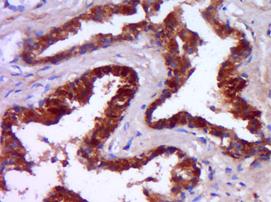 Paraformaldehyde-fixed, paraffin embedded (Human prostate); Antigen retrieval by boiling in sodium citrate buffer (pH6.0) for 15min; Block endogenous peroxidase by 3% hydrogen peroxide for 20 minutes; Blocking buffer (normal goat serum) at 37°C for 30min; Antibody incubation with (ANDR) Polyclonal Antibody, Unconjugated (SL5191R) at 1:400 overnight at 4°C, followed by operating according to SP Kit(Rabbit) (sp-0023) instructionsand DAB staining.
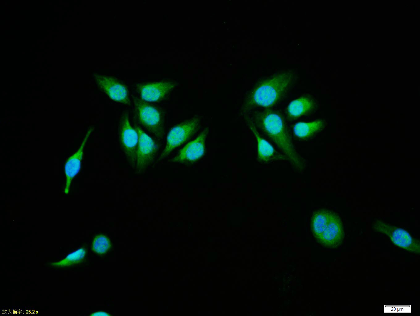 Tissue/cell:MCF7 cell; 4% Paraformaldehyde-fixed; Triton X-100 at room temperature for 20 min; Blocking buffer (normal goat serum, C-0005) at 37°C for 20 min; Antibody incubation with (phospho-Androgen Receptor (Ser213)) polyclonal Antibody, Unconjugated (SL5191R) 1:100, 90 minutes at 37°C; followed by a FITC conjugated Goat Anti-Rabbit IgG antibody at 37°C for 90 minutes, DAPI (blue, C02-04002) was used to stain the cell nuclei.
Cartpieces
Totalgoods,subtotals:¥Checkout
Bought notes(bought amounts latest0)
No one bought this product
User Comment(Total0User Comment Num)
- No comment


 +86 571 56623320
 +86 18668110335

