
Rabbit Anti-KEAP1 antibody
Cytosolic inhibitor of Nrf2; INrf 2; INrf2; Keap 1; KEAP1; KEAP1_HUMAN; Kelch like ECH associated protein 1; Kelch like protein 19; Kelch-like ECH-associated protein 1; Kelch-like protein 19; KIAA0132; KLHL 19; KLHL19; MGC10630; MGC1114; MGC20887; MGC4407
View History [Clear]
Details
Product Name KEAP1 Chinese Name 胞质接头蛋白Keap1 Alias Cytosolic inhibitor of Nrf2; INrf 2; INrf2; Keap 1; KEAP1; KEAP1_HUMAN; Kelch like ECH associated protein 1; Kelch like protein 19; Kelch-like ECH-associated protein 1; Kelch-like protein 19; KIAA0132; KLHL 19; KLHL19; MGC10630; MGC1114; MGC20887; MGC4407; MGC9454. literatures Research Area Tumour Cell biology Developmental biology Signal transduction Apoptosis transcriptional regulatory factor Kinases and Phosphatases Immunogen Species Rabbit Clonality Polyclonal React Species Human, Mouse, Rat, (predicted: Dog, Pig, Cow, Rabbit, ) Applications WB=1:500-2000 ELISA=1:5000-10000 IHC-P=1:100-500 IHC-F=1:100-500 Flow-Cyt=1ug/Test ICC=1:100 IF=1:100-500 (Paraffin sections need antigen repair)
not yet tested in other applications.
optimal dilutions/concentrations should be determined by the end user.Theoretical molecular weight 69kDa Cellular localization The nucleus cytoplasmic Form Liquid Concentration 1mg/ml immunogen KLH conjugated synthetic peptide derived from human KEAP1: 21-120/624 Lsotype IgG Purification affinity purified by Protein A Buffer Solution 0.01M TBS(pH7.4) with 1% BSA, 0.03% Proclin300 and 50% Glycerol. Storage Shipped at 4℃. Store at -20 °C for one year. Avoid repeated freeze/thaw cycles. Attention This product as supplied is intended for research use only, not for use in human, therapeutic or diagnostic applications. PubMed PubMed Product Detail This gene encodes a protein containing KELCH-1 like domains, as well as a BTB/POZ domain. Kelch-like ECH-associated protein 1 interacts with NF-E2-related factor 2 in a redox-sensitive manner and the dissociation of the proteins in the cytoplasm is followed by transportation of NF-E2-related factor 2 to the nucleus. This interaction results in the expression of the catalytic subunit of gamma-glutamylcysteine synthetase. Two alternatively spliced transcript variants encoding the same isoform have been found for this gene. [provided by RefSeq, Jul 2008].
Function:
Retains NFE2L2/NRF2 in the cytosol. Functions as substrate adapter protein for the E3 ubiquitin ligase complex formed by CUL3 and RBX1. Targets NFE2L2/NRF2 for ubiquitination and degradation by the proteasome, thus resulting in the suppression of its transcriptional activity and the repression of antioxidant response element-mediated detoxifying enzyme gene expression. May also retain BPTF in the cytosol. Targets PGAM5 for ubiquitination and degradation by the proteasome.
Subunit:
Homodimer. Forms a ternary complex with NFE2L2 and PGAM5. Interacts with the N-terminal regulatory domain of NFE2L2/NRF2. Interacts with BPTF and PTMA. Interacts with CUL3. Part of a complex that contains KEAP1, CUL3 and RBX1.
Subcellular Location:
Cytoplasm. Nucleus. Note=Shuttles between cytoplasm and nucleus.
Tissue Specificity:
Broadly expressed, with highest levels in skeletal muscle.
Similarity:
Contains 1 BACK (BTB/Kelch associated) domain.
Contains 1 BTB (POZ) domain.
Contains 6 Kelch repeats.
SWISS:
Q14145
Gene ID:
9817
Database links:
Entrez Gene: 9817 Human
Entrez Gene: 50868 Mouse
Entrez Gene: 321837 Zebrafish
Omim: 606016 Human
SwissProt: Q14145 Human
SwissProt: Q9Z2X8 Mouse
Unigene: 465870 Human
Unigene: 248266 Mouse
Unigene: 23467 Rat
Product Picture 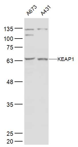 Sample:
A673(Human) Cell Lysate at 30 ug
A431(Human) Cell Lysate at 30 ug
Primary: Anti-KEAP1 (SL4900R) at 1/500 dilution
Secondary: IRDye800CW Goat Anti-Rabbit IgG at 1/20000 dilution
Predicted band size: 69 kD
Observed band size: 69 kD
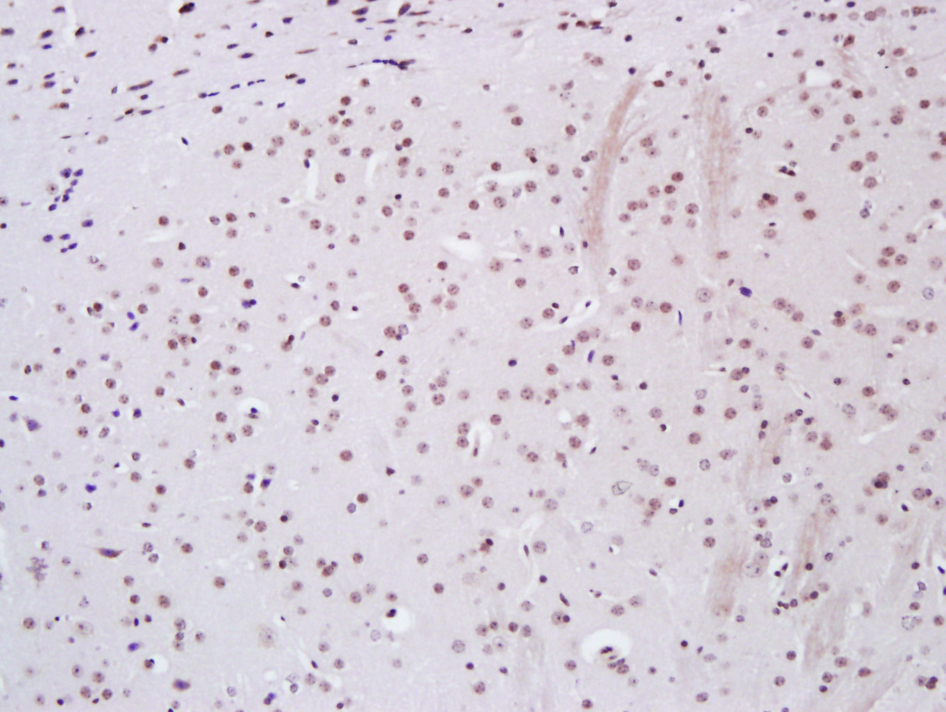 Paraformaldehyde-fixed, paraffin embedded (Mouse brain); Antigen retrieval by boiling in sodium citrate buffer (pH6.0) for 15min; Block endogenous peroxidase by 3% hydrogen peroxide for 20 minutes; Blocking buffer (normal goat serum) at 37°C for 30min; Antibody incubation with (KEAP1) Polyclonal Antibody, Unconjugated (SL4900R) at 1:400 overnight at 4°C, followed by a conjugated secondary antibody (sp-0023) for 20 minutes and DAB staining.
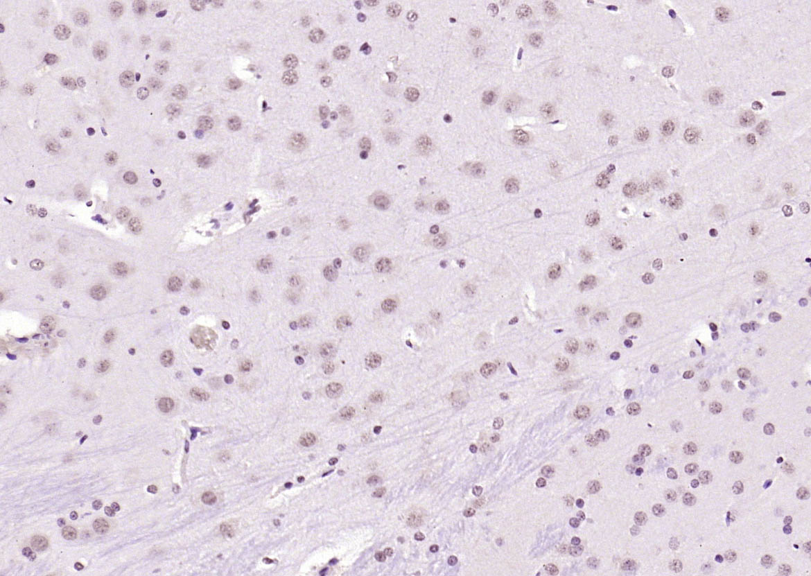 Paraformaldehyde-fixed, paraffin embedded (rat brain); Antigen retrieval by boiling in sodium citrate buffer (pH6.0) for 15min; Block endogenous peroxidase by 3% hydrogen peroxide for 20 minutes; Blocking buffer (normal goat serum) at 37°C for 30min; Antibody incubation with (KEAP1) Polyclonal Antibody, Unconjugated (SL4900R) at 1:200 overnight at 4°C, followed by operating according to SP Kit(Rabbit) (sp-0023) instructionsand DAB staining.
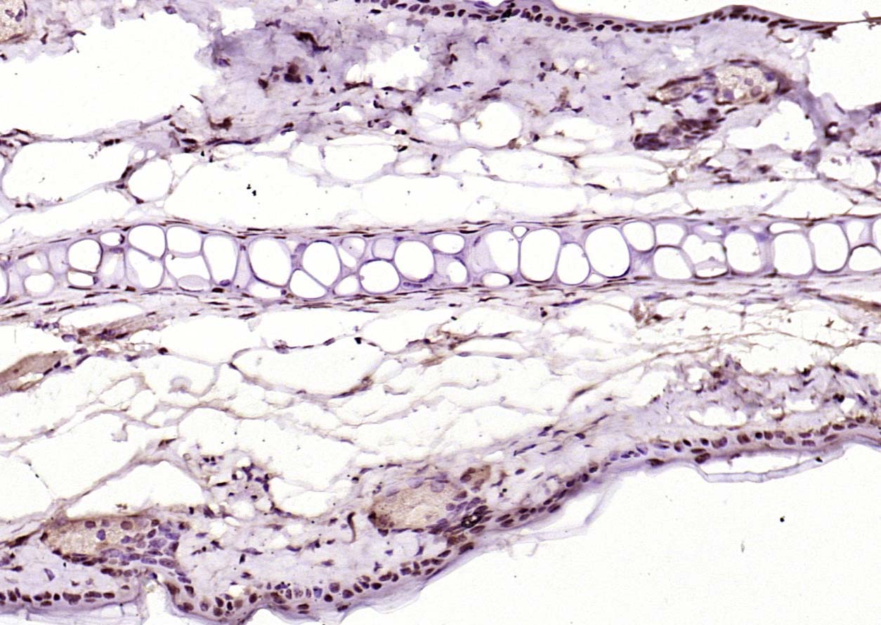 Paraformaldehyde-fixed, paraffin embedded (mouse skin); Antigen retrieval by boiling in sodium citrate buffer (pH6.0) for 15min; Block endogenous peroxidase by 3% hydrogen peroxide for 20 minutes; Blocking buffer (normal goat serum) at 37°C for 30min; Antibody incubation with (KEAP1) Polyclonal Antibody, Unconjugated (SL4900R) at 1:200 overnight at 4°C, followed by operating according to SP Kit(Rabbit) (sp-0023) instructionsand DAB staining.
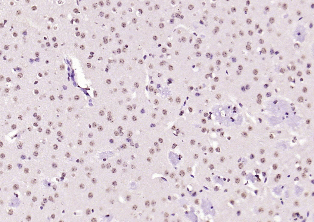 Paraformaldehyde-fixed, paraffin embedded (mouse brain); Antigen retrieval by boiling in sodium citrate buffer (pH6.0) for 15min; Block endogenous peroxidase by 3% hydrogen peroxide for 20 minutes; Blocking buffer (normal goat serum) at 37°C for 30min; Antibody incubation with (KEAP1) Polyclonal Antibody, Unconjugated (SL4900R) at 1:200 overnight at 4°C, followed by operating according to SP Kit(Rabbit) (sp-0023) instructionsand DAB staining.
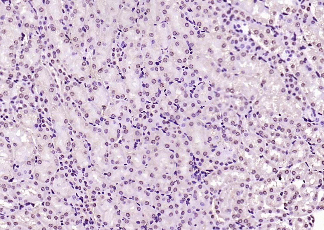 Paraformaldehyde-fixed, paraffin embedded (rat kidney); Antigen retrieval by boiling in sodium citrate buffer (pH6.0) for 15min; Block endogenous peroxidase by 3% hydrogen peroxide for 20 minutes; Blocking buffer (normal goat serum) at 37°C for 30min; Antibody incubation with (KEAP1) Polyclonal Antibody, Unconjugated (SL4900R) at 1:200 overnight at 4°C, followed by operating according to SP Kit(Rabbit) (sp-0023) instructionsand DAB staining.
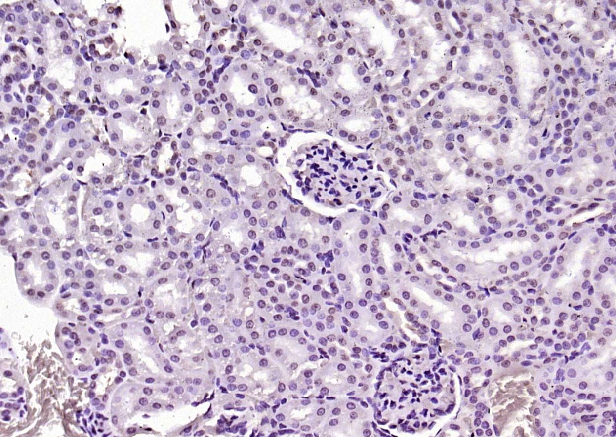 Paraformaldehyde-fixed, paraffin embedded (mouse kidney); Antigen retrieval by boiling in sodium citrate buffer (pH6.0) for 15min; Block endogenous peroxidase by 3% hydrogen peroxide for 20 minutes; Blocking buffer (normal goat serum) at 37°C for 30min; Antibody incubation with (KEAP1) Polyclonal Antibody, Unconjugated (SL4900R) at 1:200 overnight at 4°C, followed by operating according to SP Kit(Rabbit) (sp-0023) instructionsand DAB staining.
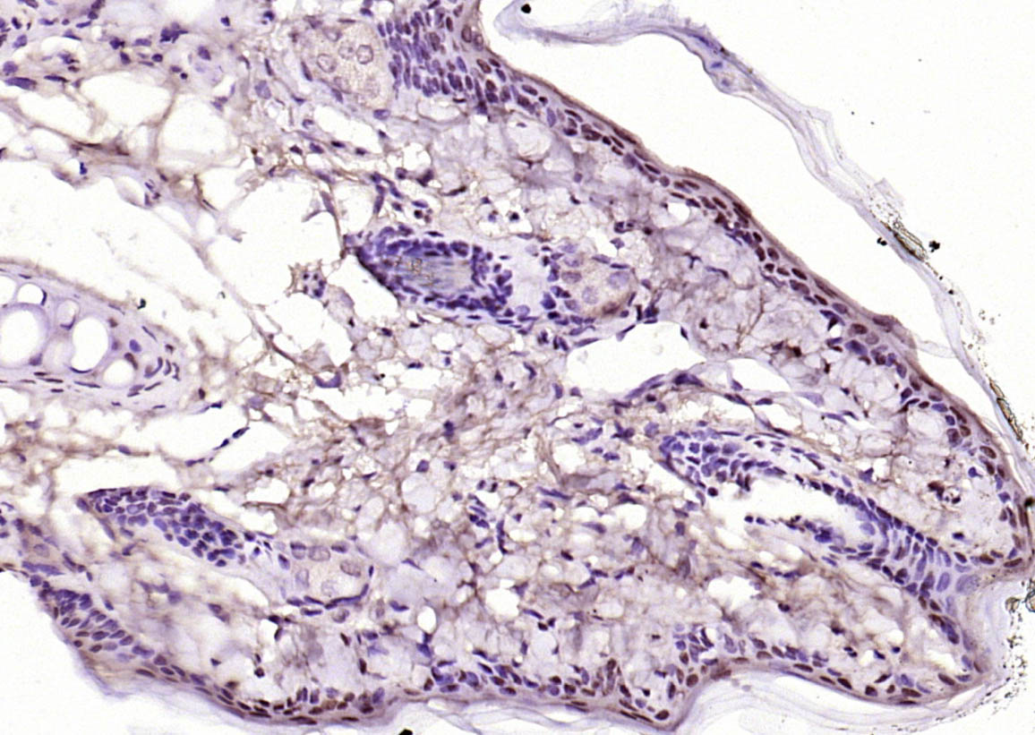 Paraformaldehyde-fixed, paraffin embedded (rat skin); Antigen retrieval by boiling in sodium citrate buffer (pH6.0) for 15min; Block endogenous peroxidase by 3% hydrogen peroxide for 20 minutes; Blocking buffer (normal goat serum) at 37°C for 30min; Antibody incubation with (KEAP1) Polyclonal Antibody, Unconjugated (SL4900R) at 1:200 overnight at 4°C, followed by operating according to SP Kit(Rabbit) (sp-0023) instructionsand DAB staining.
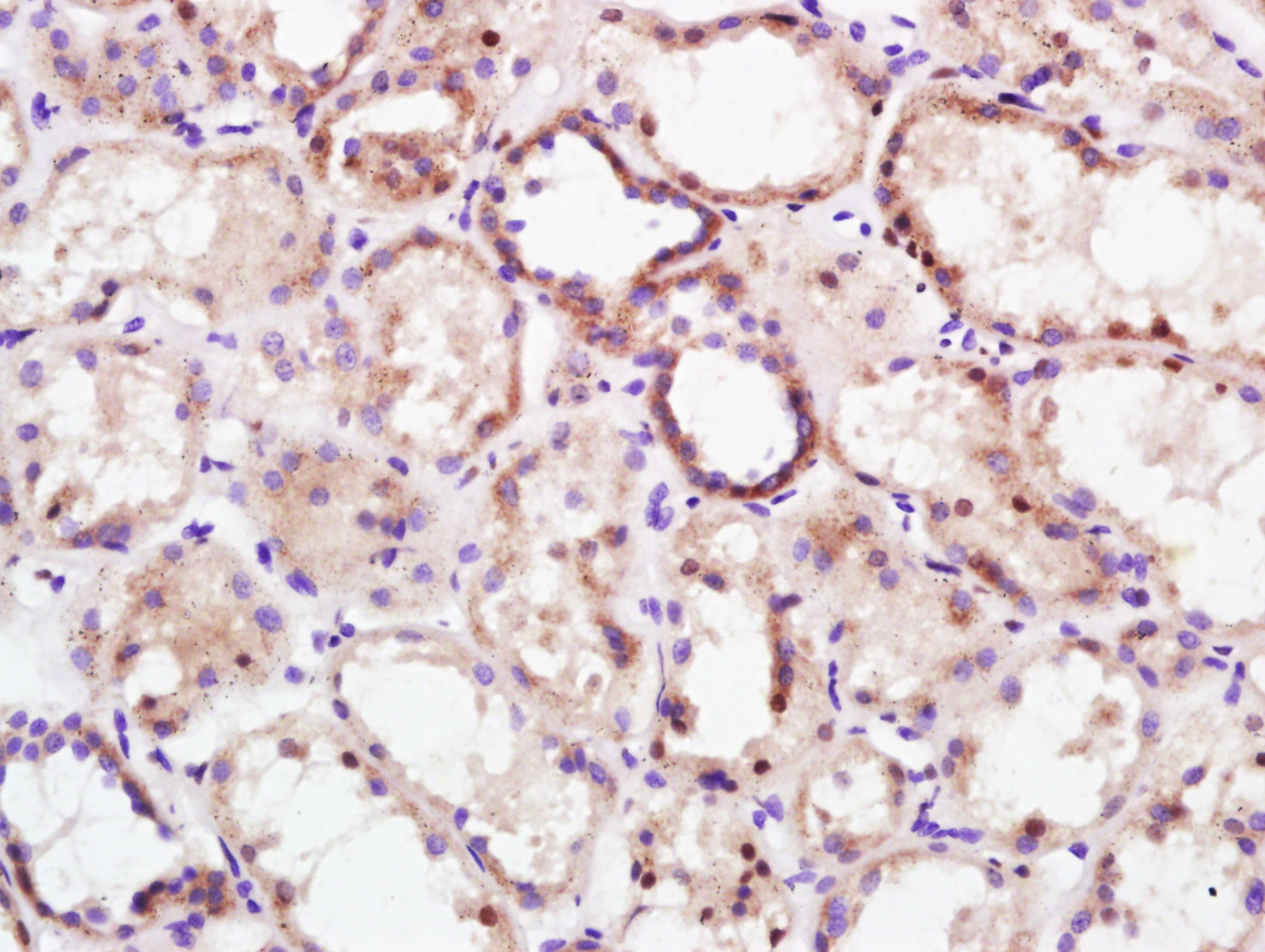 Paraformaldehyde-fixed, paraffin embedded (Human kidney); Antigen retrieval by boiling in sodium citrate buffer (pH6.0) for 15min; Block endogenous peroxidase by 3% hydrogen peroxide for 20 minutes; Blocking buffer (normal goat serum) at 37°C for 30min; Antibody incubation with (KEAP1) Polyclonal Antibody, Unconjugated (SL4900R) at 1:400 overnight at 4°C, followed by a conjugated secondary antibody (sp-0023) for 20 minutes and DAB staining.
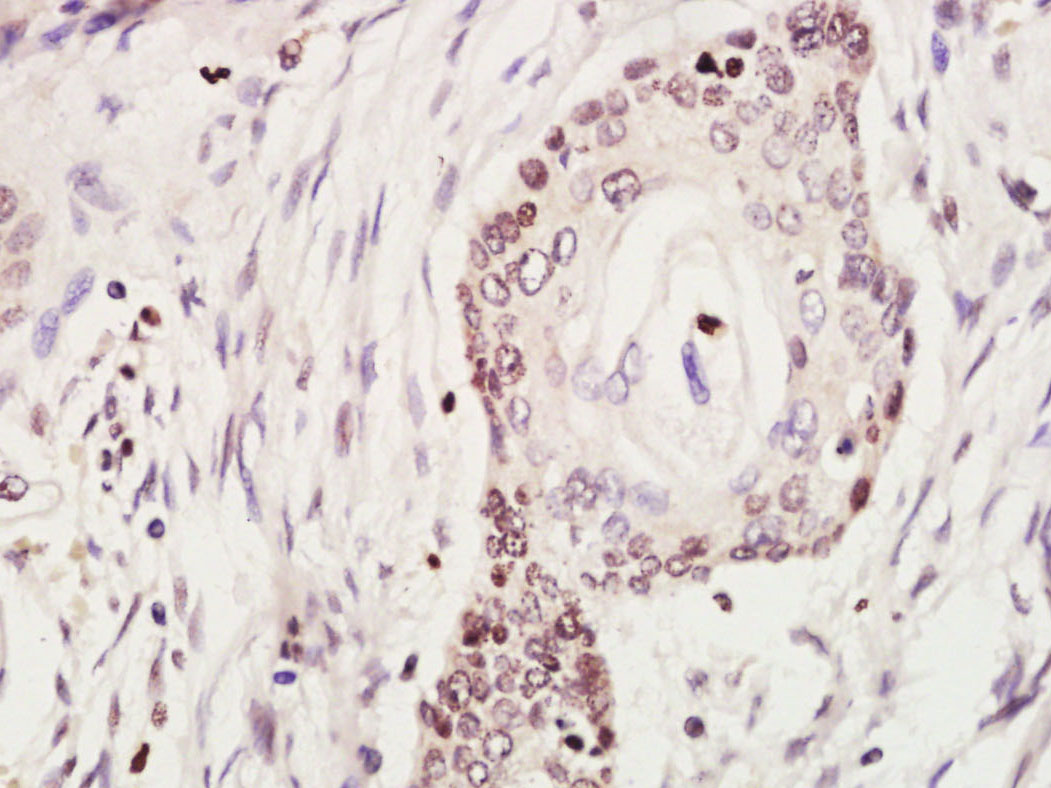 Paraformaldehyde-fixed, paraffin embedded (Human cervical carcinoma); Antigen retrieval by boiling in sodium citrate buffer (pH6.0) for 15min; Block endogenous peroxidase by 3% hydrogen peroxide for 20 minutes; Blocking buffer (normal goat serum) at 37°C for 30min; Antibody incubation with (KEAP1) Polyclonal Antibody, Unconjugated (SL4900R) at 1:500 overnight at 4°C, followed by a conjugated secondary (sp-0023) for 20 minutes and DAB staining.Paraformaldehyde-fixed, paraffin embedded (Human kidney); Antigen retrieval by boiling in sodium citrate buffer (pH6.0) for 15min; Block endogenous peroxidase by 3% hydrogen peroxide for 20 minutes; Blocking buffer (normal goat serum) at 37°C for 30min; Antibody incubation with (KEAP1) Polyclonal Antibody, Unconjugated (SL4900R) at 1:500 overnight at 4°C, followed by a conjugated secondary (sp-0023) for 20 minutes and DAB staining.Paraformaldehyde-fixed, paraffin embedded (Human colon carcinoma); Antigen retrieval by boiling in sodium citrate buffer (pH6.0) for 15min; Block endogenous peroxidase by 3% hydrogen peroxide for 20 minutes; Blocking buffer (normal goat serum) at 37°C for 30min; Antibody incubation with (KEAP1) Polyclonal Antibody, Unconjugated (SL4900R) at 1:500 overnight at 4°C, followed by a conjugated secondary (sp-0023) for 20 minutes and DAB staining.
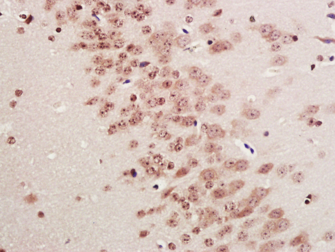 Paraformaldehyde-fixed, paraffin embedded (Mouse brain); Antigen retrieval by boiling in sodium citrate buffer (pH6.0) for 15min; Block endogenous peroxidase by 3% hydrogen peroxide for 20 minutes; Blocking buffer (normal goat serum) at 37°C for 30min; Antibody incubation with (KEAP1) Polyclonal Antibody, Unconjugated (SL4900R) at 1:500 overnight at 4°C, followed by a conjugated secondary (sp-0023) for 20 minutes and DAB staining.
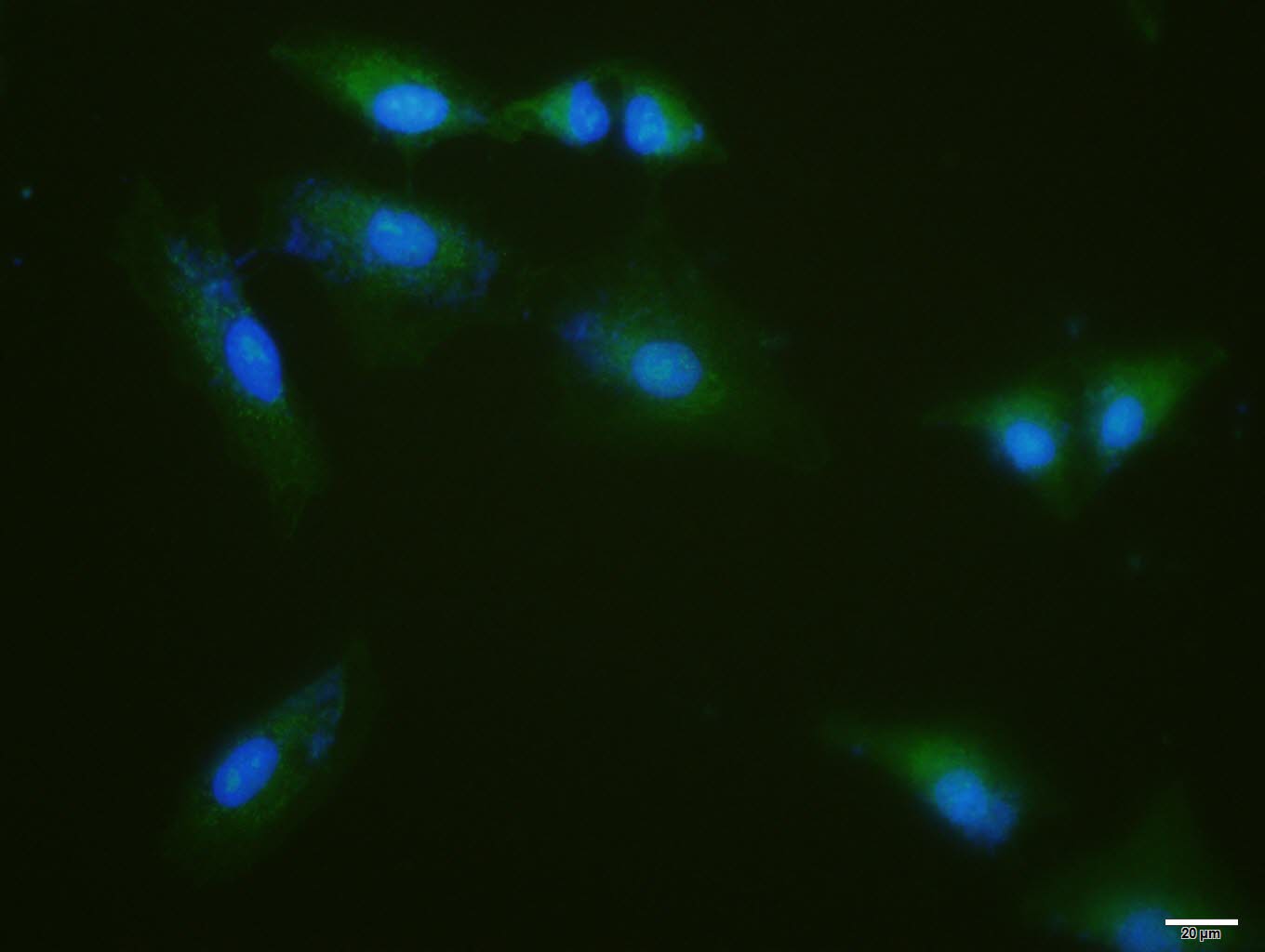 A-549cell; 4% Paraformaldehyde-fixed; Triton X-100 at room temperature for 20 min; Blocking buffer (normal goat serum, C-0005) at 37°C for 20 min; Antibody incubation with (KEAP1) polyclonal Antibody, Unconjugated (SL4900R) 1:100, 90 minutes at 37°C; followed by a conjugated Goat Anti-Rabbit IgG antibody at 37°C for 90 minutes, DAPI (blue, C02-04002) was used to stain the cell nuclei.
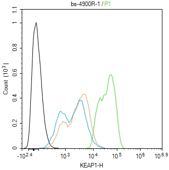 Blank control(black line):A549.
Primary Antibody (green line): Rabbit Anti-KEAP1 antibody (SL4900R)
Dilution:1ug/Test;
Secondary Antibody(white blue line): Goat anti-rabbit IgG-AF488
Dilution: 0.5ug/Test.
Isotype control(orange line): Normal Rabbit IgG
Protocol
The cells were fixed with 4% PFA (10min at room temperature)and then permeabilized with 90% ice-cold methanol for 20 min at -20℃, The cells were then incubated in 5%BSA to block non-specific protein-protein interactions for 30 min at room temperature .Cells stained with Primary Antibody for 30 min at room temperature. The secondary antibody used for 40 min at room temperature. Acquisition of 20,000 events was performed.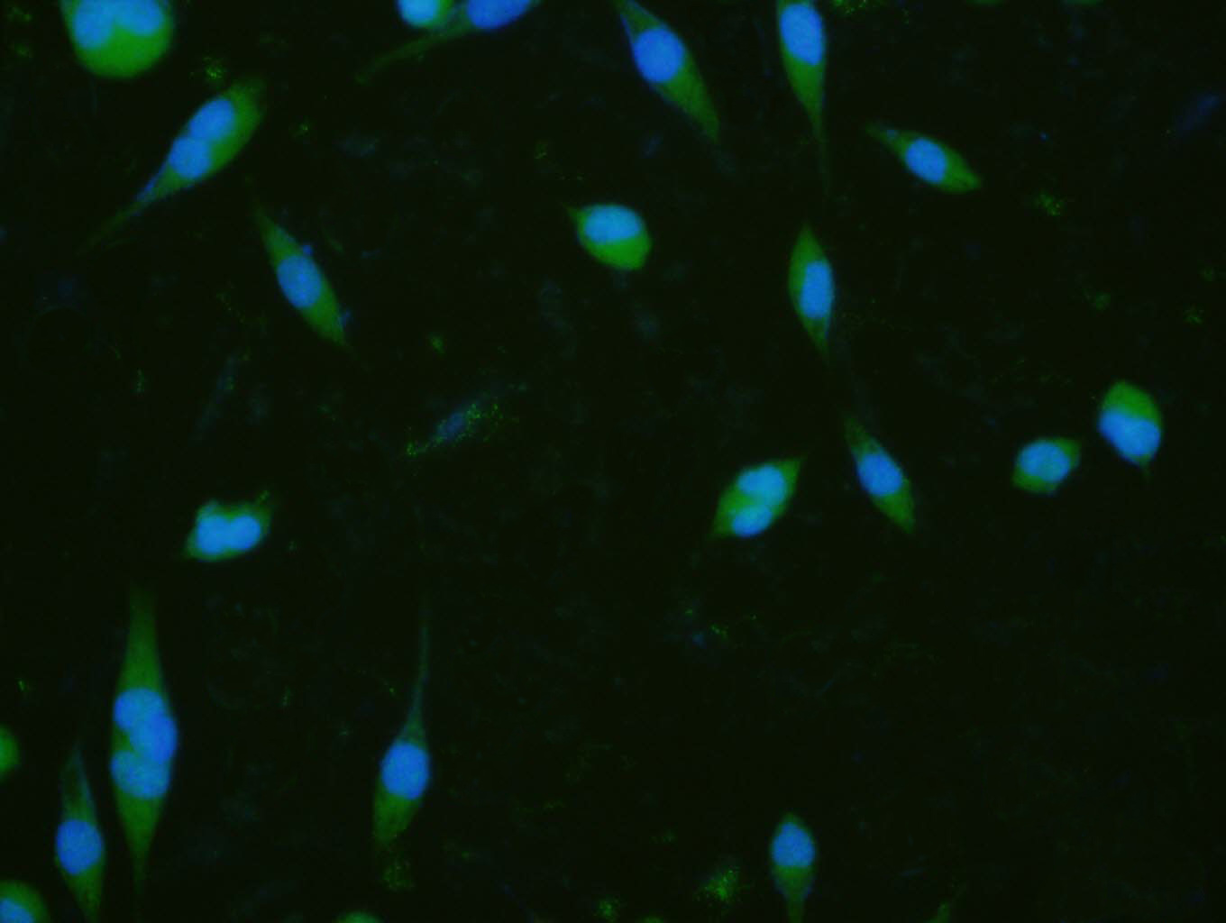 A-431 cell; 4% Paraformaldehyde-fixed; Triton X-100 at room temperature for 20 min; Blocking buffer (normal goat serum, C-0005) at 37°C for 20 min; Antibody incubation with (KEAP1) polyclonal Antibody, Unconjugated (SL4900R) 1:100, 90 minutes at 37°C; followed by a conjugated Goat Anti-Rabbit IgG antibody at 37°C for 90 minutes, DAPI (blue, C02-04002) was used to stain the cell nuclei.
Cartpieces
Totalgoods,subtotals:¥Checkout
References (0)
No References
Bought notes(bought amounts latest0)
No one bought this product
User Comment(Total0User Comment Num)
- No comment


 +86 571 56623320
 +86 18668110335

