
Rabbit Anti-LC3B antibody
Microtubule-associated protein 1 light chain 3 beta;ATG8F; Autophagy related protein LC3 B; Autophagy related ubiquitin like modifier LC3 B; Autophagy-related protein LC3 B; Autophagy-related ubiquitin-like modifier LC3 B; MAP1 light chain 3 like protein
View History [Clear]
Details
Product Name LC3B Chinese Name 自噬微管相关蛋白轻链β3抗体 Alias Microtubule-associated protein 1 light chain 3 beta; ATG8F; Autophagy related protein LC3 B; Autophagy related ubiquitin like modifier LC3 B; Autophagy-related protein LC3 B; Autophagy-related ubiquitin-like modifier LC3 B; MAP1 light chain 3 like protein 2; MAP1 light chain 3-like protein 2; MAP1A/1B light chain 3 B; MAP1A/1BLC3; MAP1A/MAP1B LC3 B; MAP1A/MAP1B light chain 3 B; MAP1ALC3; MAP1LC3B; Microtubule associated protein 1 light chain 3 beta; Microtubule associated proteins 1A/1B light chain 3B; Microtubule-associated protein 1 light chain 3 beta; Microtubule-associated proteins 1A/1B light chain 3B; MLP3B_HUMAN. MAP LC3β. literatures Research Area Tumour Cell biology Neurobiology Signal transduction Immunogen Species Rabbit Clonality Polyclonal React Species Human, Mouse, Rat, (predicted: Chicken, Cow, Danio rerio) Applications WB=1:200-1000 IHC-P=1:100-500 IF=1:100-500 (Paraffin sections need antigen repair)
not yet tested in other applications.
optimal dilutions/concentrations should be determined by the end user.Theoretical molecular weight 14kDa Cellular localization cytoplasmic The cell membrane Form Liquid Concentration 1mg/ml immunogen KLH conjugated synthetic peptide derived from mouse LC3B: 1-50/125 Lsotype IgG Purification affinity purified by Protein A Buffer Solution 0.01M TBS(pH7.4) with 1% BSA, 0.03% Proclin300 and 50% Glycerol. Storage Shipped at 4℃. Store at -20 °C for one year. Avoid repeated freeze/thaw cycles. Attention This product as supplied is intended for research use only, not for use in human, therapeutic or diagnostic applications. PubMed PubMed Product Detail Microtubule-associated proteins (MAPs) regulate microtubule stability and play critical roles in neuronal development and in maintaining the balance between neuronal plasticity and rigidity. MAP-light chain 3 beta (MAP-LC3 Beta) and MAP-light chain 3 alpha (MAP-LC3 Alpha) are subunits of both MAP1A and MAP1B. MAP-LC3 Beta, a homolog of Apg8p, is essential for autophagy and associated to the autophagosome membranes after processing. Two forms of LC3 Beta, the cytosolic LC3-I and the membrane-bound LC3-II, are produced post-translationally. LC3-I is formed by the removal of the C-terminal 22 amino acids from newly synthesized LC3 Beta, followed by the conversion of a fraction of LC3-I into LC3-II. LC3 enhances fibronectin mRNA translation in ductus arteriosus cells through association with 60S ribosomes and binding to an AU-rich element in the 3’ untranslated region of fibronectin mRNA. This facilitates sorting of fibronectin mRNA onto rough endoplasmic reticulum and translation. MAP LC3 Beta may also be involved in formation of autophagosomal vacuoles. It is expressed primarily in heart, testis, brain and skeletal muscle.
Function:
Probably involved in formation of autophagosomal vacuoles
Subunit:
3 different light chains, LC1, LC2 and LC3, can associate with MAP1A and MAP1B proteins. Interacts at microtubules with CABP1 (via EF-hands 1 and 2) but not with calmodulin. Interacts with FYCO1 (via C-terminus). Interacts with TP53INP1 and TP53INP2. Interacts with TBC1D25. Directly interacts with SQSTM1; this interaction leads to MAP1LC3B recruitment to inclusion bodies containing polyubiquitinated protein aggregates and to inclusion body degradation by autophagy.
Subcellular Location:
Cytoplasm, cytoskeleton. Endomembrane system; Lipid-anchor. Cytoplasmic vesicle, autophagosome membrane; Lipid-anchor. Note=LC3-II binds to the autophagic membranes.
Tissue Specificity:
Most abundant in heart, brain, skeletal muscle and testis. Little expression observed in liver.
Post-translational modifications:
The precursor molecule is cleaved by APG4B/ATG4B to form LC3-I. This is activated by APG7L/ATG7, transferred to ATG3 and conjugated to phospholipid to form LC3-II.
Similarity:
Belongs to the MAP1 LC3 family.
SWISS:
Q9GZQ8
Gene ID:
81631
Database links:Entrez Gene: 81631 Human
Entrez Gene: 427559 Chicken
Entrez Gene: 67443 Mouse
Omim: 609604 Human
SwissProt: Q9GZQ8 Human
SwissProt: Q9CQV6 Mouse
Unigene: 356061 Human
Unigene: 28357 Mouse
Unigene: 41412 Rat
Product Picture  Sample:
Lane 1: Mouse Cerebrum tissue lysates
Lane 2: Mouse Cerebellum tissue lysates
Lane 3: Mouse Testis tissue lysates
Lane 4: Rat Cerebrum tissue lysates
Lane 5: Rat Cerebellum tissue lysates
Lane 6: Rat Testis tissue lysates
Primary: Anti-LC3B (SL4843R) at 1/1000 dilution
Secondary: IRDye800CW Goat Anti-Rabbit IgG at 1/20000 dilution
Predicted band size: 14 kDa
Observed band size: 15,17 kDa
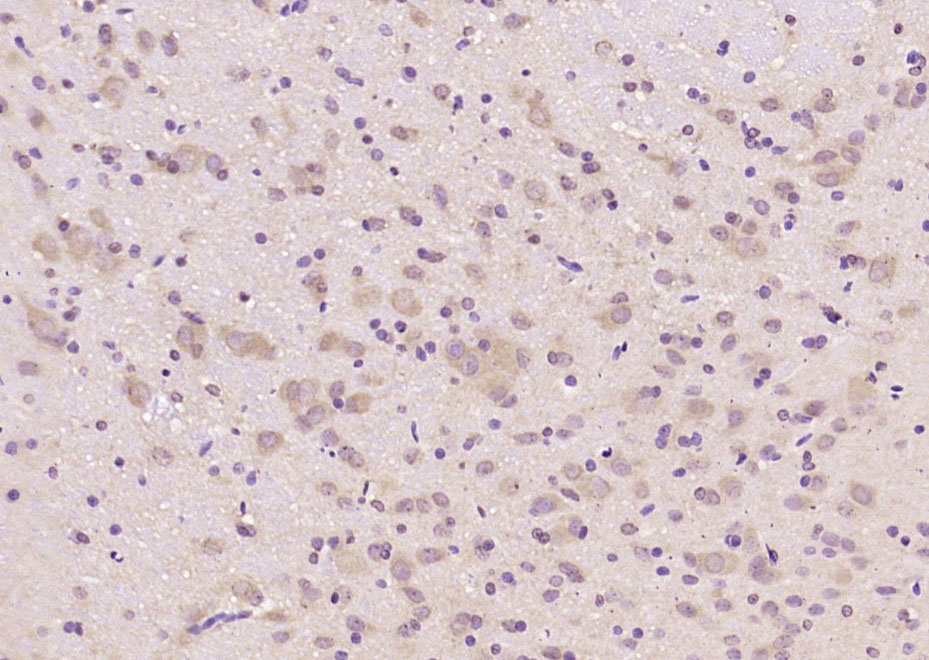 Paraformaldehyde-fixed, paraffin embedded (rat brain); Antigen retrieval by boiling in sodium citrate buffer (pH6.0) for 15min; Block endogenous peroxidase by 3% hydrogen peroxide for 20 minutes; Blocking buffer (normal goat serum) at 37°C for 30min; Antibody incubation with (LC3B) Polyclonal Antibody, Unconjugated (SL4843R) at 1:4000 overnight at 4°C, followed by operating according to SP Kit(Rabbit) (sp-0023) instructionsand DAB staining.
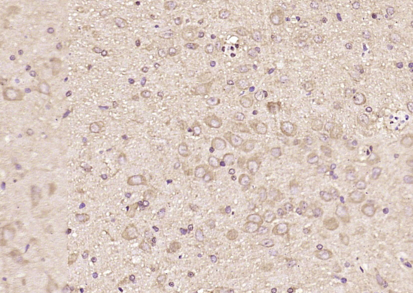 Paraformaldehyde-fixed, paraffin embedded (rat brain); Antigen retrieval by boiling in sodium citrate buffer (pH6.0) for 15min; Block endogenous peroxidase by 3% hydrogen peroxide for 20 minutes; Blocking buffer (normal goat serum) at 37°C for 30min; Antibody incubation with (LC3B) Polyclonal Antibody, Unconjugated (SL4843R) at 1:1000 overnight at 4°C, followed by operating according to SP Kit(Rabbit) (sp-0023) instructionsand DAB staining.
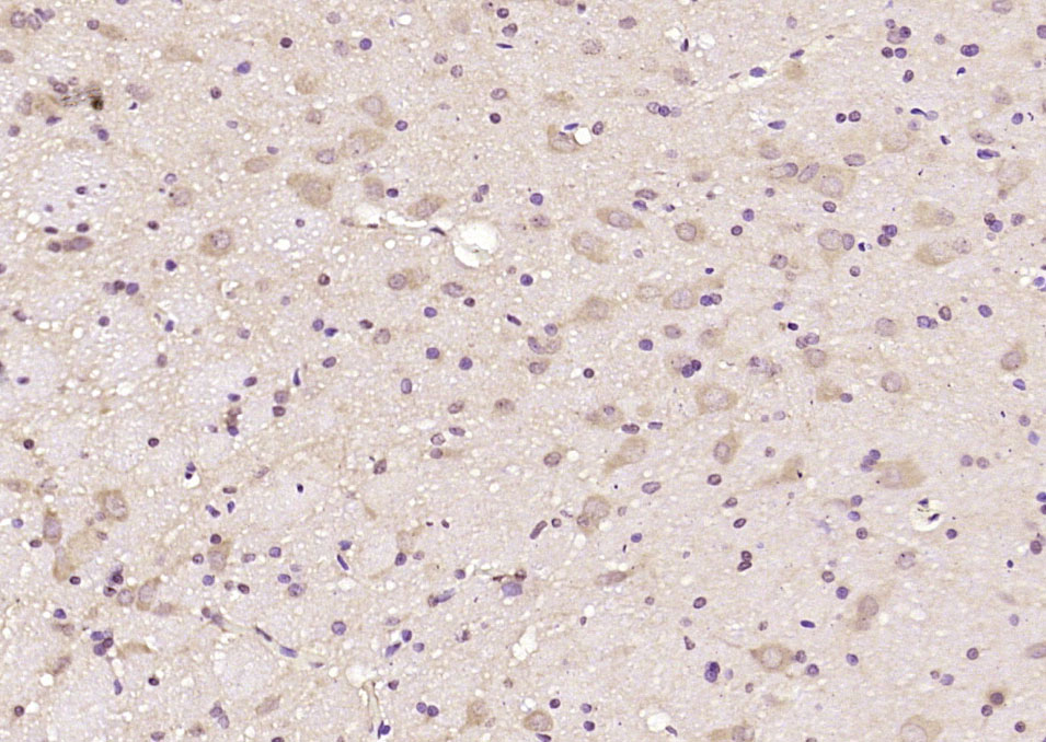 Paraformaldehyde-fixed, paraffin embedded (rat brain); Antigen retrieval by boiling in sodium citrate buffer (pH6.0) for 15min; Block endogenous peroxidase by 3% hydrogen peroxide for 20 minutes; Blocking buffer (normal goat serum) at 37°C for 30min; Antibody incubation with (LC3B) Polyclonal Antibody, Unconjugated (SL4843R) at 1:2000 overnight at 4°C, followed by operating according to SP Kit(Rabbit) (sp-0023) instructionsand DAB staining.
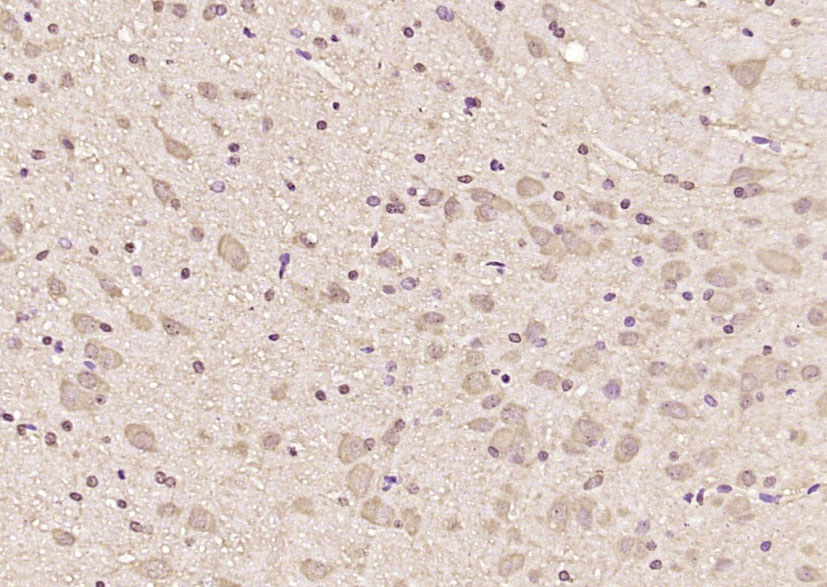 Paraformaldehyde-fixed, paraffin embedded (rat brain); Antigen retrieval by boiling in sodium citrate buffer (pH6.0) for 15min; Block endogenous peroxidase by 3% hydrogen peroxide for 20 minutes; Blocking buffer (normal goat serum) at 37°C for 30min; Antibody incubation with (LC3B) Polyclonal Antibody, Unconjugated (SL4843R) at 1:2000 overnight at 4°C, followed by operating according to SP Kit(Rabbit) (sp-0023) instructionsand DAB staining.
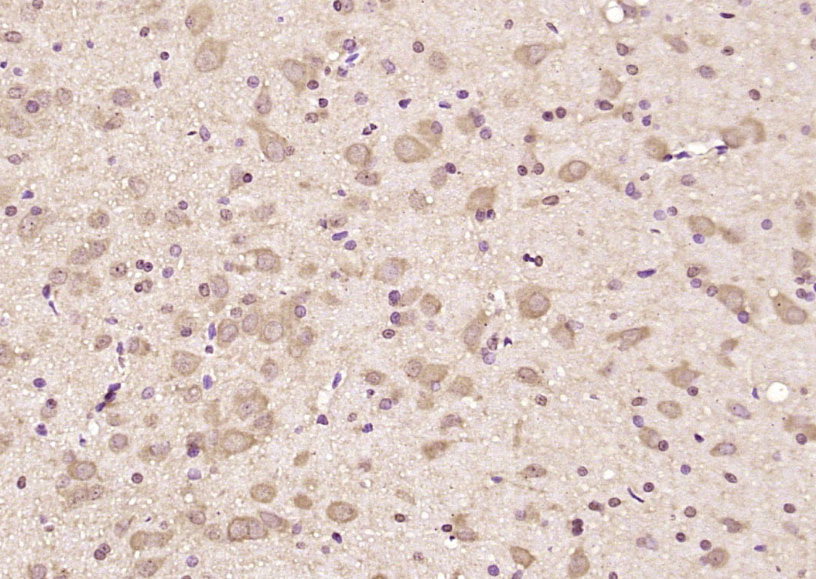 Paraformaldehyde-fixed, paraffin embedded (rat brain); Antigen retrieval by boiling in sodium citrate buffer (pH6.0) for 15min; Block endogenous peroxidase by 3% hydrogen peroxide for 20 minutes; Blocking buffer (normal goat serum) at 37°C for 30min; Antibody incubation with (LC3B) Polyclonal Antibody, Unconjugated (SL4843R) at 1:1000 overnight at 4°C, followed by operating according to SP Kit(Rabbit) (sp-0023) instructionsand DAB staining.
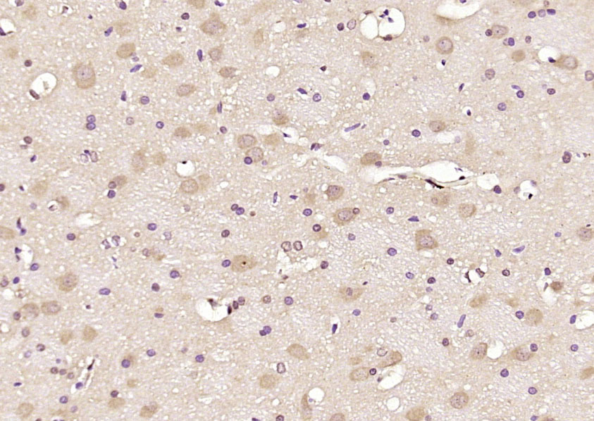 Paraformaldehyde-fixed, paraffin embedded (rat brain); Antigen retrieval by boiling in sodium citrate buffer (pH6.0) for 15min; Block endogenous peroxidase by 3% hydrogen peroxide for 20 minutes; Blocking buffer (normal goat serum) at 37°C for 30min; Antibody incubation with (LC3B) Polyclonal Antibody, Unconjugated (SL4843R) at 1:200 overnight at 4°C, followed by operating according to SP Kit(Rabbit) (sp-0023) instructionsand DAB staining.
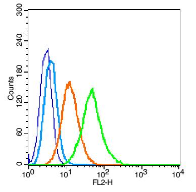 Blank control(blue): TM4 cells(fixed with 2% paraformaldehyde (10 min) , then permeabilized with 90% ice-cold methanol for 30 min on ice).
Primary Antibody:Rabbit Anti-LC3B antibody(SL4843R), Dilution: 1μg in 100 μL 1X PBS containing 0.5% BSA;
Isotype Control Antibody: Rabbit IgG(orange) ,used under the same conditions );
Secondary Antibody: Goat anti-rabbit IgG-PE(white blue), Dilution: 1:200 in 1 X PBS containing 0.5% BSA.
Cartpieces
Totalgoods,subtotals:¥Checkout
References (0)
No References
Bought notes(bought amounts latest0)
No one bought this product
User Comment(Total0User Comment Num)
- No comment


 +86 571 56623320
 +86 18668110335

