
Rabbit Anti-MLCK antibody
myosin light chain kinase; DKFZp686I10125; EC 2.7.11.18; FLJ12216; Kinase related protein; KRP; MLCK; MLCK1; MLCK108; MLCK210; MSTP083; MYLK1; Myosin light chain kinase smooth muscle and non muscle isozymes; Myosin light polypeptide kinase; OTTHUMP0000018
View History [Clear]
Details
Product Name MLCK Chinese Name 肌球蛋白轻链激酶抗体 Alias myosin light chain kinase; DKFZp686I10125; EC 2.7.11.18; FLJ12216; Kinase related protein; KRP; MLCK; MLCK1; MLCK108; MLCK210; MSTP083; MYLK1; Myosin light chain kinase smooth muscle and non muscle isozymes; Myosin light polypeptide kinase; OTTHUMP00000180642; OTTHUMP00000180643; smMLCK; Smooth muscle myosin light chain kinase; Telokin; deglutamylated form; MLCK; MYLK_HUMAN. literatures Research Area Cell biology immunology Signal transduction Kinases and Phosphatases Immunogen Species Rabbit Clonality Polyclonal React Species Human, Rat, (predicted: Mouse, Chicken, Dog, Pig, Cow, Horse, Sheep, Guinea Pig, ) Applications ELISA=1:5000-10000 IHC-P=1:100-500 IHC-F=1:100-500 Flow-Cyt=1ug/Test IF=1:100-500 (Paraffin sections need antigen repair)
not yet tested in other applications.
optimal dilutions/concentrations should be determined by the end user.Theoretical molecular weight 210kDa Cellular localization cytoplasmic Form Liquid Concentration 1mg/ml immunogen KLH conjugated synthetic peptide derived from human MLCK: 651-750/1914 Lsotype IgG Purification affinity purified by Protein A Buffer Solution 0.01M TBS(pH7.4) with 1% BSA, 0.03% Proclin300 and 50% Glycerol. Storage Shipped at 4℃. Store at -20 °C for one year. Avoid repeated freeze/thaw cycles. Attention This product as supplied is intended for research use only, not for use in human, therapeutic or diagnostic applications. PubMed PubMed Product Detail This gene, a muscle member of the immunoglobulin gene superfamily, encodes myosin light chain kinase which is a calcium/calmodulin dependent enzyme. This kinase phosphorylates myosin regulatory light chains to facilitate myosin interaction with actin filaments to produce contractile activity. This gene encodes both smooth muscle and nonmuscle isoforms. In addition, using a separate promoter in an intron in the 3' region, it encodes telokin, a small protein identical in sequence to the C-terminus of myosin light chain kinase, that is independently expressed in smooth muscle and functions to stabilize unphosphorylated myosin filaments. A pseudogene is located on the p arm of chromosome 3. Four transcript variants that produce four isoforms of the calcium/calmodulin dependent enzyme have been identified as well as two transcripts that produce two isoforms of telokin. Additional variants have been identified but lack full length transcripts. [provided by RefSeq].
Function:
Calcium/calmodulin-dependent enzyme implicated in smooth muscle contraction via phosphorylation of myosin light chains (MLC). Also regulates actin-myosin interaction through a non-kinase activty (By similarity). Implicated in the regulation of endothelial as well as vascular permeability. In the nervous system it has been shown to control the growth initiation of astrocytic processes in culture and to participate in transmitter release at synapses formed between cultured sympathetic ganglion cells. Critical participant in signaling sequences that result in fibroblast apoptosis.
Tissue Specificity:
Smooth muscle and non-muscle isozymes are expressed in a wide variety of adult and fetal tissues and in cultured endothelium with qualitative expression appearing to be neither tissue- nor development-specific. Non-muscle isoform 2 is the dominant splice variant expressed in various tissues. Telokin has been found in a wide variety of adult and fetal tissues.
Post-translational modifications:
MLCK is probably down-regulated by phosphorylation.
The C-terminus is deglutamylated by AGTPBP1/ CCP1, AGBL1/CCP4 and AGBL4/CCP6, leading to the formation of Myosin light chain kinase, smooth muscle, deglutamylated form. The consequences of C-terminal deglutamylation are unknown.
Similarity:
Belongs to the protein kinase superfamily. CAMK Ser/Thr protein kinase family.
Contains 1 fibronectin type-III domain.
Contains 9 Ig-like C2-type (immunoglobulin-like) domains.
Contains 1 protein kinase domain.
SWISS:
Q15746
Gene ID:
4638
Database links:Entrez Gene: 4638 Human
Entrez Gene: 107589 Mouse
Omim: 600922 Human
SwissProt: Q15746 Human
SwissProt: Q6PDN3 Mouse
Unigene: 477375 Human
Unigene: 33360 Mouse
Unigene: 203004 Rat
肌球蛋白轻链激酶(MLCK)表达量增多和活性升高是血管平滑肌收缩的启动因素之一。Product Picture 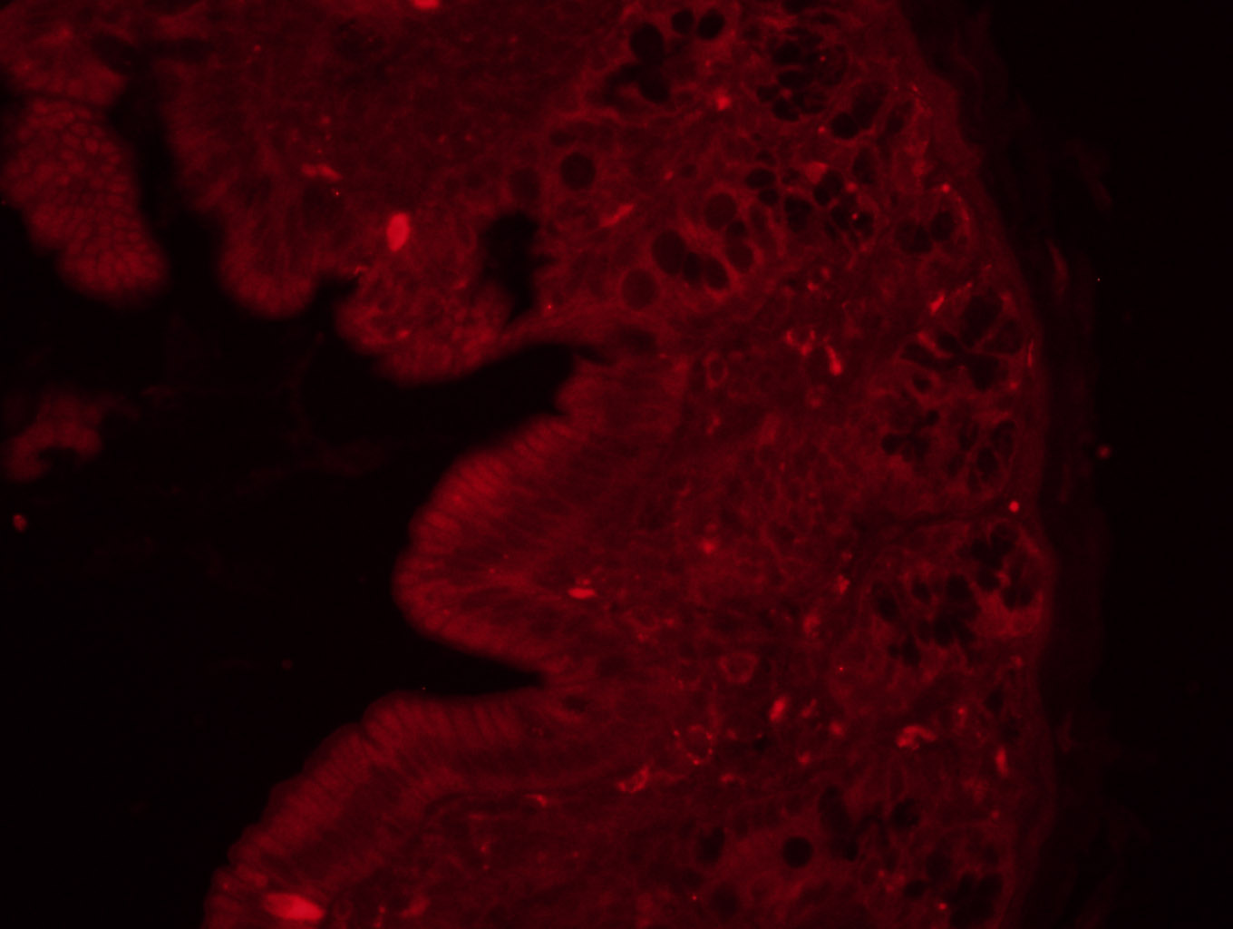 Tissue/cell: rat colon tissue;4% Paraformaldehyde-fixed and paraffin-embedded;
Antigen retrieval: citrate buffer ( 0.01M, pH 6.0 ), Boiling bathing for 15min; Blocking buffer (normal goat serum,C-0005) at 37℃ for 20 min;
Incubation: Anti-MLCK Polyclonal Antibody, Unconjugated(SL4705R) 1:200, overnight at 4°C; The secondary antibody was Goat Anti-Rabbit IgG, Cy3 conjugated(SL0295G-Cy3)used at 1:200 dilution for 40 minutes at 37°C. DAPI(5ug/ml,blue,C-0033) was used to stain the cell nuclei
 Blank control(black line):HepG2.
Primary Antibody (green line): Rabbit Anti-MLCK antibody (SL4705R)
Dilution:1ug/Test;
Secondary Antibody(white blue line): Goat anti-rabbit IgG-AF488
Dilution: 0.5ug/Test.
Isotype control(orange line): Normal Rabbit IgG
Protocol
The cells were fixed with 4% PFA (10min at room temperature)and then permeabilized with 90% ice-cold methanol for 20 min at -20℃, The cells were then incubated in 5%BSA to block non-specific protein-protein interactions for 30 min at room temperature .Cells stained with Primary Antibody for 30 min at room temperature. The secondary antibody used for 40 min at room temperature. Acquisition of 20,000 events was performed.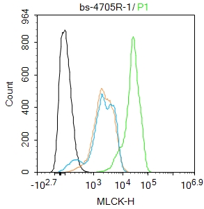 Blank control(black line):HepG2.
Primary Antibody (green line): Rabbit Anti-MLCK antibody (SL4705R)
Dilution:1ug/Test;
Secondary Antibody(white blue line): Goat anti-rabbit IgG-AF488
Dilution: 0.5ug/Test.
Isotype control(orange line): Normal Rabbit IgG
Protocol
The cells were fixed with 4% PFA (10min at room temperature)and then permeabilized with 90% ice-cold methanol for 20 min at -20℃, The cells were then incubated in 5%BSA to block non-specific protein-protein interactions for 30 min at room temperature .Cells stained with Primary Antibody for 30 min at room temperature. The secondary antibody used for 40 min at room temperature. Acquisition of 20,000 events was performed.
Cartpieces
Totalgoods,subtotals:¥Checkout
References (0)
No References
Bought notes(bought amounts latest0)
No one bought this product
User Comment(Total0User Comment Num)
- No comment


 +86 571 56623320
 +86 18668110335

