
Rabbit Anti-MAP1LC3A antibody
Microtubule-associated proteins 1B light chain 3A; Microtubule-associated proteins 1Beta light chain 3A; MAP1B LC3 A; MAP LC3 Beta; MAP-LC3 Beta; MAP1 light chain 3-like protein 1; MAP1LC3A; MLP3A_HUMAN; Microtubule-associated proteins 1A/1B light chain 3
View History [Clear]
Details
Product Name MAP1LC3A Chinese Name 自噬微管相关蛋白轻链β3抗体 Alias Microtubule-associated proteins 1B light chain 3A; Microtubule-associated proteins 1Beta light chain 3A; MAP1B LC3 A; MAP LC3 Beta; MAP-LC3 Beta; MAP1 light chain 3-like protein 1; MAP1LC3A; MLP3A_HUMAN; Microtubule-associated proteins 1A/1B light chain 3A; Autophagy-related protein LC3 A; Autophagy-related ubiquitin-like modifier LC3 A; MAP1 light chain 3-like protein 1; MAP1A/MAP1B light chain 3 A; MAP1A/MAP1B LC3 A; Microtubule-associated protein 1 light chain 3 alpha. Research Area Tumour Cell biology Neurobiology Signal transduction Autophagy Immunogen Species Rabbit Clonality Polyclonal React Species Human, (predicted: Mouse, Rat, Chicken, Dog, Pig, Cow, Horse, ) Applications ELISA=1:5000-10000 Flow-Cyt=1μg/Test
not yet tested in other applications.
optimal dilutions/concentrations should be determined by the end user.Theoretical molecular weight 14kDa Cellular localization cytoplasmic The cell membrane Form Liquid Concentration 1mg/ml immunogen KLH conjugated synthetic peptide derived from human Microtubule-associated proteins 1A/1B light chain 3A: 25-121/121 Lsotype IgG Purification affinity purified by Protein A Buffer Solution 0.01M TBS(pH7.4) with 1% BSA, 0.03% Proclin300 and 50% Glycerol. Storage Shipped at 4℃. Store at -20 °C for one year. Avoid repeated freeze/thaw cycles. Attention This product as supplied is intended for research use only, not for use in human, therapeutic or diagnostic applications. PubMed PubMed Product Detail Microtubule-associated proteins (MAPs) regulate microtubule stability and play critical roles in neuronal development and in maintaining the balance between neuronal plasticity and rigidity. MAP-light chain 3 beta (MAP-LC3 Beta) and MAP-light chain 3 alpha (MAP-LC3 alpha) are subunits of both MAP1A and MAP1B. MAP-LC3M Beta, a homolog of Apg8p, is essential for autophagy and associated to the autophagosome membranes after processing. Two forms of LC3 Beta, the cytosolic LC3-I and the membrane-bound LC3-II, are produced post-translationally. LC3-I is formed by the removal of the C-terminal 22 amino acids from newly synthesized LC3∫, followed by the conversion of a fraction of LC3-I into LC3-II. LC3 enhances fibronectin mRNA translation in ductus arteriosus cells through association with 60S ribosomes and binding to an AU-rich element in the 3’ untranslated region of fibronectin mRNA. This facilitates sorting of fibronectin mRNA onto rough endoplasmic reticulum and translation. MAP LC3 Beta may also be involved in formation of autophagosomal vacuoles. It is expressed primarily in heart, testis, brain and skeletal muscle.
Function:
Cytoplasm, cytoskeleton. Endomembrane system; Lipid-anchor. Cytoplasmic vesicle, autophagosome membrane; Lipid-anchor. Cytoplasmic vesicle, autophagosome. Note=LC3-II binds to the autophagic membranes.
Subunit:
3 different light chains, LC1, LC2 and LC3, can associate with MAP1A and MAP1B proteins. Interacts with SQSTM1. Interacts with TP53INP1 and TP53INP2.
Subcellular Location:
Cytoplasm, cytoskeleton. Endomembrane system; Lipid-anchor. Cytoplasmic vesicle,
Tissue Specificity:
Most abundant in heart, brain, liver, skeletal muscle and testis but absent in thymus and peripheral blood leukocytes.
Post-translational modifications:
The precursor molecule is cleaved by APG4B/ATG4B to form the cytosolic form, LC3-I. This is activated by APG7L/ATG7, transferred to ATG3 and conjugated to phospholipid to form the membrane-bound form, LC3-II.
Similarity:
Belongs to the MAP1 LC3 family.
SWISS:
Q9H492
Gene ID:
84557
Database links:Entrez Gene: 84557 Human
Entrez Gene: 66734 Mouse
Omim: 601242 Human
SwissProt: Q9H492 Human
SwissProt: Q91VR7 Mouse
Unigene: 632273 Human
Unigene: 196239 Mouse
Unigene: 3135 Rat
Product Picture 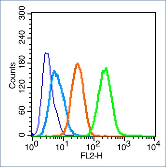 Blank control (blue line): Hela (fixed with 70% ethanol (Overmight at 4℃) and then permeabilized with 90% ice-cold methanol for 30 min at -20℃).
Primary Antibody (green line): Rabbit Anti-MAP1LC3A antibody (SL4309R),Dilution: 1μg /10^6 cells;
Isotype Control Antibody (orange line): Rabbit IgG .
Secondary Antibody (white blue line): Goat anti-rabbit IgG-PE,Dilution: 1μg /test.
Cartpieces
Totalgoods,subtotals:¥Checkout
Bought notes(bought amounts latest0)
No one bought this product
User Comment(Total0User Comment Num)
- No comment


 +86 571 56623320
 +86 18668110335

