
Rabbit Anti-Vesicle docking protein p115 antibody
P115-RhoGEF General vesicular transport factor; General vesicular transport factor p115; P115; TAP; Transcytosis associated protein; VDP; Vesicle docking protein; USO1_HUMAN.
View History [Clear]
Details
Product Name Vesicle docking protein p115 Chinese Name 囊泡对接蛋白p115抗体 Alias P115-RhoGEF General vesicular transport factor; General vesicular transport factor p115; P115; TAP; Transcytosis associated protein; VDP; Vesicle docking protein; USO1_HUMAN. Research Area immunology Signal transduction Binding protein G protein-coupled receptor Immunogen Species Rabbit Clonality Polyclonal React Species Human, Mouse, (predicted: Rat, Chicken, Pig, Cow, Horse, Sheep, ) Applications WB=1:500-2000 ELISA=1:5000-10000 IHC-P=1:100-500 IHC-F=1:100-500 IF=1:100-500 (Paraffin sections need antigen repair)
not yet tested in other applications.
optimal dilutions/concentrations should be determined by the end user.Theoretical molecular weight 108kDa Cellular localization cytoplasmic The cell membrane Form Liquid Concentration 1mg/ml immunogen KLH conjugated synthetic peptide derived from human Vesicle docking protein p115: 501-600/962 Lsotype IgG Purification affinity purified by Protein A Buffer Solution 0.01M TBS(pH7.4) with 1% BSA, 0.03% Proclin300 and 50% Glycerol. Storage Shipped at 4℃. Store at -20 °C for one year. Avoid repeated freeze/thaw cycles. Attention This product as supplied is intended for research use only, not for use in human, therapeutic or diagnostic applications. PubMed PubMed Product Detail p115 (Vesicle docking protein p115) is a peripheral membrane protein that is located on the Golgi complex. p115 exists as a homodimer with two globular heads, an extended coiled-coil tail, followed by an acidic domain at the extreme C terminus. p115 is homologous to a yeast protein, Uso1p, which is required for ER to Golgi transport. p115 likely plays an important role in vesicle transportation from the ER to the cis-Golgi comparments.
Function:
General vesicular transport factor required for intercisternal transport in the Golgi stack; it is required for transcytotic fusion and/or subsequent binding of the vesicles to the target membrane. May well act as a vesicular anchor by interacting with the target membrane and holding the vesicular and target membranes in proximity.
Subunit:
Homodimer. Dimerizes by parallel association of the tails, resulting in an elongated structure with two globular head domains side by side, and a long rod-like tail structure (Probable). Interacts with MIF.
Subcellular Location:
Cytoplasm; cytosol. Golgi apparatus membrane. Recycles between the cytosol and the Golgi apparatus during interphase. During interphase, the phosphorylated form is found exclusively in cytosol; the unphosphorylated form is associated with Golgi apparatus membranes.
Post-translational modifications:
Phosphorylated in a cell cycle-specific manner; phosphorylated in interphase but not in mitotic cells. Dephosphorylated protein associates with the Golgi membrane; phosphorylation promotes dissociation.
Similarity:
Belongs to the VDP/USO1/EDE1 family.
Contains 10 ARM repeats.
SWISS:
O60763
Gene ID:
8615
Database links:
Entrez Gene: 8615 Human
Entrez Gene: 56041 Mouse
Omim: 603344 Human
SwissProt: O60763 Human
SwissProt: Q9Z1Z0 Mouse
Unigene: 292689 Human
Unigene: 15868 Mouse
Unigene: 4746 Rat
Product Picture 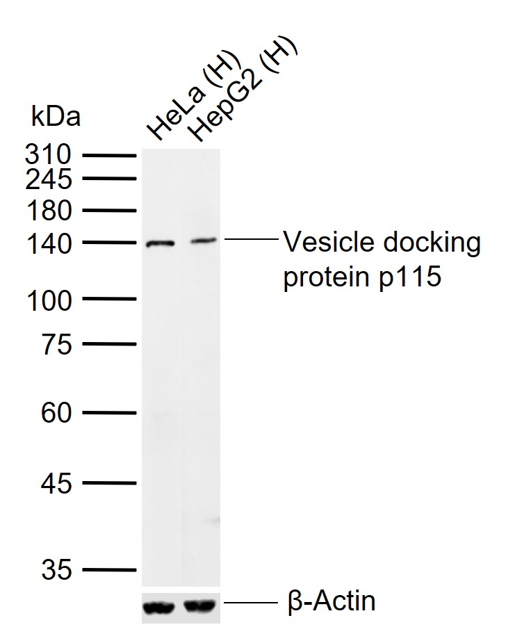 Sample:
Lane 1: Human HeLa cell lysates
Lane 2: Human HepG2 cell lysates
Primary: Anti-Vesicle docking protein p115 (SL4258R) at 1/1000 dilution
Secondary: IRDye800CW Goat Anti-Rabbit IgG at 1/20000 dilution
Predicted band size: 108 kDa
Observed band size: 140 kDa
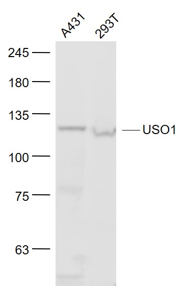 Sample:
A431(Human) Cell Lysate at 30 ug
293T(Human) Cell Lysate at 30 ug
Primary: Anti- USO1 (SL4258R) at 1/1000 dilution
Secondary: IRDye800CW Goat Anti-Rabbit IgG at 1/20000 dilution
Predicted band size: 108 kD
Observed band size: 120 kD
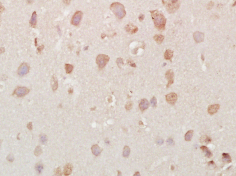 Paraformaldehyde-fixed, paraffin embedded (Mouse brain); Antigen retrieval by boiling in sodium citrate buffer (pH6.0) for 15min; Block endogenous peroxidase by 3% hydrogen peroxide for 20 minutes; Blocking buffer (normal goat serum) at 37°C for 30min; Antibody incubation with (Vesicle docking protein p115) Polyclonal Antibody, Unconjugated (SL4258R) at 1:400 overnight at 4°C, followed by operating according to SP Kit(Rabbit) (sp-0023) instructionsand DAB staining.
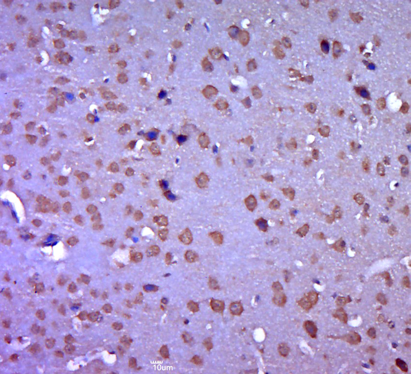 Paraformaldehyde-fixed, paraffin embedded (mouse brain tissue); Antigen retrieval by boiling in sodium citrate buffer (pH6.0) for 15min; Block endogenous peroxidase by 3% hydrogen peroxide for 20 minutes; Blocking buffer (normal goat serum) at 37°C for 30min; Antibody incubation with (Vesicle docking protein p115) Polyclonal Antibody, Unconjugated (SL4258R) at 1:400 overnight at 4°C, followed by a conjugated secondary (sp-0023) for 20 minutes and DAB staining.
Cartpieces
Totalgoods,subtotals:¥Checkout
References (0)
No References
Bought notes(bought amounts latest0)
No one bought this product
User Comment(Total0User Comment Num)
- No comment


 +86 571 56623320
 +86 18668110335

