
Rabbit Anti-Neuroligin 1 antibody
NLG 1; KIAA1070; MGC45115; NLG1; Neuroligin-1; NLGN1; NLGN1_HUMAN.
View History [Clear]
Details
Product Name Neuroligin 1 Chinese Name 突触Cell adhesion molecule1抗体 Alias NLG 1; KIAA1070; MGC45115; NLG1; Neuroligin-1; NLGN1; NLGN1_HUMAN. Research Area immunology Neurobiology Cell adhesion molecule Cell Surface Molecule Cell type markers Immunogen Species Rabbit Clonality Polyclonal React Species Human, Mouse, Rat, (predicted: Chicken, Dog, Pig, Cow, Horse, Rabbit, Sheep, Guinea Pig, ) Applications WB=1:500-2000 ELISA=1:5000-10000 Flow-Cyt=1μg/Test
not yet tested in other applications.
optimal dilutions/concentrations should be determined by the end user.Theoretical molecular weight 87kDa Cellular localization cytoplasmic The cell membrane Form Liquid Concentration 1mg/ml immunogen KLH conjugated synthetic peptide derived from human Neuroligin 1: 701-800/863 <Cytoplasmic> Lsotype IgG Purification affinity purified by Protein A Buffer Solution 0.01M TBS(pH7.4) with 1% BSA, 0.03% Proclin300 and 50% Glycerol. Storage Shipped at 4℃. Store at -20 °C for one year. Avoid repeated freeze/thaw cycles. Attention This product as supplied is intended for research use only, not for use in human, therapeutic or diagnostic applications. PubMed PubMed Product Detail Neuroligin 1 is a synaptic cell-adhesion molecule that is enriched in postsynaptic densities where it may recruit receptors, channels, and signal-transduction molecules to synaptic sites of cell adhesion. In addition, the neuroligin/beta-neurexin junction may be involved in the excitatory/inhibitory specification of CNS neuron synapses. A role for Neuroligin 1 is also suggested in autism.
Function:
Cell surface protein involved in cell-cell-interactions via its interactions with neurexin family members. Plays a role in synapse function and synaptic signal transmission, and probably mediates its effects by recruiting and clustering other synaptic proteins. May promote the initial formation of synapses, but is not essential for this. In vitro, triggers the de novo formation of presynaptic structures. May be involved in specification of excitatory synapses.
Subunit:
Interacts with NRXN1, NRXN2 and NRXN3. Interacts with NLGN3. Interacts with AIP1 and PDZRN3. Interacts (via its C-terminus) with DLG4/PSD-95 (via PDZ domain 3). Interacts with GOPC.
Subcellular Location:
Cell membrane; Single-pass type I membrane protein. Cell junction, synapse. Cell junction, synapse, postsynaptic cell membrane, postsynaptic density. Note=Enriched in synaptic plasma membranes and clustered in synaptic clefts and postsynaptic densities. Detected at dendritic spines. Colocalized with DLG4/PSD-95 and GRIN1/NMDAR1.
Tissue Specificity:
Expressed in the blood vessel walls (at protein level). Detected in brain, and at lower levels in pancreas islet beta cells.
Similarity:
Belongs to the type-B carboxylesterase/lipase family.
SWISS:
Q8N2Q7
Gene ID:
22871
Database links:Entrez Gene: 22871 Human
Entrez Gene: 192167 Mouse
Omim: 600568 Human
SwissProt: Q8N2Q7 Human
SwissProt: Q99K10 Mouse
Unigene: 478289 Human
Unigene: 316080 Mouse
Unigene: 10173 Rat
Product Picture 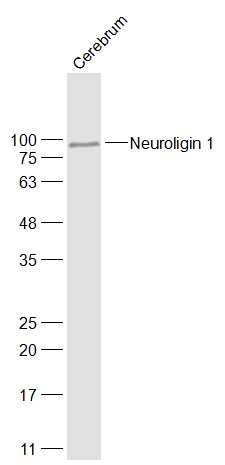 Sample:
Cerebrum (Rat) Lysate at 40 ug
Primary: Anti-Neuroligin 1 (SL4212R) at 1/1000 dilution
Secondary: IRDye800CW Goat Anti-Rabbit IgG at 1/20000 dilution
Predicted band size: 87 kD
Observed band size: 87 kD
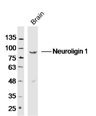 Sample: Brain (Mouse) Lysate at 40 ug
Primary: Anti-Neuroligin 1 (SL4212R) at 1/300 dilution
Secondary: IRDye800CW Goat Anti-Rabbit IgG at 1/20000 dilution
Predicted band size: 87 kD
Observed band size: 87 kD
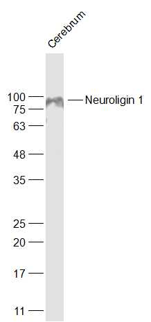 Sample:
Cerebrum (Mouse) Lysate at 40 ug
Primary: Anti-Neuroligin 1 (SL4212R) at 1/1000 dilution
Secondary: IRDye800CW Goat Anti-Rabbit IgG at 1/20000 dilution
Predicted band size: 87 kD
Observed band size: 87 kD
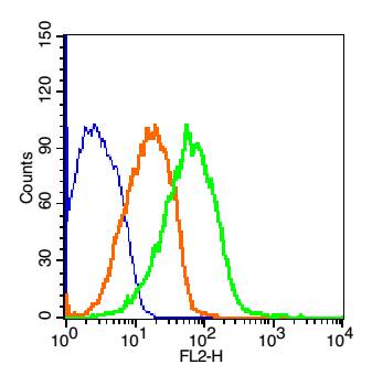
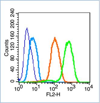 Blank control (blue line): U251 (fixed with 70% ethanol for 5 min at -20℃).
Primary Antibody (green line): Rabbit Anti- Neuroligin 1 antibody (SL4212R), Dilution: 0.2μg /10^6 cells;
Isotype Control Antibody (orange line): Rabbit IgG .
Secondary Antibody (white blue line): Goat anti-rabbit IgG-PE,Dilution: 1μg /test.
 Blank control(blue): Mouse brain cells(fixed with 2% paraformaldehyde (10 min)).
Primary Antibody: Rabbit Anti- Neuroligin 1 /PE Conjugated antibody (SL4212R /PE), Dilution: 5μg in 100 μL 1X PBS containing 0.5% BSA;
Isotype Control Antibody: Rabbit IgG/PE(orange) ,used under the same conditions.
Cartpieces
Totalgoods,subtotals:¥Checkout
References (0)
No References
Bought notes(bought amounts latest0)
No one bought this product
User Comment(Total0User Comment Num)
- No comment


 +86 571 56623320
 +86 18668110335

