
Rabbit Anti-Selenium Binding Protein 1 antibody
56 kDa selenium binding protein; 56 kDa selenium-binding protein; hSBP; LPSB; SBP; SBP1; SBP1_HUMAN; SBP56; Selenbp1; Selenbp2; Selenium binding protein 2; Selenium binding protein1; Selenium-binding protein 1; SELNBP1; SP56.
View History [Clear]
Details
Product Name Selenium Binding Protein 1 Chinese Name 硒Binding protein1抗体 Alias 56 kDa selenium binding protein; 56 kDa selenium-binding protein; hSBP; LPSB; SBP; SBP1; SBP1_HUMAN; SBP56; Selenbp1; Selenbp2; Selenium binding protein 2; Selenium binding protein1; Selenium-binding protein 1; SELNBP1; SP56. literatures Research Area Tumour immunology Neurobiology Signal transduction Immunogen Species Rabbit Clonality Polyclonal React Species Human, Mouse, Rat, (predicted: Dog, Cow, Rabbit, ) Applications WB=1:500-2000 ELISA=1:5000-10000 IHC-P=1:100-500 IHC-F=1:100-500 Flow-Cyt=2ug/Test ICC=1:100-500 IF=1:100-500 (Paraffin sections need antigen repair)
not yet tested in other applications.
optimal dilutions/concentrations should be determined by the end user.Theoretical molecular weight 52kDa Cellular localization The nucleus cytoplasmic The cell membrane Form Liquid Concentration 1mg/ml immunogen KLH conjugated synthetic peptide derived from human SBP1/Selenium Binding Protein 1: 401-472/472 Lsotype IgG Purification affinity purified by Protein A Buffer Solution 0.01M TBS(pH7.4) with 1% BSA, 0.03% Proclin300 and 50% Glycerol. Storage Shipped at 4℃. Store at -20 °C for one year. Avoid repeated freeze/thaw cycles. Attention This product as supplied is intended for research use only, not for use in human, therapeutic or diagnostic applications. PubMed PubMed Product Detail Selenium is an essential trace element that confers tolerance to toxicity arising through exposure to heavy metals or other reactive xenobiotics. Selenium exhibits potent anticarcinogenic properties, and deficiency of selenium may cause certain neurologic diseases. Both effects are attributed to selenium-binding proteins. Selenium binding protein 1 is down-regulated in lung adenocarcinoma, colorectal cander and ovarian cancer. It is two-fold upregulated in the brains of patients suffering from schizophrenia, and is therefore a biomarker for this disease.
Function:
Selenium-binding protein which may be involved in the sensing of reactive xenobiotics in the cytoplasm. May be involved in intra-Golgi protein transport.
Subunit:
Interacts with USP33.
Subcellular Location:
Nucleus. Cytoplasm, cytosol. Membrane; Peripheral membrane protein. Note=May associate with Golgi membrane. May associate with the membrane of autophagosomes.
Tissue Specificity:
Present in liver and colon (at protein level).
Post-translational modifications:
The N-terminus is blocked.
Similarity:
Belongs to the selenium-binding protein family.
SWISS:
Q13228
Gene ID:
8991
Database links:Entrez Gene: 8991 Human
Entrez Gene: 20341 Mouse
Omim: 604188 Human
SwissProt: Q13228 Human
SwissProt: P17563 Mouse
Unigene: 632460 Human
Unigene: 16617 Rat
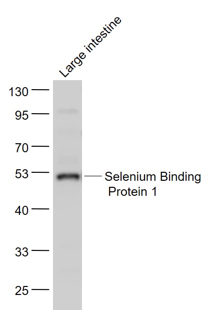
Product Picture 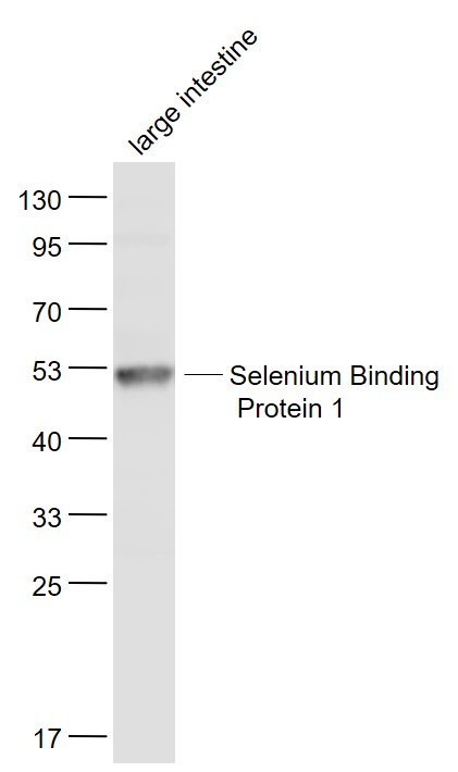 Sample:
Large intestine (Mouse) Lysate at 40 ug
Primary: Anti- Selenium Binding Protein 1 (SL4200R) at 1/1000 dilution
Secondary: IRDye800CW Goat Anti-Rabbit IgG at 1/20000 dilution
Predicted band size: 52 kD
Observed band size: 52 kD
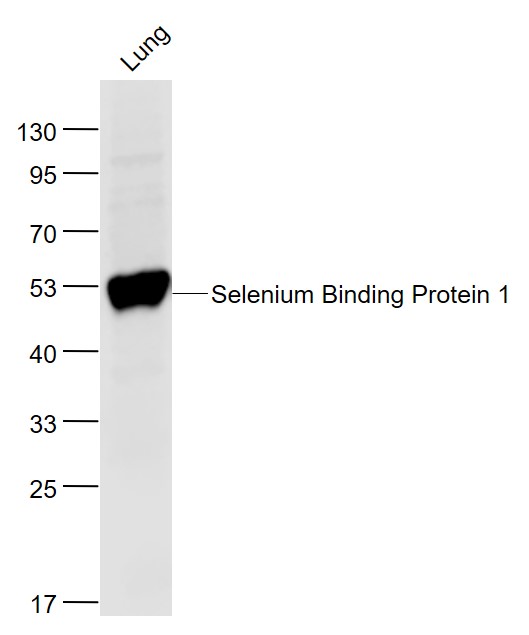 Sample:
Lung (Mouse) Lysate at 40 ug
Primary: Anti-Selenium Binding Protein 1 (SL4200R) at 1/1000 dilution
Secondary: IRDye800CW Goat Anti-Rabbit IgG at 1/20000 dilution
Predicted band size: 52 kD
Observed band size: 52 kD
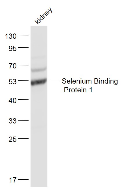 Sample:
Kidney (Mouse) Lysate at 40 ug
Primary: Anti- Selenium Binding Protein 1 (SL4200R) at 1/1000 dilution
Secondary: IRDye800CW Goat Anti-Rabbit IgG at 1/20000 dilution
Predicted band size: 52 kD
Observed band size: 52 kD
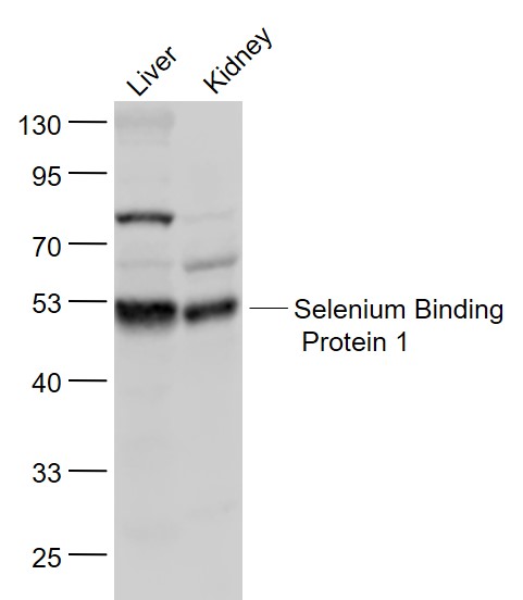 Sample:
Liver (Mouse) Lysate at 40 ug
Kidney (Mouse) Lysate at 40 ug
Primary: Anti- Selenium Binding Protein 1 (SL4200R) at 1/1000 dilution
Secondary: IRDye800CW Goat Anti-Rabbit IgG at 1/20000 dilution
Predicted band size: 52 kD
Observed band size: 52 kD
 Sample:
Large intestine (Mouse) Lysate at 40 ug
Primary: Anti- Selenium Binding Protein 1 (SL4200R) at 1/1000 dilution
Secondary: IRDye800CW Goat Anti-Rabbit IgG at 1/20000 dilution
Predicted band size: 52 kD
Observed band size: 52 kD
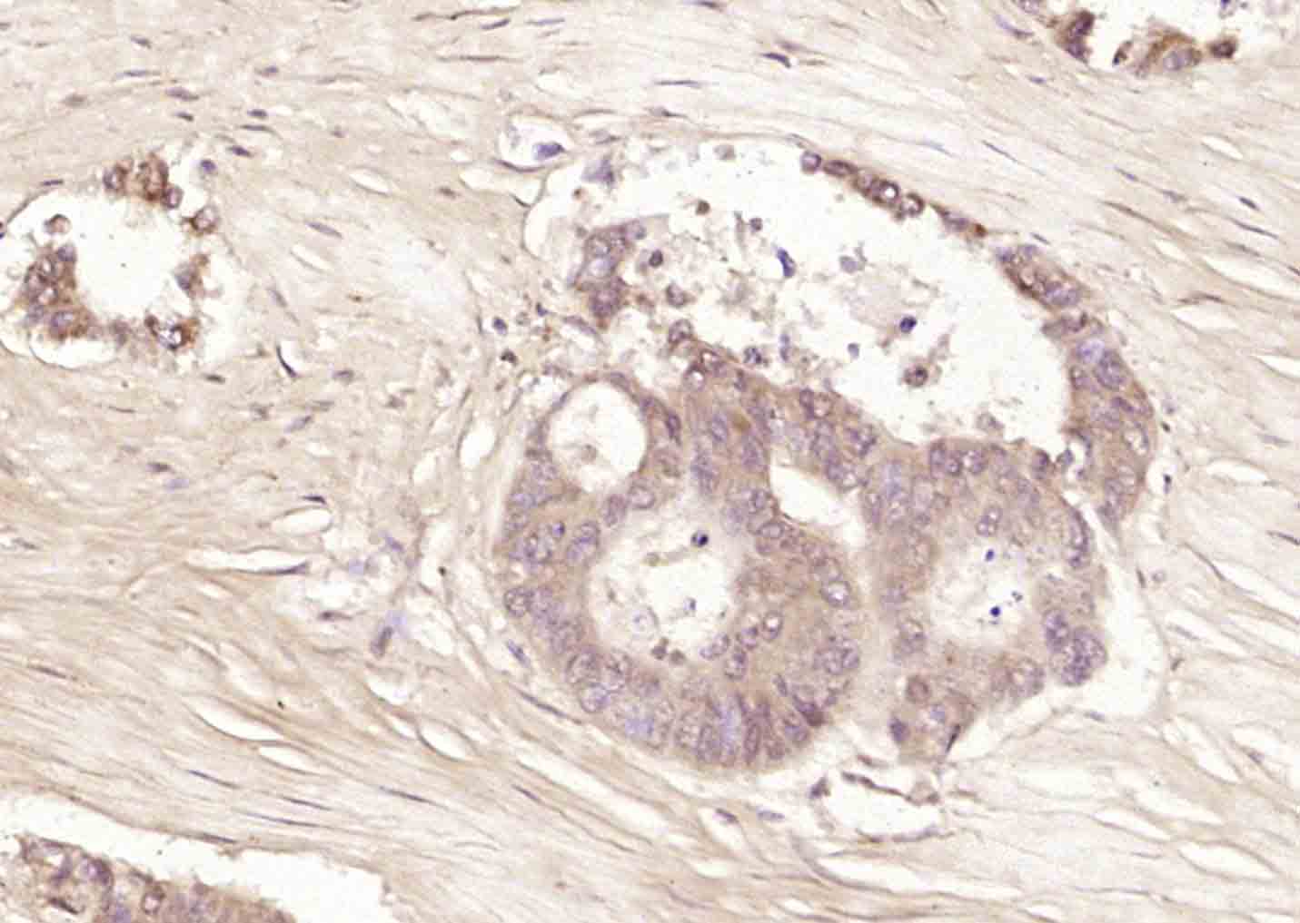 Paraformaldehyde-fixed, paraffin embedded (human cervical carcinoma); Antigen retrieval by boiling in sodium citrate buffer (pH6.0) for 15min; Block endogenous peroxidase by 3% hydrogen peroxide for 20 minutes; Blocking buffer (normal goat serum) at 37°C for 30min; Antibody incubation with (Selenium Binding Protein 1) Polyclonal Antibody, Unconjugated (SL4200R) at 1:200 overnight at 4°C, followed by operating according to SP Kit(Rabbit) (sp-0023) instructionsand DAB staining.
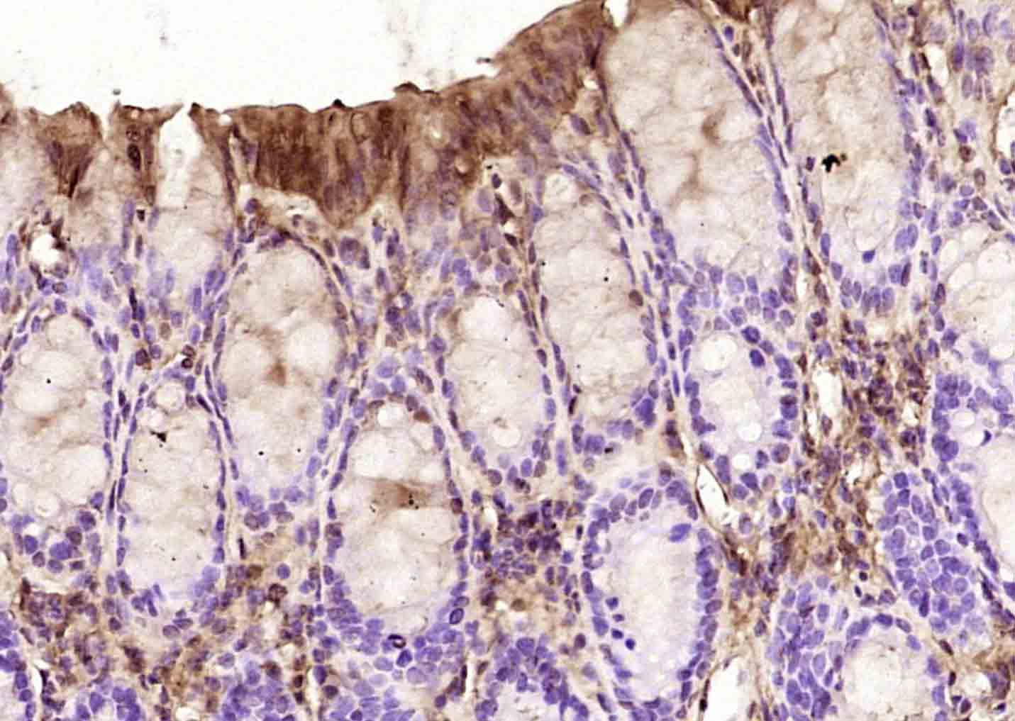 Paraformaldehyde-fixed, paraffin embedded (mouse colon); Antigen retrieval by boiling in sodium citrate buffer (pH6.0) for 15min; Block endogenous peroxidase by 3% hydrogen peroxide for 20 minutes; Blocking buffer (normal goat serum) at 37°C for 30min; Antibody incubation with (GDNF) Polyclonal Antibody, Unconjugated (SL1024R) at 1:200 overnight at 4°C, followed by operating according to SP Kit(Rabbit) (sp-0023) instructionsand DAB staining.
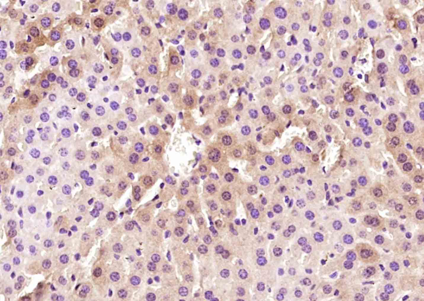 Paraformaldehyde-fixed, paraffin embedded (mouse liver); Antigen retrieval by boiling in sodium citrate buffer (pH6.0) for 15min; Block endogenous peroxidase by 3% hydrogen peroxide for 20 minutes; Blocking buffer (normal goat serum) at 37°C for 30min; Antibody incubation with (Selenium Binding Protein 1) Polyclonal Antibody, Unconjugated (SL4200R) at 1:200 overnight at 4°C, followed by operating according to SP Kit(Rabbit) (sp-0023) instructionsand DAB staining.
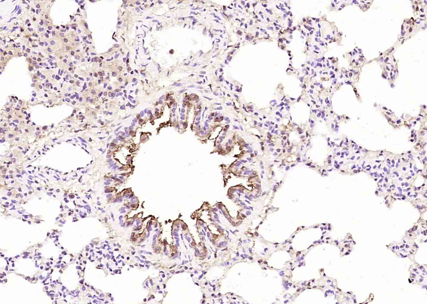 Paraformaldehyde-fixed, paraffin embedded (rat lung); Antigen retrieval by boiling in sodium citrate buffer (pH6.0) for 15min; Block endogenous peroxidase by 3% hydrogen peroxide for 20 minutes; Blocking buffer (normal goat serum) at 37°C for 30min; Antibody incubation with (Selenium Binding Protein 1) Polyclonal Antibody, Unconjugated (SL4200R) at 1:200 overnight at 4°C, followed by operating according to SP Kit(Rabbit) (sp-0023) instructionsand DAB staining.
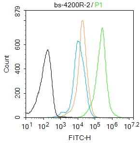 Blank control:Mouse spleen.
Primary Antibody (green line): Rabbit Anti-Selenium Binding Protein 1 antibody (SL4200R)
Dilution: 2μg /10^6 cells;
Isotype Control Antibody (orange line): Rabbit IgG .
Secondary Antibody : Goat anti-rabbit IgG-AF488R
Dilution: 1μg /test.
Protocol
The cells were fixed with 4% PFA (10min at room temperature)and then permeabilized with 90% ice-cold methanol for 20 min at-20℃. The cells were then incubated in 5%BSA to block non-specific protein-protein interactions for 30 min at room temperature .Cells stained with Primary Antibody for 30 min at room temperature. The secondary antibody used for 40 min at room temperature. Acquisition of 20,000 events was performed.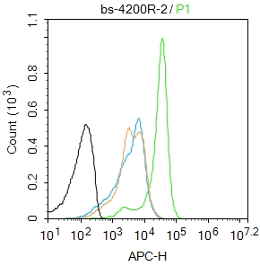 Blank control: Mouse spleen.
Primary Antibody (green line): Rabbit Anti-Selenium Binding Protein 1 antibody (SL4200R)
Dilution: 2μg /10^6 cells;
Isotype Control Antibody (orange line): Rabbit IgG .
Secondary Antibody : Goat anti-rabbit IgG-AF647
Dilution: 1μg /test.
Protocol
The cells were fixed with 4% PFA (10min at room temperature)and then permeabilized with 90% ice-cold methanol for 20 min at-20℃. The cells were then incubated in 5%BSA to block non-specific protein-protein interactions for 30 min at room temperature .Cells stained with Primary Antibody for 30 min at room temperature. The secondary antibody used for 40 min at room temperature. Acquisition of 20,000 events was performed.
Cartpieces
Totalgoods,subtotals:¥Checkout
References (0)
No References
Bought notes(bought amounts latest0)
No one bought this product
User Comment(Total0User Comment Num)
- No comment


 +86 571 56623320
 +86 18668110335

