
Rabbit Anti-MAPK4 antibody
ERK-4; ERK3; Erk3 related; ERK4; Extracellular signal regulated kinase 4; Extracellular signal-regulated kinase 4; MAP kinase 4; MAP kinase isoform p63; MAPK 4; MAPK-4; Mitogen activated protein kinase 4; Mitogen-activated protein kinase 4; MK04_HUMAN; p6
View History [Clear]
Details
Product Name MAPK4 Chinese Name 细胞外信号调节激酶4抗体 Alias ERK-4; ERK3; Erk3 related; ERK4; Extracellular signal regulated kinase 4; Extracellular signal-regulated kinase 4; MAP kinase 4; MAP kinase isoform p63; MAPK 4; MAPK-4; Mitogen activated protein kinase 4; Mitogen-activated protein kinase 4; MK04_HUMAN; p63 MAPK; p63-MAPK; p63MAPK; PRKM4. Research Area Tumour immunology Signal transduction Kinases and Phosphatases Immunogen Species Rabbit Clonality Polyclonal React Species Human, Mouse, Rat, (predicted: Dog, Pig, Cow, Horse, Rabbit, ) Applications WB=1:500-2000 ELISA=1:5000-10000 IHC-P=1:100-500 IHC-F=1:100-500 Flow-Cyt=1ug/Test IF=1:100-500 (Paraffin sections need antigen repair)
not yet tested in other applications.
optimal dilutions/concentrations should be determined by the end user.Theoretical molecular weight 66kDa Cellular localization The nucleus cytoplasmic Form Liquid Concentration 1mg/ml immunogen KLH conjugated synthetic peptide derived from human ERK5: 401-500/587 Lsotype IgG Purification affinity purified by Protein A Buffer Solution 0.01M TBS(pH7.4) with 1% BSA, 0.03% Proclin300 and 50% Glycerol. Storage Shipped at 4℃. Store at -20 °C for one year. Avoid repeated freeze/thaw cycles. Attention This product as supplied is intended for research use only, not for use in human, therapeutic or diagnostic applications. PubMed PubMed Product Detail Mitogen-activated protein kinase 4 is a member of the mitogen-activated protein kinase family. Tyrosine kinase growth factor receptors activate mitogen-activated protein kinases which then translocate into the nucleus where it phosphorylates nuclear targets. [provided by RefSeq, Jul 2008]
Function:
Atypical MAPK protein. Phosphorylates microtubule-associated protein 2 (MAP2) and MAPKAPK5. The precise role of the complex formed with MAPKAPK5 is still unclear, but the complex follows a complex set of phosphorylation events: upon interaction with atypical MAPKAPK5, ERK4/MAPK4 is phosphorylated at Ser-186 and then mediates phosphorylation and activation of MAPKAPK5, which in turn phosphorylates ERK4/MAPK4. May promote entry in the cell cycle (By similarity).
Subunit:
Homodimer. Heterodimer with ERK3/MAPK6. Interacts with (via FRIEDE motif) MAPKAPK5.
Subcellular Location:
Cytoplasm. Nucleus. Note=Translocates to the cytoplasm following interaction with MAPKAPK5.
Tissue Specificity:
High expression in heart and brain.
Post-translational modifications:
Phosphorylated at Ser-186 by PAK1, PAK2 and PAK3 resulting in catalytic activation. Phosphorylated by MAPKAPK5 at other sites.
Similarity:
Belongs to the protein kinase superfamily. CMGC Ser/Thr protein kinase family. MAP kinase subfamily.
Contains 1 protein kinase domain.
SWISS:
P31152
Gene ID:
5596
Database links:Entrez Gene: 5596 Human
Entrez Gene: 225724 Mouse
Omim: 176949 Human
SwissProt: P31152 Human
SwissProt: Q6P5G0 Mouse
Unigene: 433728 Human
Unigene: 254517 Mouse
Unigene: 146899 Rat
Product Picture 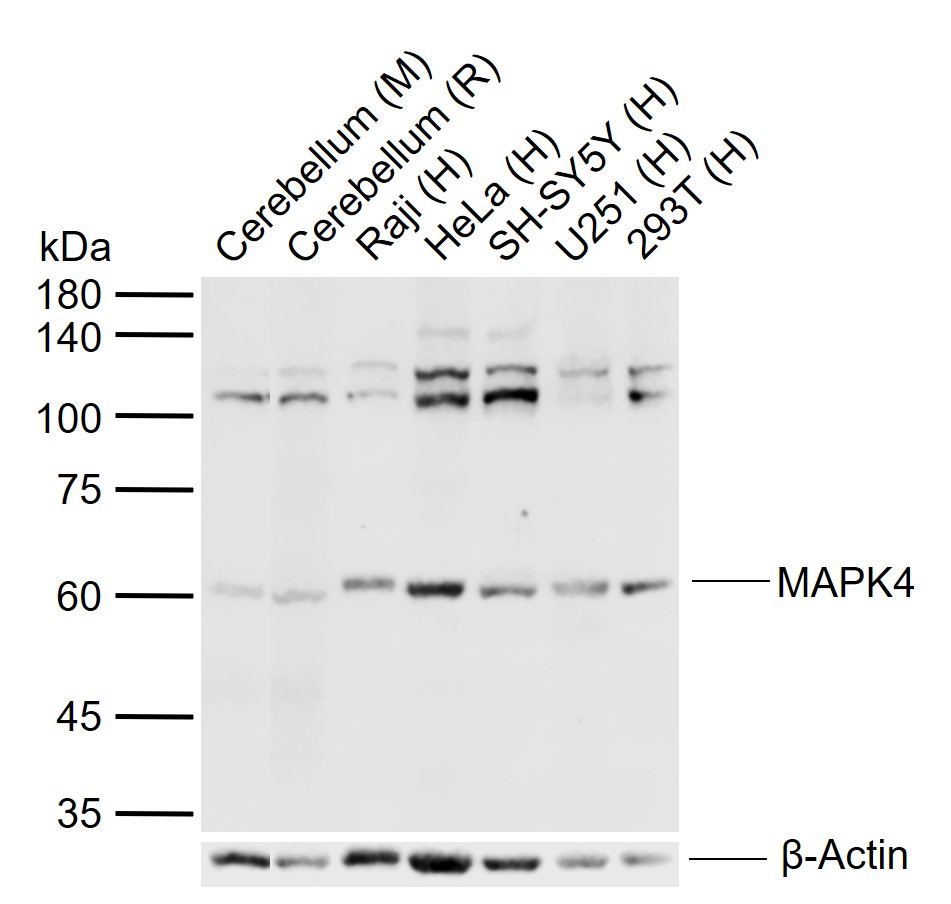 Sample:
Lane 1: Mouse Cerebellum tissue lysates
Lane 2: Rat Cerebellum tissue lysates
Lane 3: Human Raji cell lysates
Lane 4: Human HeLa cell lysates
Lane 5: Human SH-SY5Y cell lysates
Lane 6: Human U251 cell lysates
Lane 7: Human 293T cell lysates
Primary: Anti- MAPK4 (SL4131R) at 1/1000 dilution
Secondary: IRDye800CW Goat Anti-Rabbit IgG at 1/20000 dilution
Predicted band size: 66 kDa
Observed band size: 60 kDa
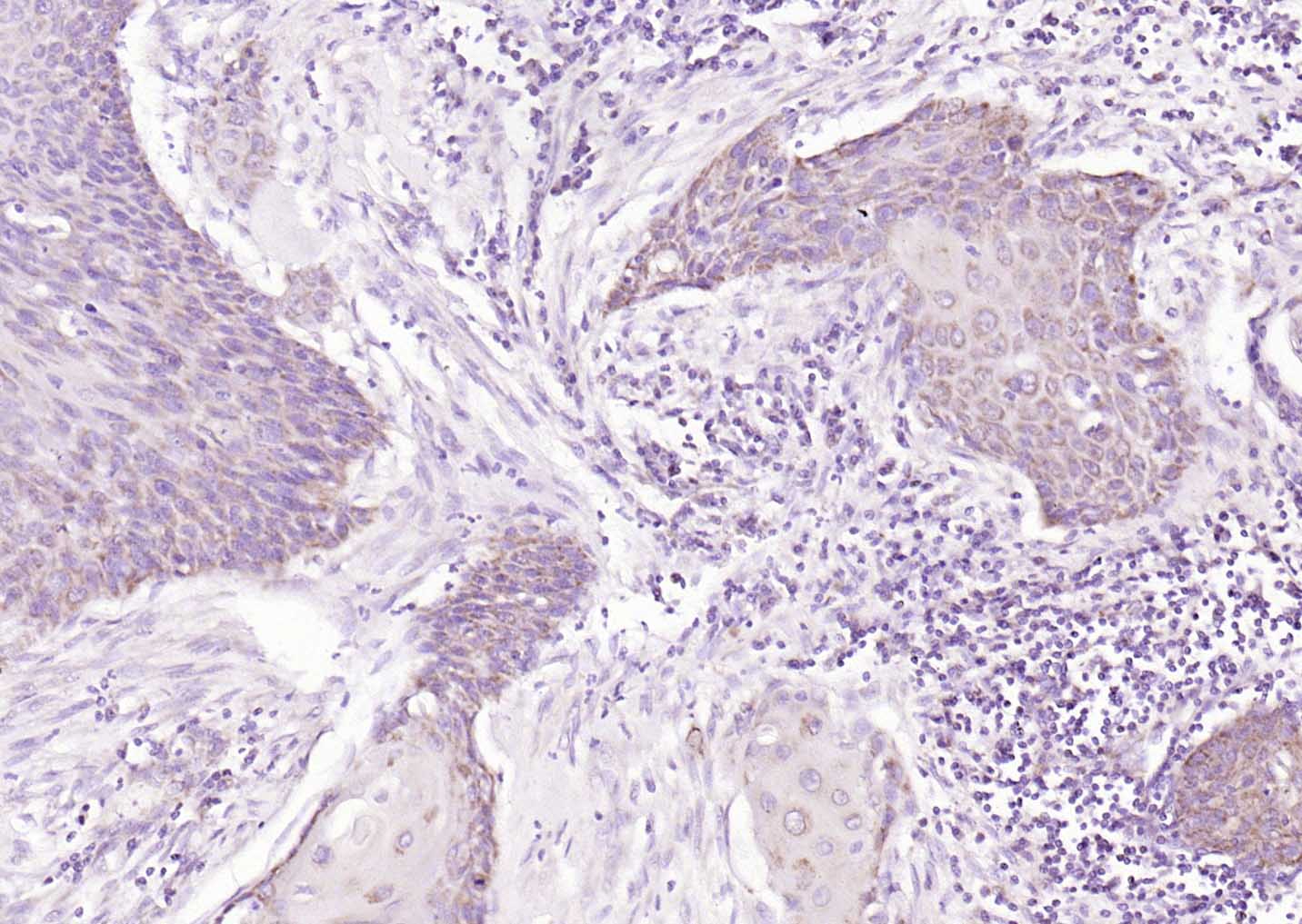 Paraformaldehyde-fixed, paraffin embedded (human cervical carcinoma); Antigen retrieval by boiling in sodium citrate buffer (pH6.0) for 15min; Block endogenous peroxidase by 3% hydrogen peroxide for 20 minutes; Blocking buffer (normal goat serum) at 37°C for 30min; Antibody incubation with (MAPK4) Polyclonal Antibody, Unconjugated (SL4131R) at 1:200 overnight at 4°C, followed by operating according to SP Kit(Rabbit) (sp-0023) instructionsand DAB staining.
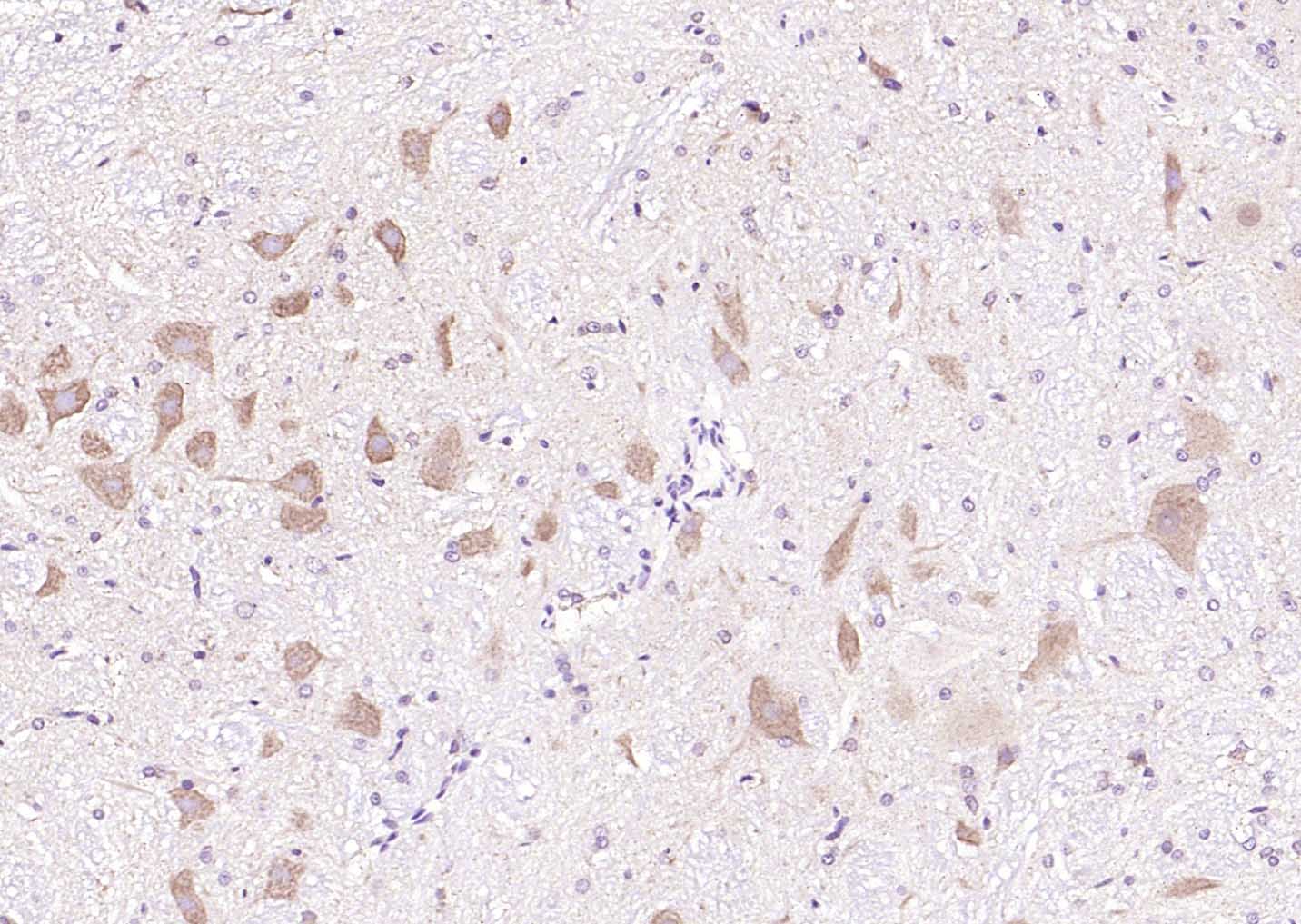 Paraformaldehyde-fixed, paraffin embedded (rat cerebellum); Antigen retrieval by boiling in sodium citrate buffer (pH6.0) for 15min; Block endogenous peroxidase by 3% hydrogen peroxide for 20 minutes; Blocking buffer (normal goat serum) at 37°C for 30min; Antibody incubation with (MAPK4) Polyclonal Antibody, Unconjugated (SL4131R) at 1:200 overnight at 4°C, followed by operating according to SP Kit(Rabbit) (sp-0023) instructionsand DAB staining.
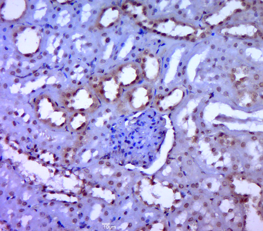 Paraformaldehyde-fixed, paraffin embedded (rat kidney tissue); Antigen retrieval by boiling in sodium citrate buffer (pH6.0) for 15min; Block endogenous peroxidase by 3% hydrogen peroxide for 20 minutes; Blocking buffer (normal goat serum) at 37°C for 30min; Antibody incubation with (ERK4) Polyclonal Antibody, Unconjugated (SL4131R) at 1:400 overnight at 4°C, followed by a conjugated secondary (sp-0023) for 20 minutes and DAB staining.
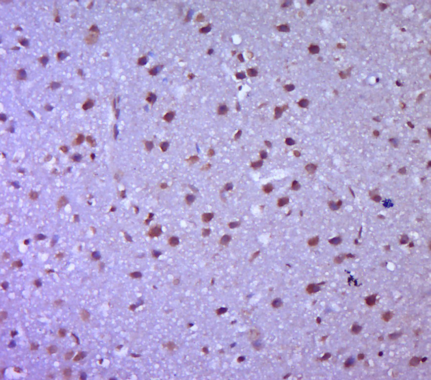 Paraformaldehyde-fixed, paraffin embedded (rat brain tissue); Antigen retrieval by boiling in sodium citrate buffer (pH6.0) for 15min; Block endogenous peroxidase by 3% hydrogen peroxide for 20 minutes; Blocking buffer (normal goat serum) at 37°C for 30min; Antibody incubation with (ERK4) Polyclonal Antibody, Unconjugated (SL4131R) at 1:400 overnight at 4°C, followed by a conjugated secondary (sp-0023) for 20 minutes and DAB staining.
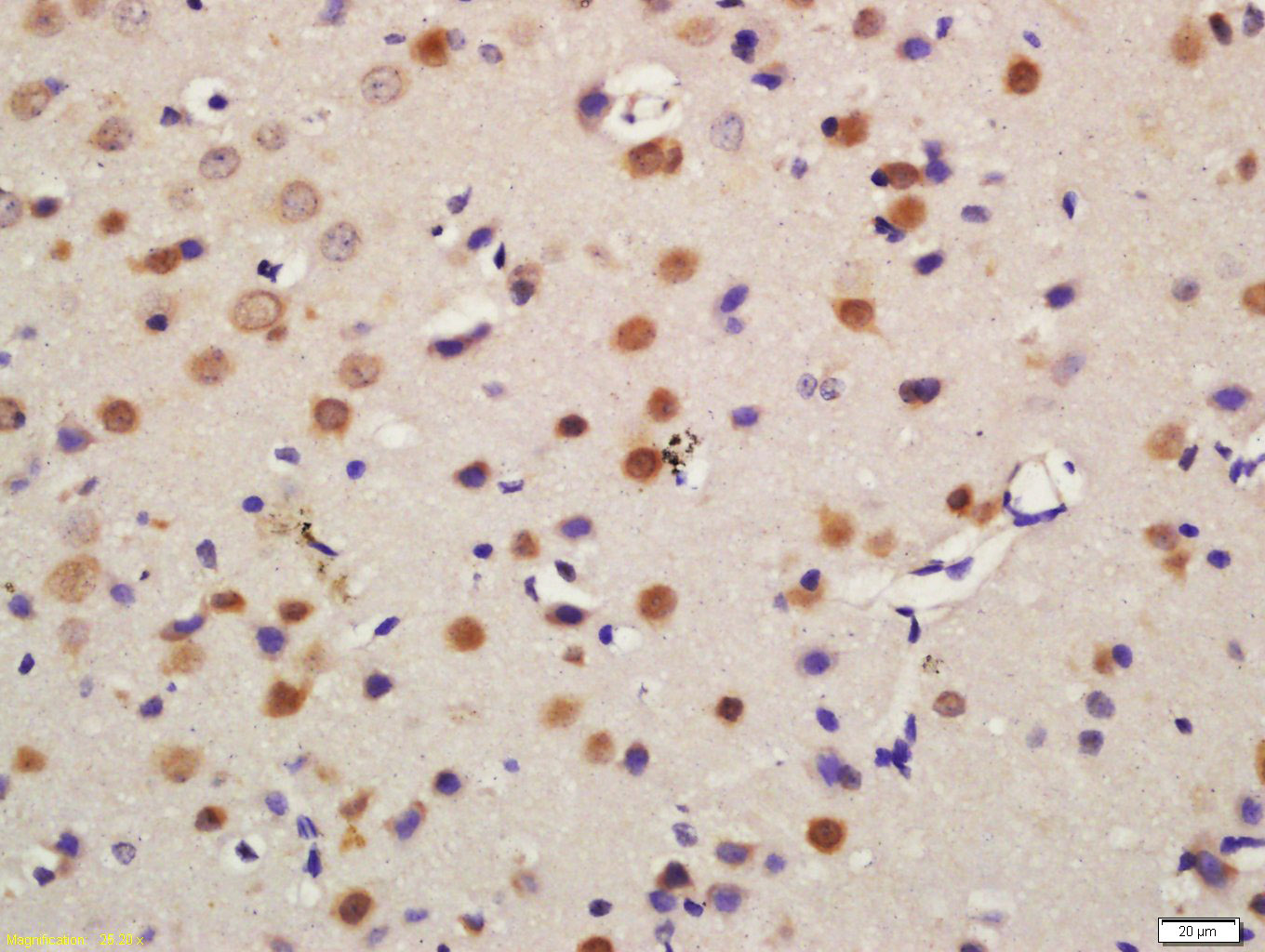 Tissue/cell: mouse brain tissue; 4% Paraformaldehyde-fixed and paraffin-embedded;
Antigen retrieval: citrate buffer ( 0.01M, pH 6.0 ), Boiling bathing for 15min; Block endogenous peroxidase by 3% Hydrogen peroxide for 30min; Blocking buffer (normal goat serum,C-0005) at 37℃ for 20 min;
Incubation: Anti-ERK4 Polyclonal Antibody, Unconjugated(SL4131R) 1:200, overnight at 4°C, followed by conjugation to the secondary antibody(SP-0023) and DAB(C-0010) staining
 Tissue/cell: rat brain tissue; 4% Paraformaldehyde-fixed and paraffin-embedded;
Antigen retrieval: citrate buffer ( 0.01M, pH 6.0 ), Boiling bathing for 15min; Block endogenous peroxidase by 3% Hydrogen peroxide for 30min; Blocking buffer (normal goat serum,C-0005) at 37℃ for 20 min;
Incubation: Anti-ERK4 Polyclonal Antibody, Unconjugated(SL4131R) 1:200, overnight at 4°C, followed by conjugation to the secondary antibody(SP-0023) and DAB(C-0010) staining
 Paraformaldehyde-fixed, paraffin embedded (mouse cerebellum); Antigen retrieval by boiling in sodium citrate buffer (pH6.0) for 15min; Block endogenous peroxidase by 3% hydrogen peroxide for 20 minutes; Blocking buffer (normal goat serum) at 37°C for 30min; Antibody incubation with (MAPK4) Polyclonal Antibody, Unconjugated (SL4131R) at 1:200 overnight at 4°C, followed by operating according to SP Kit(Rabbit) (sp-0023) instructionsand DAB staining.
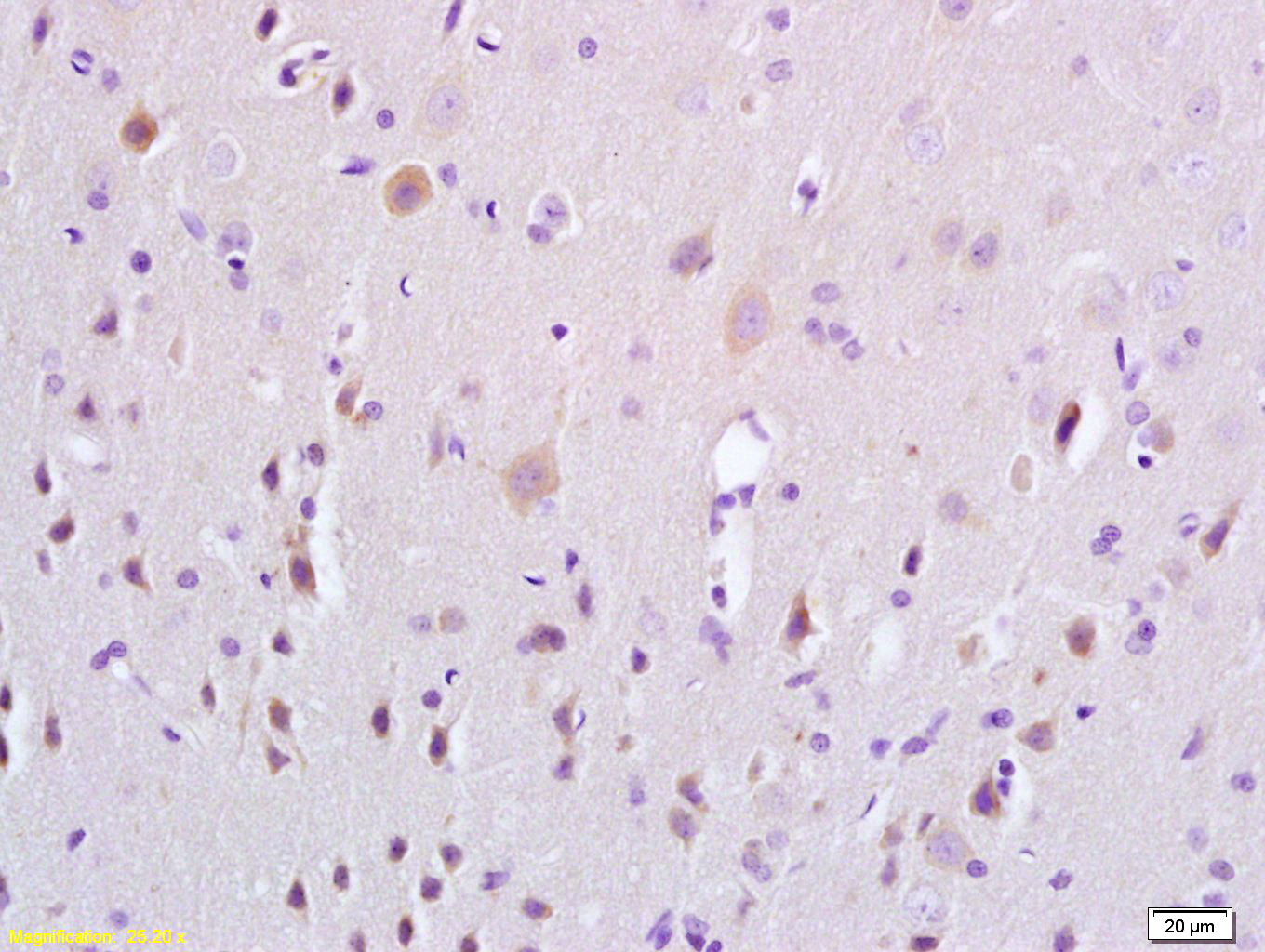
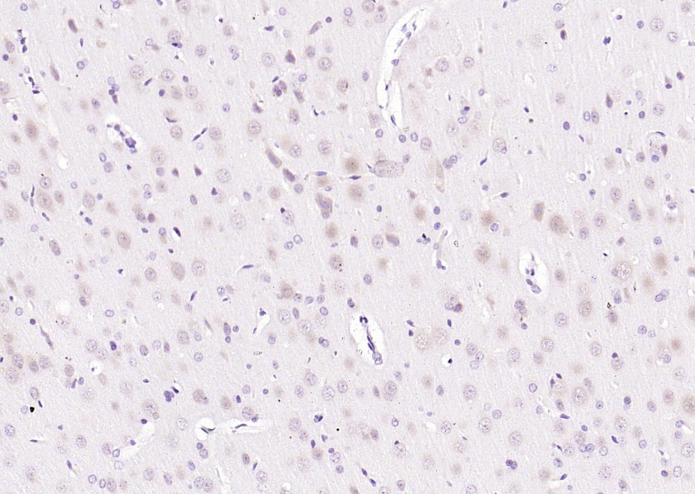 Paraformaldehyde-fixed, paraffin embedded (rat brain); Antigen retrieval by boiling in sodium citrate buffer (pH6.0) for 15min; Block endogenous peroxidase by 3% hydrogen peroxide for 20 minutes; Blocking buffer (normal goat serum) at 37°C for 30min; Antibody incubation with (MAPK4) Polyclonal Antibody, Unconjugated (SL4131R) at 1:200 overnight at 4°C, followed by operating according to SP Kit(Rabbit) (sp-0023) instructionsand DAB staining.
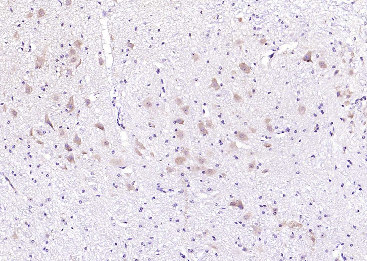
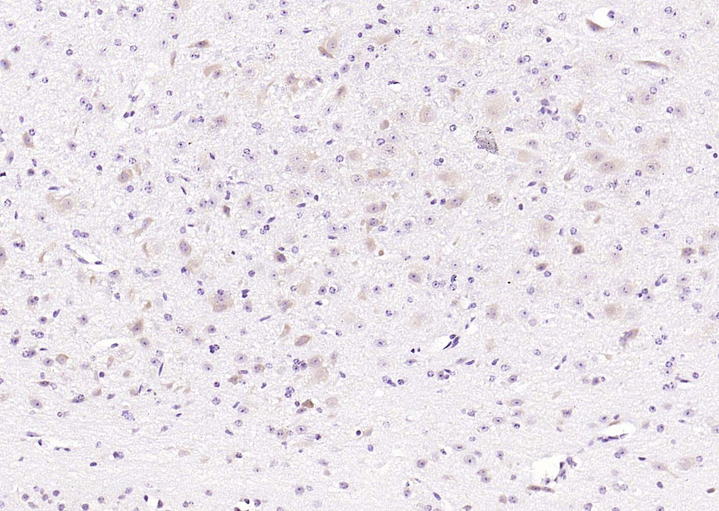 Paraformaldehyde-fixed, paraffin embedded (mouse brain); Antigen retrieval by boiling in sodium citrate buffer (pH6.0) for 15min; Block endogenous peroxidase by 3% hydrogen peroxide for 20 minutes; Blocking buffer (normal goat serum) at 37°C for 30min; Antibody incubation with (MAPK4) Polyclonal Antibody, Unconjugated (SL4131R) at 1:200 overnight at 4°C, followed by operating according to SP Kit(Rabbit) (sp-0023) instructionsand DAB staining.
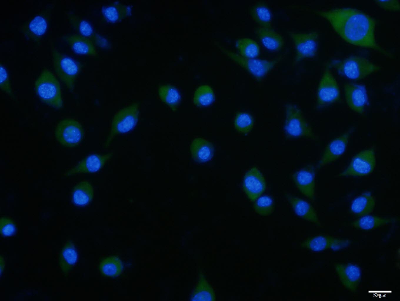 SH-SY5Y cell; 4% Paraformaldehyde-fixed; Triton X-100 at room temperature for 20 min; Blocking buffer (normal goat serum, C-0005) at 37°C for 20 min; Antibody incubation with (MAPK4) polyclonal Antibody, Unconjugated (SL4131R) 1:100, 90 minutes at 37°C; followed by a conjugated Goat Anti-Rabbit IgG antibody at 37°C for 90 minutes, DAPI (blue, C02-04002) was used to stain the cell nuclei.
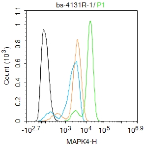 Blank control(black line):Hela.
Primary Antibody (green line): Rabbit Anti-MAPK4 antibody (SL4131R)
Dilution:1ug/Test;
Secondary Antibody(white blue line): Goat anti-rabbit IgG-AF488
Dilution: 0.5ug/Test.
Isotype control(orange line): Normal Rabbit IgG
Protocol
The cells were fixed with 4% PFA (10min at room temperature)and then permeabilized with 90% ice-cold methanol for 20 min at -20℃, The cells were then incubated in 5%BSA to block non-specific protein-protein interactions for 30 min at room temperature .Cells stained with Primary Antibody for 30 min at room temperature. The secondary antibody used for 40 min at room temperature. Acquisition of 20,000 events was performed.
Cartpieces
Totalgoods,subtotals:¥Checkout
References (0)
No References
Bought notes(bought amounts latest0)
No one bought this product
User Comment(Total0User Comment Num)
- No comment


 +86 571 56623320
 +86 18668110335

