
Rabbit Anti-phospho-E2F1 (Ser364)antibody
E2F1 (phospho S364); E2F1 (phospho Ser364); p-E2F1 (S364); p-E2F1 (Ser364); E2F 1; E2F transcription factor 1; E2F-1; E2f1 E2F transcription factor 1; KIAA4009; mKIAA4009; OTTHUMP00000030661; PBR 3; PBR3; PRB binding protein E2F 1; PRB-binding protein E2F
View History [Clear]
Details
Product Name phospho-E2F1 (Ser364) Chinese Name 磷酸化转录因子E2F-1抗体 Alias E2F1 (phospho S364); E2F1 (phospho Ser364); p-E2F1 (S364); p-E2F1 (Ser364); E2F 1; E2F transcription factor 1; E2F-1; E2f1 E2F transcription factor 1; KIAA4009; mKIAA4009; OTTHUMP00000030661; PBR 3; PBR3; PRB binding protein E2F 1; PRB-binding protein E2F-1; RBAP 1; RBAP-1; RBAP1; RBBP 3; RBBP-3; RBBP3; RBP 3; RBP3; Retinoblastoma associated protein 1; Retinoblastoma binding protein 3; Retinoblastoma-associated protein 1; Retinoblastoma-binding protein 3; Transcription factor E2F1; E2F1_HUMAN. Product Type Phosphorylated anti Research Area Tumour Cell biology immunology transcriptional regulatory factor Immunogen Species Rabbit Clonality Polyclonal React Species Human, (predicted: Mouse, Rat, ) Applications WB=1:500-2000 ELISA=1:5000-10000 IHC-P=1:100-500 IHC-F=1:100-500 Flow-Cyt=1ug/Test ICC=1:100 IF=1:100-500 (Paraffin sections need antigen repair)
not yet tested in other applications.
optimal dilutions/concentrations should be determined by the end user.Theoretical molecular weight 48kDa Cellular localization The nucleus Form Liquid Concentration 1mg/ml immunogen KLH conjugated Synthesised phosphopeptide derived from human E2F1 around the phosphorylation site of Ser364: MG(p-S)LR Lsotype IgG Purification affinity purified by Protein A Buffer Solution 0.01M TBS(pH7.4) with 1% BSA, 0.03% Proclin300 and 50% Glycerol. Storage Shipped at 4℃. Store at -20 °C for one year. Avoid repeated freeze/thaw cycles. Attention This product as supplied is intended for research use only, not for use in human, therapeutic or diagnostic applications. PubMed PubMed Product Detail E2F's are DNA binding proteins, which associate with negative regulators, such as the retinoblastoma p107 protein, resulting in an altered rate of gene transcription. The E2F proteins contain several evolutionally conserved domains found in most members of the family. These domains include a DNA binding domain, a dimerization domain which determines interaction with the differentiation regulated transcription factor proteins (DP), a transactivation domain enriched in acidic amino acids, and a tumor suppressor protein association domain which is embedded within the transactivation domain. This protein and another 2 members, E2F2 and E2F3, have an additional cyclin binding domain. E2F1 is proposed to be involved in several cellular processes that range from tumor suppressor, cell progression and oncogenesis. E2F1 overexpression can also drive cells into apoptosis.
Subunit:
Component of the DRTF1/E2F transcription factor complex. Forms heterodimers with DP family members. The E2F-1 complex binds specifically hypophosphorylated retinoblastoma protein RB1. During the cell cycle, RB1 becomes phosphorylated in mid-to-late G1 phase, detaches from the DRTF1/E2F complex, rendering E2F transcriptionally active. Viral oncoproteins, notably E1A, T-antigen and HPV E7, are capable of sequestering RB protein, thus releasing the active complex. Interacts with TRRAP, which probably mediates its interaction with histone acetyltransferase complexes, leading to transcription activation. Binds TOPBP1 and EAPP. Interacts with ARID3A. Interacts with TRIM28; the interaction inhibits E2F1 acetylation through recruiting HDAC1 and represses its transcriptional activity. Interaction with KAT2B; the interaction acetylates E2F1 enhancing its DNA-binding and transcriptional activity. Interacts with BIRC2/c-IAP1 (via BIR domains). Interacts with human cytomegalovirus/HHV-5 protein UL123.
Subcellular Location:
Nucleus.
Post-translational modifications:
Phosphorylated by CDK2 and cyclin A-CDK2 in the S-phase.
Similarity:
Belongs to the E2F/DP family.
SWISS:
Q01094
Gene ID:
1869
Database links:Entrez Gene: 1869 Human
Entrez Gene: 13555 Mouse
Omim: 189971 Human
SwissProt: Q01094 Human
SwissProt: Q61501 Mouse
Unigene: 654393 Human
Unigene: 18036 Mouse
Unigene: 72471 Rat
E2F1—属于调节性转录因子E2F家族。有学者认为:E2F-1既可作为癌基因起作用,又可作为抑癌基因起作用。其不同可能由细胞中其他生长促进或抑制性蛋白质水平和(或)活性决定,同时与细胞所处环境及器官特异性有关。在控制细胞周期和Tumour抑制基因蛋白的活性方面起关键作用。Product Picture 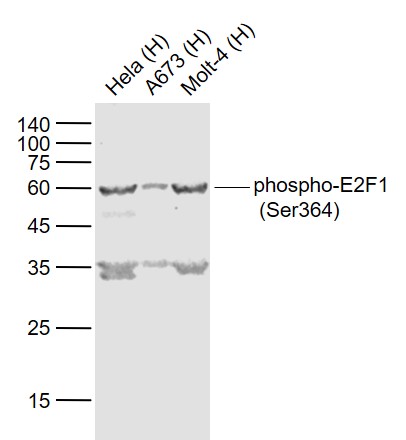 Sample:
Lane 1: Hela (Human) Cell Lysate at 30 ug
Lane 2: A673 (Human) Cell Lysate at 30 ug
Lane 3: Molt-4 (Human) Cell Lysate at 30 ug
Primary: Anti-phospho-E2F1 (Ser364) (SL4076R) at 1/1000 dilution
Secondary: IRDye800CW Goat Anti-Rabbit IgG at 1/20000 dilution
Predicted band size: 55-60 kD
Observed band size: 60 kD
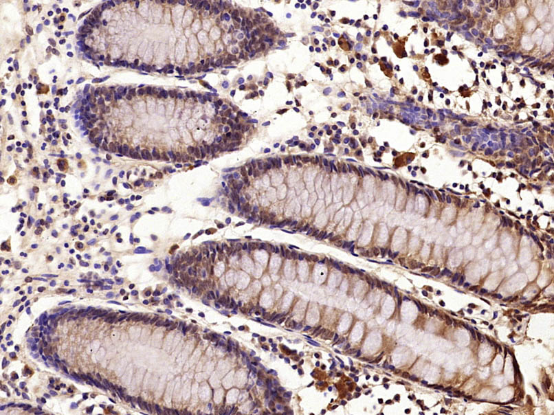 Paraformaldehyde-fixed, paraffin embedded (Human colon carcinoma); Antigen retrieval by microwave in sodium citrate buffer (pH6.0) ; Block endogenous peroxidase by 3% hydrogen peroxide for 30 minutes; Blocking buffer (3% BSA) at RT for 30min; Antibody incubation with (phospho-E2F1 (Ser364)) Polyclonal Antibody, Unconjugated (SL4076R) at 1:400 overnight at 4°C, followed by conjugation to the secondary antibody (labeled with HRP)and DAB staining.
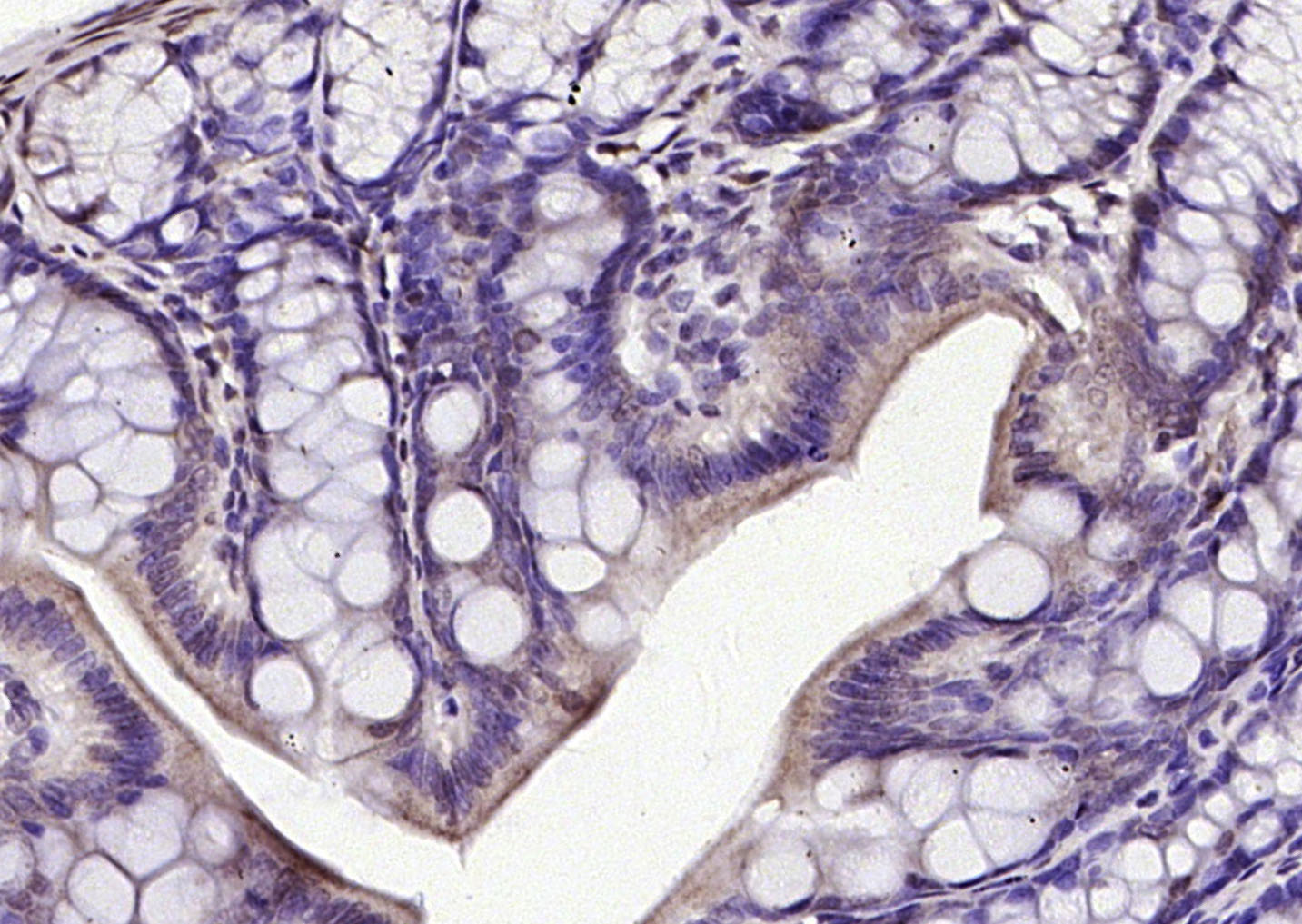 Paraformaldehyde-fixed, paraffin embedded (rat colon); Antigen retrieval by boiling in sodium citrate buffer (pH6.0) for 15min; Block endogenous peroxidase by 3% hydrogen peroxide for 20 minutes; Blocking buffer (normal goat serum) at 37°C for 30min; Antibody incubation with (phospho-E2F1 (Ser364)) Polyclonal Antibody, Unconjugated (SL4076R) at 1:200 overnight at 4°C, followed by operating according to SP Kit(Rabbit) (sp-0023) instructionsand DAB staining.
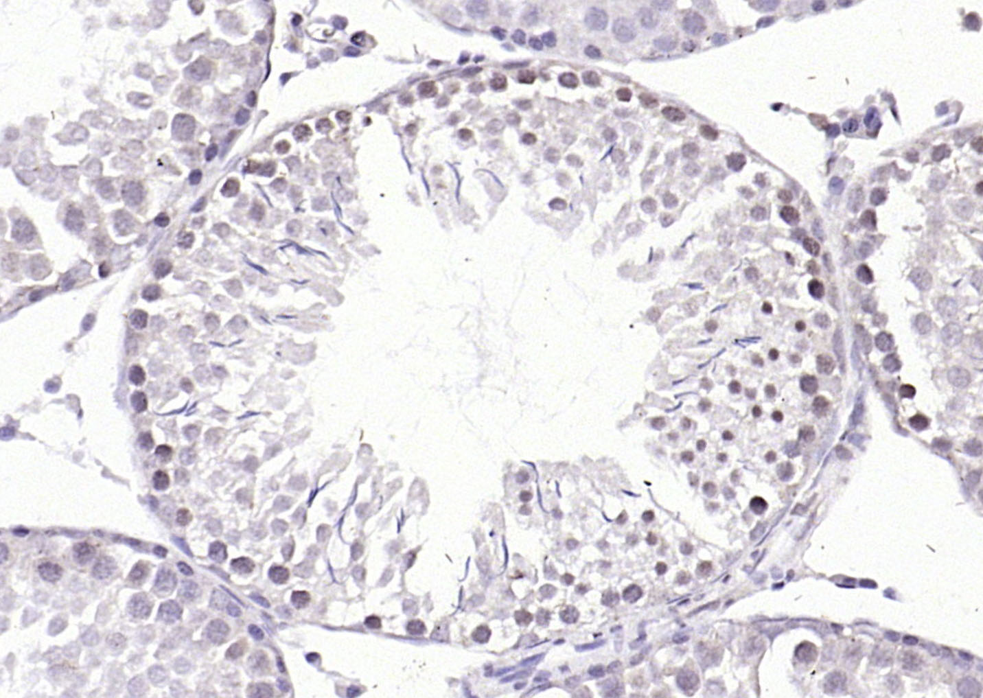 Paraformaldehyde-fixed, paraffin embedded (rat testis); Antigen retrieval by boiling in sodium citrate buffer (pH6.0) for 15min; Block endogenous peroxidase by 3% hydrogen peroxide for 20 minutes; Blocking buffer (normal goat serum) at 37°C for 30min; Antibody incubation with (phospho-E2F1 (Ser364)) Polyclonal Antibody, Unconjugated (SL4076R) at 1:200 overnight at 4°C, followed by operating according to SP Kit(Rabbit) (sp-0023) instructionsand DAB staining.
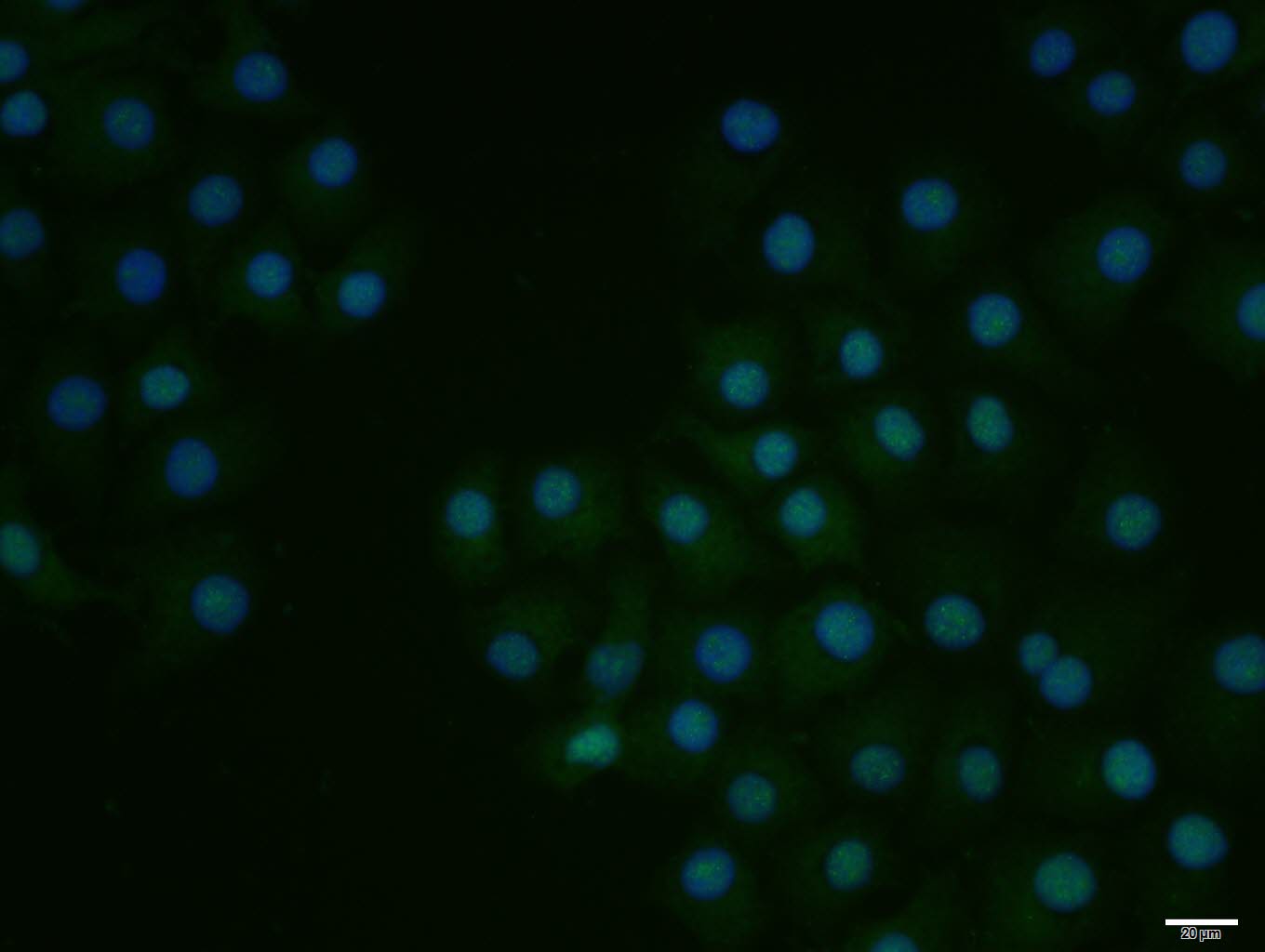 HepG2 cell; 4% Paraformaldehyde-fixed; Triton X-100 at room temperature for 20 min; Blocking buffer (normal goat serum, C-0005) at 37°C for 20 min; Antibody incubation with (phospho-E2F1 (Ser364)) polyclonal Antibody, Unconjugated (SL4076R) 1:100, 90 minutes at 37°C; followed by a conjugated Goat Anti-Rabbit IgG antibody at 37°C for 90 minutes, DAPI (blue, C02-04002) was used to stain the cell nuclei.
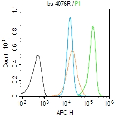 Blank control (Black line):Molt4 (Black).
Primary Antibody (green line): Rabbit Anti-phospho-E2F1 (Ser364) antibody (SL4076R)
Dilution: 1μg /10^6 cells;
Isotype Control Antibody (orange line): Rabbit IgG .
Secondary Antibody (white blue line): Goat anti-rabbit IgG-AF647
Dilution: 1μg /test.
Protocol
The cells were fixed with 4% PFA (10min at room temperature)and then permeabilized with 90% ice-cold methanol for 20 min at room temperature. The cells were then incubated in 5%BSA to block non-specific protein-protein interactions for 30 min at room temperature .Cells stained with Primary Antibody for 30 min at room temperature. The secondary antibody used for 40 min at room temperature. Acquisition of 20,000 events was performed.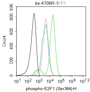 Blank control:HepG2.
Primary Antibody (green line): Rabbit Anti-phospho-E2F1 (Ser364) antibody (SL4076R)
Dilution: 1ug/Test;
Secondary Antibody : Goat anti-rabbit IgG-FITC
Dilution: 0.5ug/Test.
Protocol
The cells were fixed with 4% PFA (10min at room temperature)and then permeabilized with 0.1% PBST for 20 min at room temperature.The cells were then incubated in 5%BSA to block non-specific protein-protein interactions for 30 min at room temperature .Cells stained with Primary Antibody for 30 min at room temperature. The secondary antibody used for 40 min at room temperature. Acquisition of 20,000 events was performed.
Cartpieces
Totalgoods,subtotals:¥Checkout
References (0)
No References
Bought notes(bought amounts latest0)
No one bought this product
User Comment(Total0User Comment Num)
- No comment


 +86 571 56623320
 +86 18668110335

