
Rabbit Anti-Phospho-CDK1 (Thr161)antibody
Phospho-cdc2 (Thr161); CDK1 (Phospho Thr161); CDK1 (Phospho T161); Cdc 2; Cdc2; CDC28A; CDK 1; CDK1_HUMAN; CDKN1; CELL CYCLE CONTROLLER CDC2; Cell division control protein 2; Cell division control protein 2 homolog; Cell division cycle 2 G1 to S and G2 to
View History [Clear]
Details
Product Name Phospho-CDK1 (Thr161) Chinese Name 磷酸化周期素依赖激酶2抗体 Alias Phospho-cdc2 (Thr161); CDK1 (Phospho Thr161); CDK1 (Phospho T161); Cdc 2; Cdc2; CDC28A; CDK 1; CDK1_HUMAN; CDKN1; CELL CYCLE CONTROLLER CDC2; Cell division control protein 2; Cell division control protein 2 homolog; Cell division cycle 2 G1 to S and G2 to M; Cell division protein kinase 1; Cell Divsion Cycle 2 Protein; Cyclin Dependent Kinase 1; Cyclin-dependent kinase 1; DKFZp686L20222; MGC111195; p34 Cdk1; p34 protein kinase; P34CDC2. literatures Product Type Phosphorylated anti Research Area Tumour Cell biology immunology Signal transduction transcriptional regulatory factor Kinases and Phosphatases Immunogen Species Rabbit Clonality Polyclonal React Species Human, (predicted: Mouse, Rat, Chicken, Pig, Cow, Horse, Rabbit, Sheep, ) Applications ELISA=1:5000-10000 IHC-P=1:100-500 IHC-F=1:100-500 IF=1:100-500 (Paraffin sections need antigen repair)
not yet tested in other applications.
optimal dilutions/concentrations should be determined by the end user.Theoretical molecular weight 34kDa Cellular localization The nucleus cytoplasmic Form Liquid Concentration 1mg/ml immunogen KLH conjugated Synthesised phosphopeptide derived from human cdc2 around the phosphorylation site of Thr161: VY(p-T)HE Lsotype IgG Purification affinity purified by Protein A Buffer Solution 0.01M TBS(pH7.4) with 1% BSA, 0.03% Proclin300 and 50% Glycerol. Storage Shipped at 4℃. Store at -20 °C for one year. Avoid repeated freeze/thaw cycles. Attention This product as supplied is intended for research use only, not for use in human, therapeutic or diagnostic applications. PubMed PubMed Product Detail The cell division control protein cdc2, also known as cyclin dependent kinase 1 (Cdk1) or p34/cdk1, plays a key role in the control of the eukaryotic cell cycle, where it is required for entry into S phase and mitosis. Cdc2 exists as a complex with both cyclin A and cyclin B. The best characterized of these associations is the Cdc2 p34 cyclin B complex, which is required for the G2 to M phase transition. Activation of Cdc2 is controlled at several steps including cyclin binding and phosphorylation of threonine 161. However, the critical regulatory step in activating cdc2 during progression into mitosis appears to be dephosphorylation of Tyr15 and Tyr14. Phosphorylation at Tyr15 and inhibition of Cdc2 is carried out by WEE1 and MIK protein kinases while Tyr15 dephosphorylation and activation of Cdc2 is carried out by the cdc25 phosphatase. The isoform CDC2deltaT is found in breast cancer tissues. Furthermore, cdc2/Cdk1 is a key mediator of neuronal cell death in brain development and degeneration.
Function:
Plays a key role in the control of the eukaryotic cell cycle by modulating the centrosome cycle as well as mitotic onset; promotes G2-M transition, and regulates G1 progress and G1-S transition via association with multiple interphase cyclins. Required in higher cells for entry into S-phase and mitosis. Phosphorylates PARVA/actopaxin, APC, AMPH, APC, BARD1, Bcl-xL/BCL2L1, BRCA2, CALD1, CASP8, CDC7, CDC20, CDC25A, CDC25C, CC2D1A, CSNK2 proteins/CKII, FZR1/CDH1, CDK7, CEBPB, CHAMP1, DMD/dystrophin, EEF1 proteins/EF-1, EZH2, KIF11/EG5, EGFR, FANCG, FOS, GFAP, GOLGA2/GM130, GRASP1, UBE2A/hHR6A, HIST1H1 proteins/histone H1, HMGA1, HIVEP3/KRC, LMNA, LMNB, LMNC, LBR, LATS1, MAP1B, MAP4, MARCKS, MCM2, MCM4, MKLP1, MYB, NEFH, NFIC, NPC/nuclear pore complex, PITPNM1/NIR2, NPM1, NCL, NUCKS1, NPM1/numatrin, ORC1, PRKAR2A, EEF1E1/p18, EIF3F/p47, p53/TP53, NONO/p54NRB, PAPOLA, PLEC/plectin, RB1, UL40/R2, RAB4A, RAP1GAP, RCC1, RPS6KB1/S6K1, KHDRBS1/SAM68, ESPL1, SKI, BIRC5/survivin, STIP1, TEX14, beta-tubulins, MAPT/TAU, NEDD1, VIM/vimentin, TK1, FOXO1, RUNX1/AML1 and RUNX2. CDK1/CDC2-cyclin-B controls pronuclear union in interphase fertilized eggs. Essential for early stages of embryonic development. During G2 and early mitosis, CDC25A/B/C-mediated dephosphorylation activates CDK1/cyclin complexes which phosphorylate several substrates that trigger at least centrosome separation, Golgi dynamics, nuclear envelope breakdown and chromosome condensation. Once chromosomes are condensed and aligned at the metaphase plate, CDK1 activity is switched off by WEE1- and PKMYT1-mediated phosphorylation to allow sister chromatid separation, chromosome decondensation, reformation of the nuclear envelope and cytokinesis. Inactivated by PKR/EIF2AK2- and WEE1-mediated phosphorylation upon DNA damage to stop cell cycle and genome replication at the G2 checkpoint thus facilitating DNA repair. Reactivated after successful DNA repair through WIP1-dependent signaling leading to CDC25A/B/C-mediated dephosphorylation and restoring cell cycle progression. In proliferating cells, CDK1-mediated FOXO1 phosphorylation at the G2-M phase represses FOXO1 interaction with 14-3-3 proteins and thereby promotes FOXO1 nuclear accumulation and transcription factor activity, leading to cell death of postmitotic neurons. The phosphorylation of beta-tubulins regulates microtubule dynamics during mitosis. NEDD1 phosphorylation promotes PLK1-mediated NEDD1 phosphorylation and subsequent targeting of the gamma-tubulin ring complex (gTuRC) to the centrosome, an important step for spindle formation. In addition, CC2D1A phosphorylation regulates CC2D1A spindle pole localization and association with SCC1/RAD21 and centriole cohesion during mitosis. The phosphorylation of Bcl-xL/BCL2L1 after prolongated G2 arrest upon DNA damage triggers apoptosis. In contrast, CASP8 phosphorylation during mitosis prevents its activation by proteolysis and subsequent apoptosis. This phosphorylation occurs in cancer cell lines, as well as in primary breast tissues and lymphocytes. EZH2 phosphorylation promotes H3K27me3 maintenance and epigenetic gene silencing. CALD1 phosphorylation promotes Schwann cell migration during peripheral nerve regeneration.
Subunit:
Forms a stable but non-covalent complex with a regulatory subunit and with a cyclin. Interacts with cyclins-B (CCNB1, CCNB2 and CCNB3) to form a serine/threonine kinase holoenzyme complex also known as maturation promoting factor (MPF). The cyclin subunit imparts substrate specificity to the complex. Can also form CDK1-cylin-D and CDK1-cyclin-E complexes that phosphorylate RB1 in vitro. Binds to RB1 and other transcription factors such as FOXO1 and RUNX2. Promotes G2-M transition when in complex with a cyclin-B. Interacts with DLGAP5. Binds to the CDK inhibitors CDKN1A/p21 and CDKN1B/p27. Isoform 2 is unable to complex with cyclin-B1 and also fails to bind to CDKN1A/p21. Interacts with catalytically active CCNB1 and RALBP1 during mitosis to form an endocytotic complex during interphase. Associates with cyclins-A and B1 during S-phase in regenerating hepatocytes. Interacts with FANCC. Interacts with CEP63; this interaction recruits CDK1 to centrosomes.
Subcellular Location:
Nucleus. Cytoplasm. Mitochondrion. Cytoplasm, cytoskeleton, centrosome. Note=Cytoplasmic during the interphase. Reversibly translocated from cytoplasm to nucleus when phosphorylated before G2-M transition when associated with cyclin-B1. Accumulates in mitochondria in G2-arrested cells upon DNA-damage.
Tissue Specificity:
Isoform 2 is found in breast cancer tissues.
Post-translational modifications:
Phosphorylation at Thr-161 by CAK/CDK7 activates kinase activity. Phosphorylation at Thr-14 and Tyr-15 by PKMYT1 prevents nuclear translocation. Phosphorylation at Tyr-15 by WEE1 and WEE2 inhibits the protein kinase activity and acts as a negative regulator of entry into mitosis (G2 to M transition). Phosphorylation by PKMYT1 and WEE1 takes place during mitosis to keep CDK1-cyclin-B complexes inactive until the end of G2. By the end of G2, PKMYT1 and WEE1 are inactivated, but CDC25A and CDC25B are activated. Dephosphorylation by active CDC25A and CDC25B at Thr-14 and Tyr-15, leads to CDK1 activation at the G2-M transition. Phosphorylation at Tyr-15 by WEE2 during oogenesis is required to maintain meiotic arrest in oocytes during the germinal vesicle (GV) stage, a long period of quiescence at dictyate prophase I, leading to prevent meiotic reentry. Phosphorylation by WEE2 is also required for metaphase II exit during egg activation to ensure exit from meiosis in oocytes and promote pronuclear formation. Phosphorylated at Tyr-4 by PKR/EIF2AK2 upon genotoxic stress. This phosphorylation triggers CDK1 polyubiquitination and subsequent proteolysis, thus leading to G2 arrest. In response to UV irradiation, phosphorylation at Tyr-15 by PRKCD activates the G2/M DNA damage checkpoint.
Polyubiquitinated upon genotoxic stress.
Similarity:
Belongs to the protein kinase superfamily. CMGC Ser/Thr protein kinase family. CDC2/CDKX subfamily.
Contains 1 protein kinase domain.
SWISS:
P06493
Gene ID:
983
Database links:Entrez Gene: 983 Human
Entrez Gene: 12534 Mouse
Entrez Gene: 380246 Xenopus laevis
Omim: 116940 Human
SwissProt: P13863 Chicken
SwissProt: P06493 Human
SwissProt: P11440 Mouse
SwissProt: P24033 Xenopus laevis
Unigene: 334562 Human
Unigene: 281367 Mouse
Unigene: 6934 Rat
Cdc2蛋白激酶也被称作Cyclin依赖性激酶1 (cyclin-dependent kinase 1, CDK1)或M期促进因子(M-phase promoting factor, MPF),是Cyclin依赖性激酶家族成员,参与真核细胞周期的调控。Cdc2-cyclin B活化后可启动有丝分裂,其活化受到严格调控。Cdc2-cyclin B是丝氨酸/苏氨酸蛋白酶,由催化亚基Cdc2和正向调节亚基cyclin B (B1 异构体) 组成。Cdc2与CyclinB的结合对激酶的激活是非常重要的。在G2期,Cdc2-cyclin B处于非活化状态,这是因为其两个负向调控位点T14和T15分别被抑制蛋白激酶Myt1和Wee1磷酸化。在G2后期,T14和T15两个位点在Cdc25C蛋白磷酸酶作用下去磷酸化,使Cdc2-cyclin B复合体活化,从而引发有丝分裂的启始。Product Picture 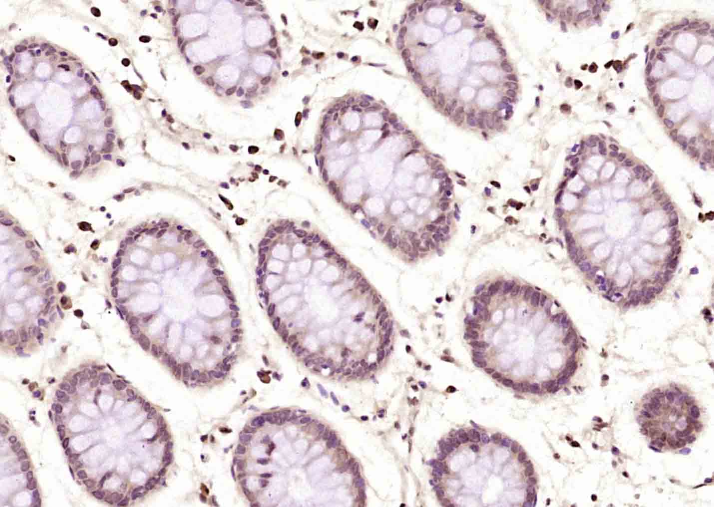 Paraformaldehyde-fixed, paraffin embedded (human colon carcinoma); Antigen retrieval by boiling in sodium citrate buffer (pH6.0) for 15min; Block endogenous peroxidase by 3% hydrogen peroxide for 20 minutes; Blocking buffer (normal goat serum) at 37°C for 30min; Antibody incubation with (Phospho-CDK1 (Thr161)) Polyclonal Antibody, Unconjugated (SL3481R) at 1:200 overnight at 4°C, followed by operating according to SP Kit(Rabbit) (sp-0023) instructionsand DAB staining.
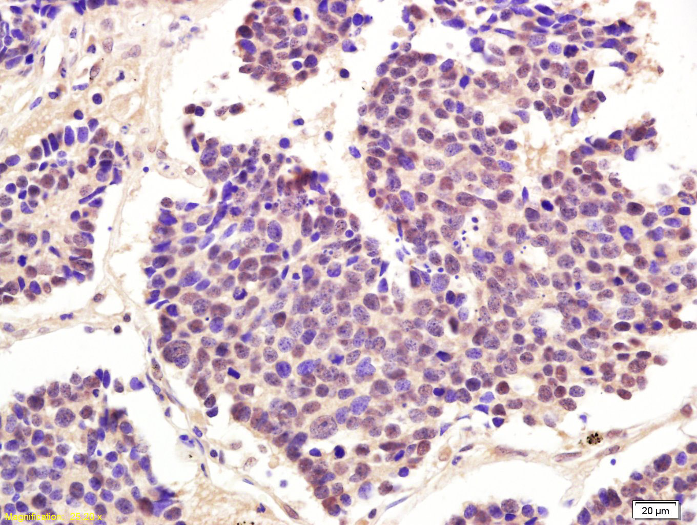 Tissue/cell: human lung carcinoma; 4% Paraformaldehyde-fixed and paraffin-embedded;
Antigen retrieval: citrate buffer ( 0.01M, pH 6.0 ), Boiling bathing for 15min; Block endogenous peroxidase by 3% Hydrogen peroxide for 30min; Blocking buffer (normal goat serum,C-0005) at 37℃ for 20 min;
Incubation: Anti-Phospho-cdc2(Thr161) Polyclonal Antibody, Unconjugated(SL3481R) 1:200, overnight at 4°C, followed by conjugation to the secondary antibody(SP-0023) and DAB(C-0010) staining
Cartpieces
Totalgoods,subtotals:¥Checkout
Bought notes(bought amounts latest0)
No one bought this product
User Comment(Total0User Comment Num)
- No comment


 +86 571 56623320
 +86 18668110335

