
Rabbit Anti-Phospho-Tie2 (Ser1119)antibody
Tie-2; Tie2; Tek; Angiopoietin-1 receptor; Tyrosine-protein kinase receptor TIE-2; hTIE2; Tyrosine-protein kinase receptor TEK; Tunica interna endothelial cell kinase; p140 TEK; Angiopoietin 1 receptor; CD202b; CD202b antigen; Endothelial tyrosine kinase
View History [Clear]
Details
Product Name Phospho-Tie2 (Ser1119) Chinese Name 磷酸化血管生成素受体2抗体 Alias Tie-2; Tie2; Tek; Angiopoietin-1 receptor; Tyrosine-protein kinase receptor TIE-2; hTIE2; Tyrosine-protein kinase receptor TEK; Tunica interna endothelial cell kinase; p140 TEK; Angiopoietin 1 receptor; CD202b; CD202b antigen; Endothelial tyrosine kinase; Endothelium specific receptor tyrosine kinase 2; hTIE 2; Hyk; Soluble TIE2 variant 1; Soluble TIE2 variant 2; tek tyrosine kinase; TEK tyrosine kinase endothelial; tek tyrosine kinase, endothelial; TIE 2; TIE2_HUMAN; Tunica interna endothelial cell kinase; Tyrosine kinase with Ig and EGF homology domains 2; Tyrosine protein kinase receptor TEK; Tyrosine protein kinase receptor TIE 2; Tyrosine-protein kinase receptor TIE-2; Venous malformations multiple cutaneous and mucosal; VMCM 1; VMCM; VMCM1; CD202b. Product Type Phosphorylated anti Research Area Tumour Cardiovascular Signal transduction Stem cells Growth factors and hormones Kinases and Phosphatases Immunogen Species Rabbit Clonality Polyclonal React Species Human, (predicted: Mouse, Rat, Chicken, Dog, Pig, Cow, Horse, Rabbit, Sheep, ) Applications ELISA=1:5000-10000 IHC-P=1:100-500 IHC-F=1:100-500 IF=1:100-500 (Paraffin sections need antigen repair)
not yet tested in other applications.
optimal dilutions/concentrations should be determined by the end user.Theoretical molecular weight 124kDa Cellular localization cytoplasmic The cell membrane Secretory protein Form Liquid Concentration 1mg/ml immunogen KLH conjugated Synthesised phosphopeptide derived from human Tie2 around the phosphorylation site of Ser1119: DC(p-S)AE Lsotype IgG Purification affinity purified by Protein A Buffer Solution 0.01M TBS(pH7.4) with 1% BSA, 0.03% Proclin300 and 50% Glycerol. Storage Shipped at 4℃. Store at -20 °C for one year. Avoid repeated freeze/thaw cycles. Attention This product as supplied is intended for research use only, not for use in human, therapeutic or diagnostic applications. PubMed PubMed Product Detail The TEK receptor tyrosine kinase is expressed almost exclusively in endothelial cells in mice, rats, and humans. This receptor possesses a unique extracellular domain containing 2 immunoglobulin-like loops separated by 3 epidermal growth factor-like repeats that are connected to 3 fibronectin type III-like repeats. The ligand for the receptor is angiopoietin-1. Defects in TEK are associated with inherited venous malformations; the TEK signaling pathway appears to be critical for endothelial cell-smooth muscle cell communication in venous morphogenesis.TEK is closely related to the TIE receptor tyrosine kinase.
Function:
Tyrosine-protein kinase that acts as cell-surface receptor for ANGPT1, ANGPT2 and ANGPT4 and regulates angiogenesis, endothelial cell survival, proliferation, migration, adhesion and cell spreading, reorganization of the actin cytoskeleton, but also maintenance of vascular quiescence. Has anti-inflammatory effects by preventing the leakage of proinflammatory plasma proteins and leukocytes from blood vessels. Required for normal angiogenesis and heart development during embryogenesis. Required for post-natal hematopoiesis. After birth, activates or inhibits angiogenesis, depending on the context. Inhibits angiogenesis and promotes vascular stability in quiescent vessels, where endothelial cells have tight contacts. In quiescent vessels, ANGPT1 oligomers recruit TEK to cell-cell contacts, forming complexes with TEK molecules from adjoining cells, and this leads to preferential activation of phosphatidylinositol 3-kinase and the AKT1 signaling cascades. In migrating endothelial cells that lack cell-cell adhesions, ANGT1 recruits TEK to contacts with the extracellular matrix, leading to the formation of focal adhesion complexes, activation of PTK2/FAK and of the downstream kinases MAPK1/ERK2 and MAPK3/ERK1, and ultimately to the stimulation of sprouting angiogenesis. ANGPT1 signaling triggers receptor dimerization and autophosphorylation at specific tyrosine residues that then serve as binding sites for scaffold proteins and effectors. Signaling is modulated by ANGPT2 that has lower affinity for TEK, can promote TEK autophosphorylation in the absence of ANGPT1, but inhibits ANGPT1-mediated signaling by competing for the same binding site. Signaling is also modulated by formation of heterodimers with TIE1, and by proteolytic processing that gives rise to a soluble TEK extracellular domain. The soluble extracellular domain modulates signaling by functioning as decoy receptor for angiopoietins. TEK phosphorylates DOK2, GRB7, GRB14, PIK3R1; SHC1 and TIE1.
Subunit:
Tyrosine-protein kinase that acts as cell-surface receptor for ANGPT1, ANGPT2 and ANGPT4 and regulates angiogenesis, endothelial cell survival, proliferation, migration, adhesion and cell spreading, reorganization of the actin cytoskeleton, but also maintenance of vascular quiescence. Has anti-inflammatory effects by preventing the leakage of proinflammatory plasma proteins and leukocytes from blood vessels. Required for normal angiogenesis and heart development during embryogenesis. Required for post-natal hematopoiesis. After birth, activates or inhibits angiogenesis, depending on the context. Inhibits angiogenesis and promotes vascular stability in quiescent vessels, where endothelial cells have tight contacts. In quiescent vessels, ANGPT1 oligomers recruit TEK to cell-cell contacts, forming complexes with TEK molecules from adjoining cells, and this leads to preferential activation of phosphatidylinositol 3-kinase and the AKT1 signaling cascades. In migrating endothelial cells that lack cell-cell adhesions, ANGT1 recruits TEK to contacts with the extracellular matrix, leading to the formation of focal adhesion complexes, activation of PTK2/FAK and of the downstream kinases MAPK1/ERK2 and MAPK3/ERK1, and ultimately to the stimulation of sprouting angiogenesis. ANGPT1 signaling triggers receptor dimerization and autophosphorylation at specific tyrosine residues that then serve as binding sites for scaffold proteins and effectors. Signaling is modulated by ANGPT2 that has lower affinity for TEK, can promote TEK autophosphorylation in the absence of ANGPT1, but inhibits ANGPT1-mediated signaling by competing for the same binding site. Signaling is also modulated by formation of heterodimers with TIE1, and by proteolytic processing that gives rise to a soluble TEK extracellular domain. The soluble extracellular domain modulates signaling by functioning as decoy receptor for angiopoietins. TEK phosphorylates DOK2, GRB7, GRB14, PIK3R1; SHC1 and TIE1.
Subcellular Location:
Cell membrane; Single-pass type I membrane protein. Cell junction. Cell junction, focal adhesion. Cytoplasm, cytoskeleton. Secreted.
Tissue Specificity:
Detected in umbilical vein endothelial cells. Proteolytic processing gives rise to a soluble extracellular domain that is detected in blood plasma (at protein level). Predominantly expressed in endothelial cells and their progenitors, the angioblasts. Has been directly found in placenta and lung, with a lower level in umbilical vein endothelial cells, brain and kidney.
Post-translational modifications:
Proteolytic processing leads to the shedding of the extracellular domain (soluble TIE-2 alias sTIE-2).
Autophosphorylated on tyrosine residues in response to ligand binding. Autophosphorylation occurs in trans, i.e. one subunit of the dimeric receptor phosphorylates tyrosine residues on the other subunit. Autophosphorylation occurs in a sequential manner, where Tyr-992 in the kinase activation loop is phosphorylated first, followed by autophosphorylation at Tyr-1108 and at additional tyrosine residues. ANGPT1-induced phosphorylation is impaired during hypoxia, due to increased expression of ANGPT2. Phosphorylation is important for interaction with GRB14, PIK3R1 and PTPN11. Phosphorylation at Tyr-1102 is important for interaction with SHC1, GRB2 and GRB7. Phosphorylation at Tyr-1108 is important for interaction with DOK2 and for coupling to downstream signal transduction pathways in endothelial cells. Dephosphorylated by PTPRB.
Ubiquitinated. The phosphorylated receptor is ubiquitinated and internalized, leading to its degradation.
DISEASE:
Defects in TEK are a cause of dominantly inherited venous malformations (VMCM) [MIM:600195]; an error of vascular morphogenesis characterized by dilated, serpiginous channels.
Note=May play a role in a range of diseases with a vascular component, including neovascularization of tumors, psoriasis and inflammation.
Similarity:
Belongs to the protein kinase superfamily. Tyr protein kinase family. Tie subfamily.
Contains 3 EGF-like domains.
Contains 3 fibronectin type-III domains.
Contains 2 Ig-like C2-type (immunoglobulin-like)domains.
Contains 1 protein kinase domain.
SWISS:
Q02763
Gene ID:
7010
Database links:Entrez Gene: 7010 Human
Entrez Gene: 21687 Mouse
Omim: 600221 Human
SwissProt: Q02763 Human
SwissProt: Q02858 Mouse
Unigene: 89640 Human
Unigene: 14313 Mouse
Tie2 是血管内皮特异性的酪氨酸激酶型受体, 主要表达在肺血管内皮以及卵泡、创口肉芽组织等血管内皮. 在血管发育中起重要的调节作用.Product Picture 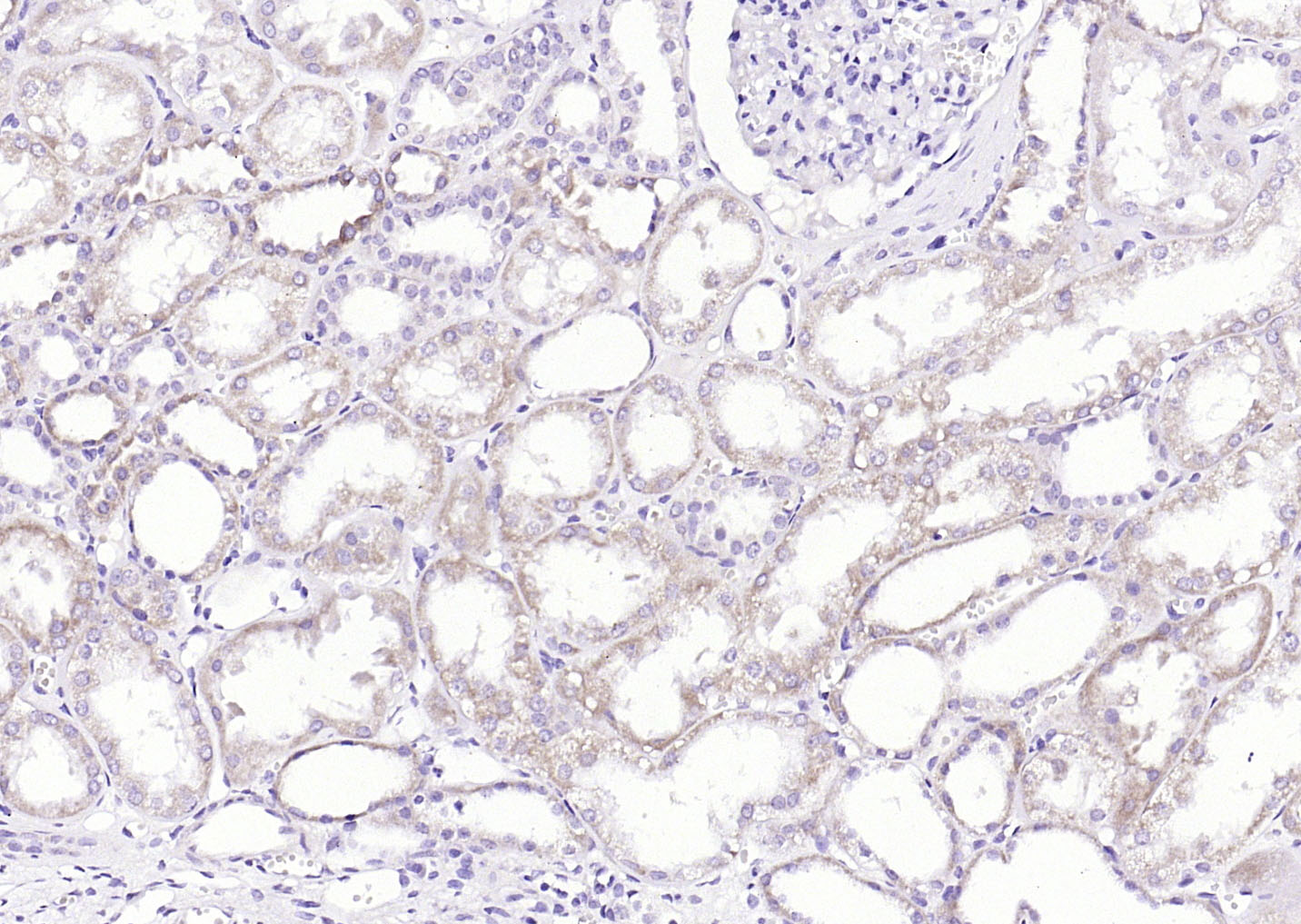 Paraformaldehyde-fixed, paraffin embedded (Human kidney); Antigen retrieval by boiling in sodium citrate buffer (pH6.0) for 15min; Block endogenous peroxidase by 3% hydrogen peroxide for 20 minutes; Blocking buffer (normal goat serum) at 37°C for 30min; Antibody incubation with (Phospho-Tie2 (Ser1119)) Polyclonal Antibody, Unconjugated (SL3448R) at 1:200 overnight at 4°C, followed by operating according to SP Kit(Rabbit) (sp-0023) instructionsand DAB staining.
Cartpieces
Totalgoods,subtotals:¥Checkout
Bought notes(bought amounts latest0)
No one bought this product
User Comment(Total0User Comment Num)
- No comment


 +86 571 56623320
 +86 18668110335

