
Rabbit Anti-Phospho-TAK1 (Thr187)antibody
TAK1 (phospho T187); TAK1(Phospho Thr187); TAK1(Phospho T187); MAP3K7; Mitogen-activated protein kinase kinase kinase 7; Transforming growth factor-beta-activated kinase 1; TGF-beta-activated kinase 1; MAP3K 7; MAPKKK7; Mitogen activated protein kinase ki
View History [Clear]
Details
Product Name Phospho-TAK1 (Thr187) Chinese Name 磷酸化转化生长因子β活化激酶1 Alias TAK1 (phospho T187); TAK1(Phospho Thr187); TAK1(Phospho T187); MAP3K7; Mitogen-activated protein kinase kinase kinase 7; Transforming growth factor-beta-activated kinase 1; TGF-beta-activated kinase 1; MAP3K 7; MAPKKK7; Mitogen activated protein kinase kinase kinase 7; TAK1; TGF beta activated kinase 1; TGF1a; Transforming growth factor beta activated kinase 1; M3K7_HUMAN; Map3k7; MEKK7; TGF-beta-activated kinase 1; TGF1a; Transforming growth factor-beta-activated kinase 1. literatures Product Type Phosphorylated anti Research Area Tumour immunology Signal transduction Apoptosis transcriptional regulatory factor Kinases and Phosphatases Immunogen Species Rabbit Clonality Polyclonal React Species Human, Mouse, Rat, (predicted: Chicken, Pig, Cow, Horse, Rabbit, ) Applications WB=1:500-2000 ELISA=1:5000-10000 IHC-P=1:100-500 IHC-F=1:100-500 Flow-Cyt=1μg/Test IF=1:100-500 (Paraffin sections need antigen repair)
not yet tested in other applications.
optimal dilutions/concentrations should be determined by the end user.Theoretical molecular weight 67kDa Cellular localization cytoplasmic The cell membrane Form Liquid Concentration 1mg/ml immunogen KLH conjugated Synthesised phosphopeptide derived from human TAK1 around the phosphorylation site of Thr187: HM(p-T)NN Lsotype IgG Purification affinity purified by Protein A Buffer Solution 0.01M TBS(pH7.4) with 1% BSA, 0.03% Proclin300 and 50% Glycerol. Storage Shipped at 4℃. Store at -20 °C for one year. Avoid repeated freeze/thaw cycles. Attention This product as supplied is intended for research use only, not for use in human, therapeutic or diagnostic applications. PubMed PubMed Product Detail The protein encoded by this gene is a member of the serine/threonine protein kinase family. This kinase mediates the signaling transduction induced by TGF beta and morphogenetic protein (BMP), and controls a variety of cell functions including transcription regulation and apoptosis. In response to IL-1, this protein forms a kinase complex including TRAF6, MAP3K7P1/TAB1 and MAP3K7P2/TAB2; this complex is required for the activation of nuclear factor kappa B. This kinase can also activate MAPK8/JNK, MAP2K4/MKK4, and thus plays a role in the cell response to environmental stresses. Four alternatively spliced transcript variants encoding distinct isoforms have been reported. [provided by RefSeq, Jul 2008]
Function:
Component of a protein kinase signal transduction cascade. Mediator of TRAF6 and TGF-beta signal transduction. Activates IKBKB and MAPK8 in response to TRAF6 signaling. Stimulates NF-kappa-B activation and the p38 MAPK pathway. In osmotic stress signaling, plays a major role in the activation of MAPK8/JNK, but not that of NF-kappa-B.
Subunit:
Binds both upstream activators and downstream substrates in multimolecular complexes. Interacts with TAB1/MAP3K7IP1 and TAB2/MAP3K7IP2. Identified in the TRIKA2 complex composed of MAP3K7, TAB1/MAP3K7IP1 and TAB2/MAP3K7IP2. Interacts with PPM1L. Interaction with PP2A and PPP6C leads to its repressed activity. Interacts with TRAF6 and TAB1/MAP3K7IP1; during IL-1 signaling. Interacts with TAOK1 and TAOK2; interaction with TAOK2 interferEs with MAP3K7 interaction with IKKA, thus preventing NF-kappa-B activation. Interacts with WDR34 (via WD domains). Interacts with RBCK1. Interacts with CYLD.
Subcellular Location:
Cytoplasm. Cell membrane; Peripheral membrane protein; Cytoplasmic side. Note=Although the majority of MAP3K7/TAK1 is found in the cytosol, when complexed with TAB1/MAP3K7IP1 and TAB2/MAP3K7IP2, it is also localized at the cell membrane.
Tissue Specificity:
Isoform 1A is the most abundant in ovary, skeletal muscle, spleen and blood mononuclear cells. Isoform 1B is highly expressed in brain, kidney and small intestine. Isoform 1C is the major form in prostate. Isoform 1D is the less abundant form.
Post-translational modifications:
Association with TAB1/MAP3K7IP1 promotes autophosphorylation and subsequent activation. Association with TAB2/MAP3K7IP2, itself associated with free unanchored Lys-63 polyubiquitin chain, promotes autophosphorylation and subsequent activation of MAP3K7. Dephosphorylation at Thr-187 by PP2A and PPP6C leads to inactivation.
Ubiquitinated, leading to proteasomal degradation. Requires 'Lys-63'-linked polyubiquitination for autophosphorylation and subsequent activation. 'Lys-63'-linked ubiquitination does not lead to proteasomal degradation. Deubiquitinated by CYLD, a protease that selectively cleaves 'Lys-63'-linked ubiquitin chains.
Similarity:
Belongs to the protein kinase superfamily. STE Ser/Thr protein kinase family. MAP kinase kinase kinase subfamily.
Contains 1 protein kinase domain.
SWISS:
O43318
Gene ID:
6885
Database links:Entrez Gene: 6885 Human
Entrez Gene: 26409 Mouse
Omim: 602614 Human
SwissProt: O43318 Human
SwissProt: Q62073 Mouse
Unigene: 722892 Human
Unigene: 258589 Mouse
Unigene: 24019 Rat
Product Picture 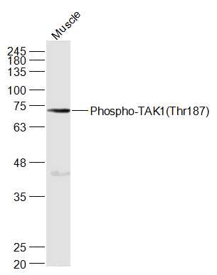 Sample:
Muscle (Mouse) Lysate at 40 ug
Primary: Anti-Phospho-TAK1(Thr187) (SL3438R) at 1/1000 dilution
Secondary: IRDye800CW Goat Anti-Rabbit IgG at 1/20000 dilution
Predicted band size: 67 kD
Observed band size: 70 kD
 Sample:
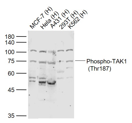
Lane 1: MCF-7 (Human) Cell Lysate at 30 ug
Lane 2: Hela (Human) Cell Lysate at 30 ug
Lane 3: A431 (Human) Cell Lysate at 30 ug
Lane 4: 293T (Human) Cell Lysate at 30 ug
Lane 5: K562 (Human) Cell Lysate at 30 ug
Primary:
Anti-Phospho-TAK1 (Thr187) (SL3438R) at 1/1000 dilution
Secondary: IRDye800CW Goat Anti-Rabbit IgG at 1/20000 dilution
Predicted band size: 78 kD
Observed band size: 75 kD
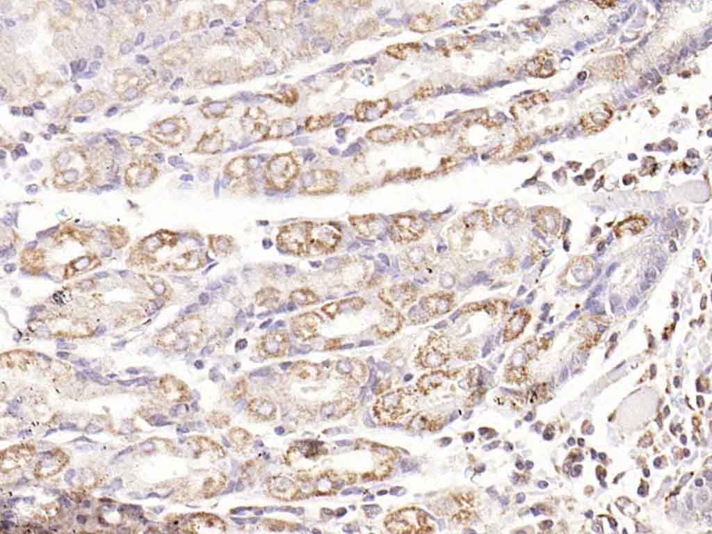 Paraformaldehyde-fixed, paraffin embedded (human gastric carcinoma); Antigen retrieval by boiling in sodium citrate buffer (pH6.0) for 15min; Block endogenous peroxidase by 3% hydrogen peroxide for 20 minutes; Blocking buffer (normal goat serum) at 37°C for 30min; Antibody incubation with (Phospho-TAK1 (Thr187)) Polyclonal Antibody, Unconjugated (SL3438R) at 1:200 overnight at 4°C, followed by operating according to SP Kit(Rabbit) (sp-0023) instructionsand DAB staining.
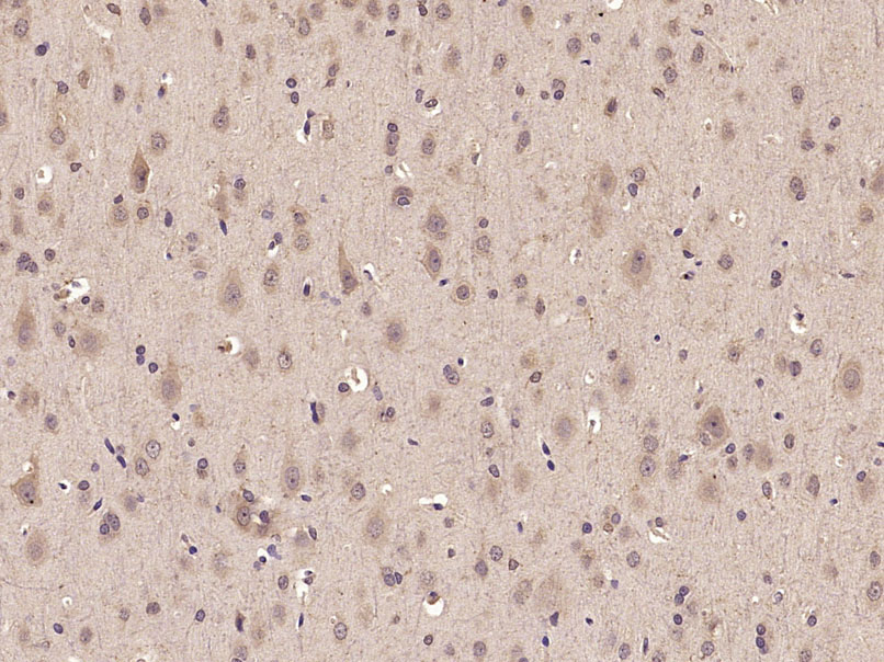 Paraformaldehyde-fixed, paraffin embedded (Rat brain); Antigen retrieval by microwave in sodium citrate buffer (pH6.0) ; Block endogenous peroxidase by 3% hydrogen peroxide for 30 minutes; Blocking buffer (3% BSA) at RT for 30min; Antibody incubation with (Phospho-TAK1(Thr187)) Polyclonal Antibody, Unconjugated (SL3438R) at 1:400 overnight at 4℃, followed by conjugation to the secondary antibody (labeled with HRP)and DAB staining.
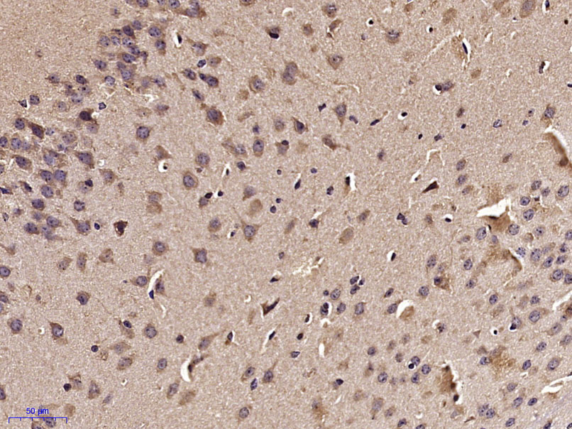 Paraformaldehyde-fixed, paraffin embedded (Mouse brain); Antigen retrieval by microwave in sodium citrate buffer (pH6.0) ; Block endogenous peroxidase by 3% hydrogen peroxide for 30 minutes; Blocking buffer (3% BSA) at RT for 30min; Antibody incubation with (Phospho-TAK1(Thr187)) Polyclonal Antibody, Unconjugated (SL3438R) at 1:400 overnight at 4℃, followed by conjugation to the secondary antibody (labeled with HRP)and DAB staining.
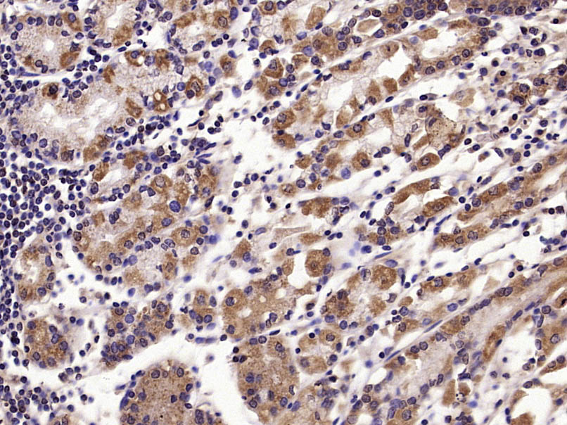 Paraformaldehyde-fixed, paraffin embedded (Human stomach); Antigen retrieval by microwave in sodium citrate buffer (pH6.0) ; Block endogenous peroxidase by 3% hydrogen peroxide for 30 minutes; Blocking buffer (3% BSA) at RT for 30min; Antibody incubation with (Phospho-TAK1(Thr187)) Polyclonal Antibody, Unconjugated (SL3438R) at 1:400 overnight at 4℃, followed by conjugation to the secondary antibody (labeled with HRP)and DAB staining.
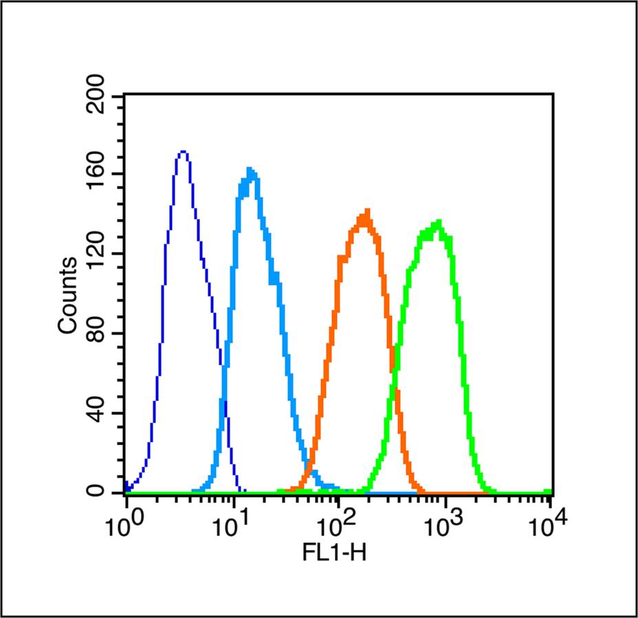 Blank control (Black line): Raji (Black).
Primary Antibody (green line): Rabbit Anti-Phospho-TAK1(Thr187) antibody (SL3438R)
Dilution: 1μg /10^6 cells;
Isotype Control Antibody (orange line): Rabbit IgG .
Secondary Antibody (white blue line): Goat anti-rabbit IgG-PE
Dilution: 1μg /test.
Protocol
The cells were fixed with 70% ice-cold methanol overnight at 4℃ and then permeabilized with 0.1% PBS-Tween for 20 min at room temperature (The cells were fixed with 2% paraformaldehyde (10 min) , then permeabilized with 90% ice-cold methanol for 20 min on ice.). Cells stained with Primary Antibody for 30 min at room temperature. The cells were then incubated in 1 X PBS/2%BSA/10% goat serum to block non-specific protein-protein interactions followed by the antibody for 15 min at room temperature. The secondary antibody used for 40 min at room temperature. Acquisition of 20,000 events was performed.
Cartpieces
Totalgoods,subtotals:¥Checkout
Bought notes(bought amounts latest0)
No one bought this product
User Comment(Total0User Comment Num)
- No comment


 +86 571 56623320
 +86 18668110335

