
Rabbit Anti-Phospho-MDM2 (Ser166)antibody
MDM2 (phospho S166); p-MDM2 (phospho S166); MDM2(Phospho Ser166); MDM2 (phospho S166); Double minute 2 protein; Hdm 2; HDM2; MDM 2; Mdm2 transformed 3T3 cell double minute 2 p53 binding protein (mouse) binding protein 104kDa; MDM2BP; Mouse Double Minute 2
View History [Clear]
Details
Product Name Phospho-MDM2 (Ser166) Chinese Name 磷酸化双微体2癌基因抗体 Alias MDM2 (phospho S166); p-MDM2 (phospho S166); MDM2(Phospho Ser166); MDM2 (phospho S166); Double minute 2 protein; Hdm 2; HDM2; MDM 2; Mdm2 transformed 3T3 cell double minute 2 p53 binding protein (mouse) binding protein 104kDa; MDM2BP; Mouse Double Minute 2; MTBP; Murine Double Minute Chromosome 2; Oncoprotein Mdm2; p53 Binding Protein Mdm2; Ubiquitin protein ligase E3 Mdm2. Product Type Phosphorylated anti Research Area Tumour Cell biology Apoptosis Cyclin transcriptional regulatory factor Epigenetics Immunogen Species Rabbit Clonality Polyclonal React Species Human, (predicted: Mouse, Rat, Horse, Rabbit, ) Applications IHC-P=1:100-500 IHC-F=1:100-500 Flow-Cyt=1μg/Test ICC=1:100 IF=1:100-500 (Paraffin sections need antigen repair)
not yet tested in other applications.
optimal dilutions/concentrations should be determined by the end user.Theoretical molecular weight 55kDa Cellular localization The nucleus cytoplasmic Form Liquid Concentration 1mg/ml immunogen KLH conjugated synthesised phosphopeptide derived from human MDM2 around the phosphorylation site of Ser166: AI(p-S)ET Lsotype IgG Purification affinity purified by Protein A Buffer Solution 0.01M TBS(pH7.4) with 1% BSA, 0.03% Proclin300 and 50% Glycerol. Storage Shipped at 4℃. Store at -20 °C for one year. Avoid repeated freeze/thaw cycles. Attention This product as supplied is intended for research use only, not for use in human, therapeutic or diagnostic applications. PubMed PubMed Product Detail Inhibits TP53/p53- and TP73/p73-mediated cell cyclearrest and apoptosis by binding its transcriptional activation domain. Functions as a ubiquitin ligase E3, in the presence of E1 and E2, toward p53 and itself. Permits the nuclear export of p53 and targets it for proteasome-mediated proteolysis. Binds p53, p73, ARF(P14), ribosomal protein L5 and specifically to RNA. Can interact also with retinoblastoma protein(RB), E1A-associated protein EP300 and the E2F1 transcription factor. Forms a ternary complex with TP53/p53 and WWOX. Interacts with CDKN2AIP, MTBP, TRBG1 and USP7. Isoform Mdm2-F does not interact with TP53/p53. Interacts with PYHIN1. Interacts with, and ubiquitinates HIV-1 Tat. Belongs to the MDM2/MDM4 family.
Function:
E3 ubiquitin-protein ligase that mediates ubiquitination of p53/TP53, leading to its degradation by the proteasome. Inhibits p53/TP53- and p73/TP73-mediated cell cycle arrest and apoptosis by binding its transcriptional activation domain. Also acts as an ubiquitin ligase E3 toward itself and ARRB1. Permits the nuclear export of p53/TP53. Promotes proteasome-dependent ubiquitin-independent degradation of retinoblastoma RB1 protein. nhibits DAXX-mediated apoptosis by inducing its ubiquitination and degradation. Component of the TRIM28/KAP1-MDM2-p53/TP53 complex involved in stabilizing p53/TP53. Also component of the TRIM28/KAP1-ERBB4-MDM2 complex which links growth factor and DNA damage response pathways. Mediates ubiquitination and subsequent proteasome degradation of DYRK2 in nucleus. Ubiquitinates IGF1R and promotes it to proteasomal degradation.
Subunit:
Binds p53/TP53, TP73/p73, ARF/P14, PML, RBL5 and RP11, and specifically to RNA. Can interact with RB1, E1A-associated protein EP300 and the E2F1 transcription factor. Forms a ternary complex with p53/TP53 and WWOX. Interacts with CDKN2AIP, MTBP, RFWD3, TBRG1, USP7, PYHIN1, UBXN6, and RBBP6. Isoform Mdm2-F does not interact with p53/TP53. Interacts with and ubiquitinates HIV-1 Tat. Interacts with ARRB1 and ARRB2. Interacts (isoform 2) with PSMA3. Found in a trimeric complex with MDM2, MDM4 and UPB2. Interacts with USP2 (via N-terminus and C-terminus). Interacts with MDM4. Part of a complex with MDM2, DAXX, RASSF1 and USP7. Part of a complex with DAXX, MDM2 and USP7. Interacts directly with DAXX and USP7. Interacts (via C-terminus) with RASSF1 isoform A (via N-terminus); the interaction is independent of TP53. Interacts with APEX1; leading to its ubiquitination and degradation. Interacts with RYBP; this inhibits ubiquitination of TP53. Identified in a complex with RYBP and p53/TP53. Also component of the TRIM28/KAP1-MDM2-p53/TP53 complex involved in regulating p53/TP53 stabilization and activity. Binds directly both p53/TP53 and TRIM28. Component of the TRIM28/KAP1-ERBB4-MDM2 complex involved in connecting growth factor responses with DNA damage. Interacts directly with both TRIM28 and ERBB4 in the complex. Interacts with DYRK2. Interacts with IGF1R. Interacts with TRIM13; the interaction ubiquitinates MDM2 leading to its proteasomal degradation.
Subcellular Location:
Nucleus, nucleoplasm. Cytoplasm. Nucleus, nucleolus. Note=Expressed predominantly in the nucleoplasm. Interaction with ARF(P14) results in the localization of both proteins to the nucleolus. The nucleolar localization signals in both ARF(P14) and MDM2 may be necessary to allow efficient nucleolar localization of both proteins. Colocalizes with RASSF1 isoform A in the nucleus.
Tissue Specificity:
Ubiquitous. Isoform Mdm2-A, isoform Mdm2-B, isoform Mdm2-C, isoform Mdm2-D, isoform Mdm2-E, isoform Mdm2-F and isoform Mdm2-G are observed in a range of cancers but absent in normal tissues.
Post-translational modifications:
Phosphorylated in response to ionizing radiation in an ATM-dependent manner. Phosphorylation on Ser-166 by SGK1 activates ubiquitination of p53/TP53.
Auto-ubiquitinated; which leads to proteasomal degradation. Also ubiquitinated by TRIM13. Deubiquitinated by USP2 leads to its accumulation and increases deubiquitination and degradation of p53/TP53. Deubiquitinated by USP7 leading to its stabilization.
DISEASE:
Note=Seems to be amplified in certain tumors (including soft tissue sarcomas, osteosarcomas and gliomas). A higher frequency of splice variants lacking p53 binding domain sequences was found in late-stage and high-grade ovarian and bladder carcinomas. Four of the splice variants show loss of p53 binding.
Similarity:
Belongs to the MDM2/MDM4 family.
Contains 1 RanBP2-type zinc finger.
Contains 1 RING-type zinc finger.
Contains 1 SWIB domain.
SWISS:
Q00987
Gene ID:
4193
Database links:Entrez Gene: 4193 Human
Entrez Gene: 17246 Mouse
Omim: 164785 Human
SwissProt: Q00987 Human
SwissProt: P23804 Mouse
Unigene: 484551 Human
Unigene: 22670 Mouse
Unigene: 447669 Mouse
Unigene: 91829 Rat
Mdm2基因在Tumour发生、发展的过程中起着重要作用,Mdm2蛋白过度表达与p53基因突变有重要的关联性。Mdm2基因在Tumour发生、发展的过程中起着重要作用。Product Picture 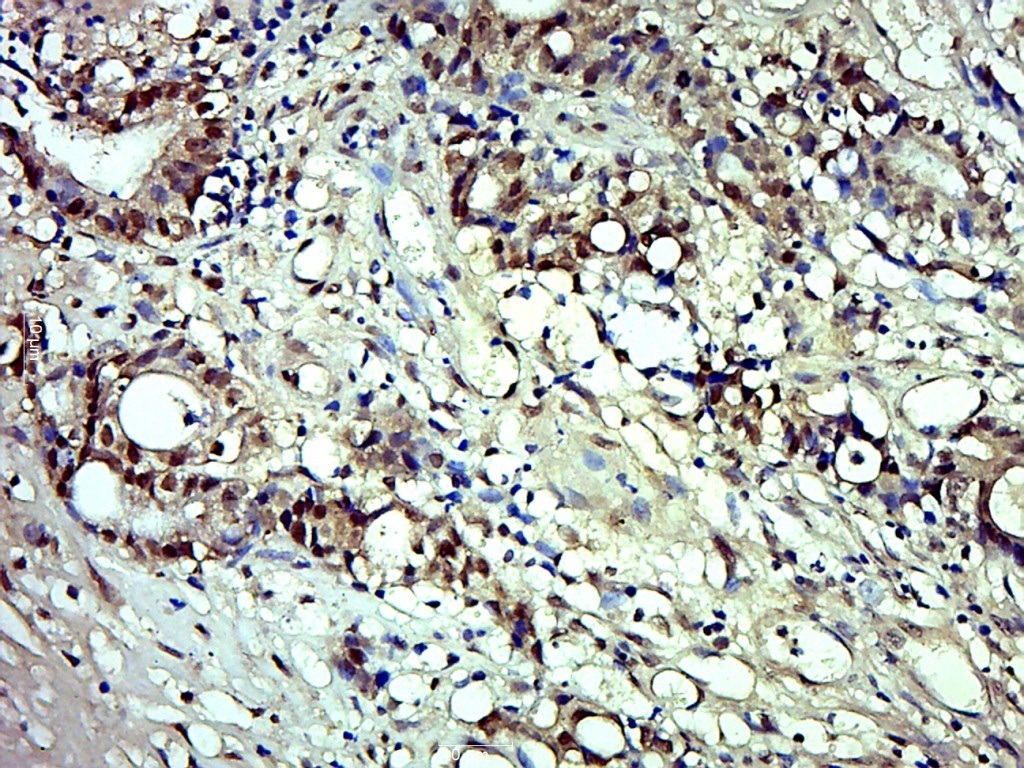 Paraformaldehyde-fixed, paraffin embedded (Human colon carcinoma); Antigen retrieval by boiling in sodium citrate buffer (pH6.0) for 15min; Block endogenous peroxidase by 3% hydrogen peroxide for 20 minutes; Blocking buffer (normal goat serum) at 37°C for 30min; Antibody incubation with (Phospho-MDM2 (Ser166)) Polyclonal Antibody, Unconjugated (SL3266R) at 1:500 overnight at 4°C, followed by a conjugated secondary (sp-0023) for 20 minutes and DAB staining.
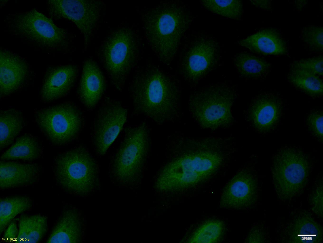 HepG2 cell; 4% Paraformaldehyde-fixed; Triton X-100 at room temperature for 20 min; Blocking buffer (normal goat serum, C-0005) at 37°C for 20 min; Antibody incubation with (Phospho-MDM2 (Ser166)) polyclonal Antibody, Unconjugated (SL3266R) 1:100, 90 minutes at 37°C; followed by a conjugated Goat Anti-Rabbit IgG antibody at 37°C for 90 minutes, DAPI (blue, C02-04002) was used to stain the cell nuclei.
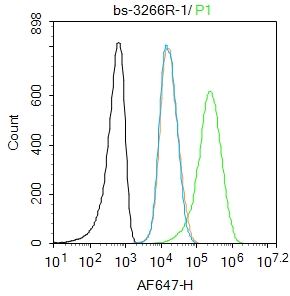 Blank control:Molt4.
Primary Antibody (green line): Rabbit Anti-Phospho-MDM2 (Ser166) antibody (SL3266R)
Dilution: 1μg /10^6 cells;
Isotype Control Antibody (orange line): Rabbit IgG .
Secondary Antibody : Goat anti-rabbit IgG-AF647
Dilution: 1μg /test.
Protocol
The cells were fixed with 4% PFA (10min at room temperature)and then permeabilized with 90% ice-cold methanol for 20 min at-20℃. The cells were then incubated in 5%BSA to block non-specific protein-protein interactions for 30 min at room temperature .Cells stained with Primary Antibody for 30 min at room temperature. The secondary antibody used for 40 min at room temperature. Acquisition of 20,000 events was performed.
Cartpieces
Totalgoods,subtotals:¥Checkout
Bought notes(bought amounts latest0)
No one bought this product
User Comment(Total0User Comment Num)
- No comment


 +86 571 56623320
 +86 18668110335

