
Rabbit Anti-phospho-JAK2 (Tyr221)antibody
JAK2 (phospho Y221); p-JAK2 (phospho Tyr221); Tyrosine protein kinase JAK2; JAK 2; JAK-2; JAK2; JAK2_HUMAN; Janus Activating Kinase 2; Janus Kinase 2; JTK 10; JTK10; OTTHUMP00000043260; Tyrosine-protein kinase JAK2; Tyrosine protein kinase JAK2.
View History [Clear]
Details
Product Name phospho-JAK2 (Tyr221) Chinese Name 磷酸化蛋白酪氨酸激酶JAK-2抗体 Alias JAK2 (phospho Y221); p-JAK2 (phospho Tyr221); Tyrosine protein kinase JAK2; JAK 2; JAK-2; JAK2; JAK2_HUMAN; Janus Activating Kinase 2; Janus Kinase 2; JTK 10; JTK10; OTTHUMP00000043260; Tyrosine-protein kinase JAK2; Tyrosine protein kinase JAK2. literatures Product Type Phosphorylated anti Research Area Tumour Cell biology immunology Chromatin and nuclear signals Signal transduction Kinases and Phosphatases Epigenetics Immunogen Species Rabbit Clonality Polyclonal React Species Human, Zebrafish, (predicted: Mouse, Rat, Dog, Pig, Horse, ) Applications ELISA=1:5000-10000 IHC-P=1:100-500 IHC-F=1:100-500 Flow-Cyt=1ug/Test ICC=1:100 IF=1:100-500 (Paraffin sections need antigen repair)
not yet tested in other applications.
optimal dilutions/concentrations should be determined by the end user.Theoretical molecular weight 131kDa Cellular localization The nucleus Form Liquid Concentration 1mg/ml immunogen KLH conjugated synthesised phosphopeptide derived from human JNK2 around the phosphorylation site of Tyr221: IQD(p-Y)HI Lsotype IgG Purification affinity purified by Protein A Buffer Solution 0.01M TBS(pH7.4) with 1% BSA, 0.03% Proclin300 and 50% Glycerol. Storage Shipped at 4℃. Store at -20 °C for one year. Avoid repeated freeze/thaw cycles. Attention This product as supplied is intended for research use only, not for use in human, therapeutic or diagnostic applications. PubMed PubMed Product Detail JAK2 (Janus Activating Kinase 2) is a tyrosine kinase of the non-receptor type, that associates with the intracellular domains of cytokine receptors; JAK2 is the predominant JAK kinase activated in response to several growth factors and cytokines such as IL-3, GM-CSF and erythropoietin; it has been found to be constitutively associated with the prolactin receptor and is required for responses to gamma interferon. Ligand binding to a variety of cell surface receptors (e.g., cytokine, growth factor, GPCRs) leads to an association of those receptors with JAK proteins, which are then activated via phosphorylation on tyrosines 1007 and 1008 in the kinase activation loop. Activated JAK proteins phosphorylate and activate STAT (signal transducers and activators of transcription) proteins, which then dimerize and translocate to the nucleus. Once in the nucleus, STAT proteins bind to DNA and modify the transcription of various genes.
Function:
Non-receptor tyrosine kinase involved in various processes such as cell growth, development, differentiation or histone modifications. Mediates essential signaling events in both innate and adaptive immunity. In the cytoplasm, plays a pivotal role in signal transduction via its association with type I receptors such as growth hormone (GHR), prolactin (PRLR), leptin (LEPR), erythropoietin (EPOR), thrombopoietin (THPO); or type II receptors including IFN-alpha, IFN-beta, IFN-gamma and multiple interleukins. Following ligand-binding to cell surface receptors, phosphorylates specific tyrosine residues on the cytoplasmic tails of the receptor, creating docking sites for STATs proteins. Subsequently, phosphorylates the STATs proteins once they are recruited to the receptor. Phosphorylated STATs then form homodimer or heterodimers and translocate to the nucleus to activate gene transcription. For example, cell stimulation with erythropoietin (EPO) during erythropoiesis leads to JAK2 autophosphorylation, activation, and its association with erythropoietin receptor (EPOR) that becomes phosphorylated in its cytoplasmic domain. Then, STAT5 (STAT5A or STAT5B) is recruited, phosphorylated and activated by JAK2. Once activated, dimerized STAT5 translocates into the nucleus and promotes the transcription of several essential genes involved in the modulation of erythropoiesis. In addition, JAK2 mediates angiotensin-2-induced ARHGEF1 phosphorylation. Plays a role in cell cycle by phosphorylating CDKN1B. Cooperates with TEC through reciprocal phosphorylation to mediate cytokine-driven activation of FOS transcription. In the nucleus, plays a key role in chromatin by specifically mediating phosphorylation of 'Tyr-41' of histone H3 (H3Y41ph), a specific tag that promotes exclusion of CBX5 (HP1 alpha) from chromatin.
Subunit:
Interacts with EPOR, LYN, SIRPA, SH2B1 and TEC. Interacts with IL23R, SKB1 and STAM2.
Subcellular Location:
Endomembrane system; Peripheral membrane protein. Cytoplasm. Nucleus.
Tissue Specificity:
Ubiquitously expressed throughout most tissues.
Post-translational modifications:
Autophosphorylated, leading to regulate its activity. Leptin promotes phosphorylation on tyrosine residues, including phosphorylation on Tyr-813. Autophosphorylation on Tyr-119 in response to EPO down-regulates its kinase activity. Autophosphorylation on Tyr-868, Tyr-966 and Tyr-972 in response to growth hormone (GH) are required for maximal kinase activity. Also phosphorylated by TEC.
DISEASE:
Note=Chromosomal aberrations involving JAK2 are found in both chronic and acute forms of eosinophilic, lymphoblastic and myeloid leukemia. Translocation t(8;9)(p22;p24) with PCM1 links the protein kinase domain of JAK2 to the major portion of PCM1. Translocation t(9;12)(p24;p13) with ETV6.
Defects in JAK2 are a cause of susceptibility to Budd-Chiari syndrome (BDCHS) [MIM:600880]. A syndrome caused by obstruction of hepatic venous outflow involving either the hepatic veins or the terminal segment of the inferior vena cava. Obstructions are generally caused by thrombosis and lead to hepatic congestion and ischemic necrosis. Clinical manifestations observed in the majority of patients include hepatomegaly, right upper quadrant pain and abdominal ascites. Budd-Chiari syndrome is associated with a combination of disease states including primary myeloproliferative syndromes and thrombophilia due to factor V Leiden, protein C deficiency and antithrombin III deficiency. Budd-Chiari syndrome is a rare but typical complication in patients with polycythemia vera.
Similarity:
Belongs to the protein kinase superfamily. Tyr protein kinase family. JAK subfamily.
Contains 1 FERM domain.
Contains 1 protein kinase domain.
Contains 1 SH2 domain.
SWISS:
O60674
Gene ID:
3717
Database links:Entrez Gene: 3717 Human
Entrez Gene: 16452 Mouse
GenBank: NP_004963 Human
Omim: 147796 Human
SwissProt: O60674 Human
SwissProt: Q62120 Mouse
Unigene: 656213 Human
Unigene: 275839 Mouse
Unigene: 18909 Rat
Product Picture 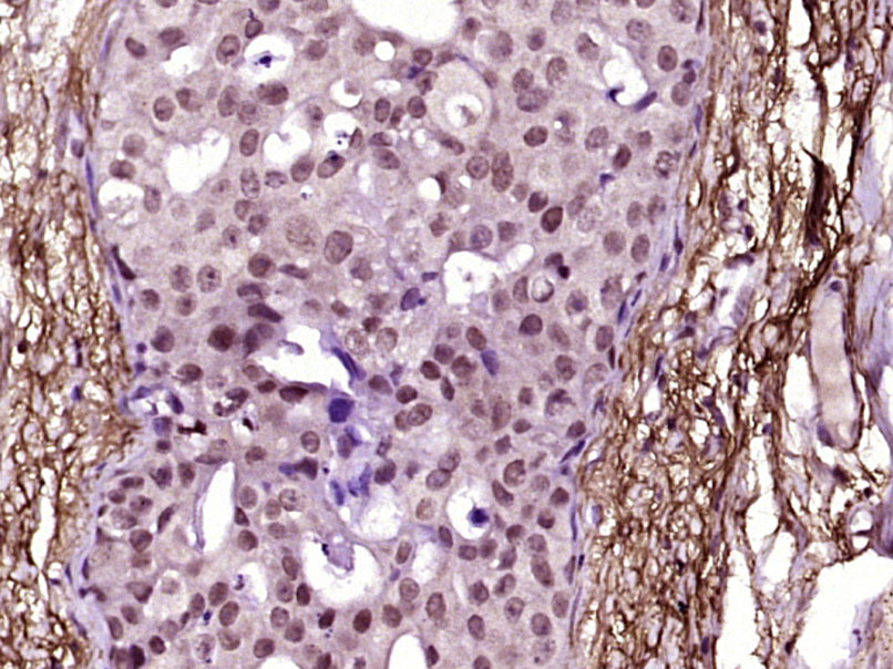 Paraformaldehyde-fixed, paraffin embedded (Human breast carcinoma); Antigen retrieval by boiling in sodium citrate buffer (pH6.0) for 15min; Block endogenous peroxidase by 3% hydrogen peroxide for 20 minutes; Blocking buffer (normal goat serum) at 37°C for 30min; Antibody incubation with (phospho-JAK2(Tyr221)) Polyclonal Antibody, Unconjugated (SL3206R) at 1:400 overnight at 4°C, followed by operating according to SP Kit(Rabbit) (sp-0023) instructionsand DAB staining.
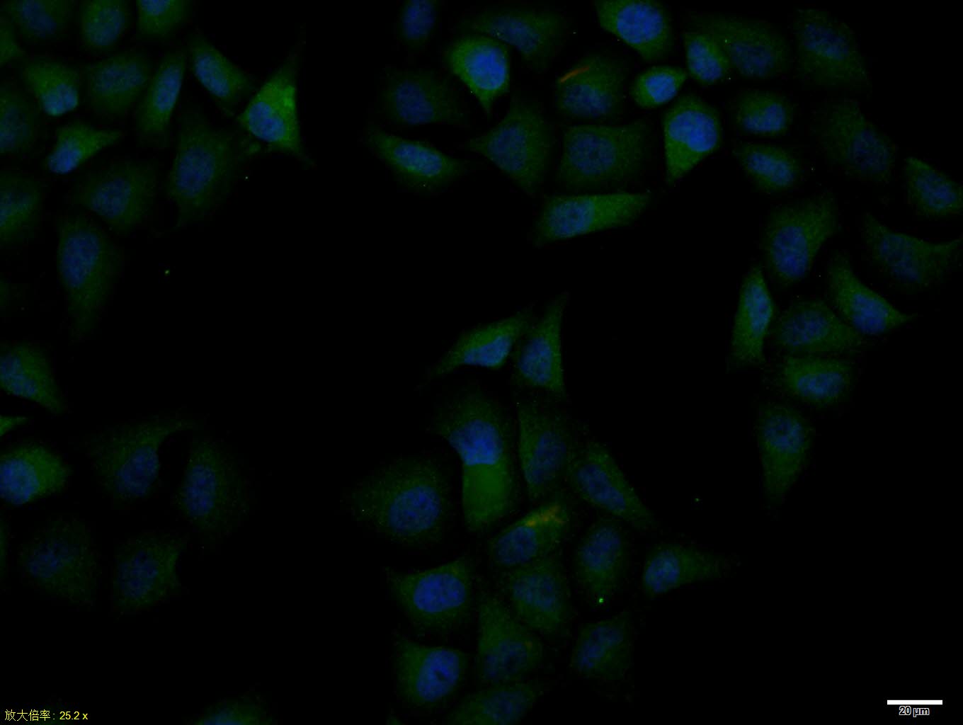 Hela cell; 4% Paraformaldehyde-fixed; Triton X-100 at room temperature for 20 min; Blocking buffer (normal goat serum, C-0005) at 37°C for 20 min; Antibody incubation with (phospho-JAK2 (Tyr221)) polyclonal Antibody, Unconjugated (SL3206R) 1:100, 90 minutes at 37°C; followed by a conjugated Goat Anti-Rabbit IgG antibody at 37°C for 90 minutes, DAPI (blue, C02-04002) was used to stain the cell nuclei.
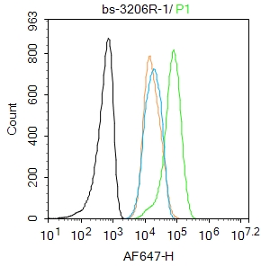 Blank control:HL-60
Primary Antibody (green line): Rabbit Anti-phospho-JAK2 (Tyr221) antibody (SL3206R)
Dilution: 1μg /10^6 cells;
Isotype Control Antibody (orange line): Rabbit IgG .
Secondary Antibody : Goat anti-rabbit IgG-AF647
Dilution: 1μg /test.
Protocol
The cells were fixed with 4% PFA (10min at room temperature)and then permeabilized with 90% ice-cold methanol for 20 min at-20℃.The cells were then incubated in 5%BSA to block non-specific protein-protein interactions for 30 min at room temperature .Cells stained with Primary Antibody for 30 min at room temperature. The secondary antibody used for 40 min at room temperature. Acquisition of 20,000 events was performed.
Cartpieces
Totalgoods,subtotals:¥Checkout
References (0)
No References
Bought notes(bought amounts latest0)
No one bought this product
User Comment(Total0User Comment Num)
- No comment


 +86 571 56623320
 +86 18668110335

