
Rabbit Anti-Phospho-IRF7 (Ser471 + Ser472)antibody
Interferon regulatory factor 7; Interferon regulatory factor 7H; IRF 7; IRF 7A; IRF 7H; IRF7A; IRF7; IRF7H.
View History [Clear]
Details
Product Name Phospho-IRF7 (Ser471 + Ser472) Chinese Name 磷酸化Interferon调节因子7抗体 Alias Interferon regulatory factor 7; Interferon regulatory factor 7H; IRF 7; IRF 7A; IRF 7H; IRF7A; IRF7; IRF7H. literatures Product Type Phosphorylated anti Research Area Tumour Signal transduction Apoptosis transcriptional regulatory factor Immunogen Species Rabbit Clonality Polyclonal React Species Human, Mouse, Rat, (predicted: Pig, Cow, Horse, ) Applications WB=1:500-2000 ELISA=1:5000-10000 IHC-P=1:100-500 ICC=1:100 (Paraffin sections need antigen repair)
not yet tested in other applications.
optimal dilutions/concentrations should be determined by the end user.Theoretical molecular weight 54kDa Cellular localization The nucleus cytoplasmic Form Liquid Concentration 1mg/ml immunogen KLH conjugated synthesised phosphopeptide derived from human IRF7 around the phosphorylation site of Ser471/472: GV(p-S)(p-S)LD Lsotype IgG Purification affinity purified by Protein A Buffer Solution 0.01M TBS(pH7.4) with 1% BSA, 0.03% Proclin300 and 50% Glycerol. Storage Shipped at 4℃. Store at -20 °C for one year. Avoid repeated freeze/thaw cycles. Attention This product as supplied is intended for research use only, not for use in human, therapeutic or diagnostic applications. PubMed PubMed Product Detail IRF7 encodes interferon regulatory factor 7, a member of the interferon regulatory transcription factor (IRF) family. IRF7 has been shown to play a role in the transcriptional activation of virus-inducible cellular genes, including interferon beta chain genes. Inducible expression of IRF7 is largely restricted to lymphoid tissue. Multiple IRF7 transcript variants have been identified, although the functional consequences of these have not yet been established. [provided by RefSeq, Jul 2008]
Function:
Transcriptional activator. Binds to the interferon-stimulated response element (ISRE) in IFN promoters and in the Q promoter (Qp) of EBV nuclear antigen 1 (EBNA1). Functions as a molecular switch for antiviral activity. Activated by phosphorylation in response to infection. Activation leads to nuclear retention, DNA binding, and derepression of transactivation ability.
Subunit:
Homodimer.
Subcellular Location:
Nucleus. Cytoplasm. The phosphorylated and active form accumulates selectively in the nucleus.
Tissue Specificity:
Expressed predominantly in spleen, thymus and peripheral blood leukocytes.
Post-translational modifications:
In response to a viral infection, phosphorylated on the C-terminal serine cluster. Phosphorylation, and subsequent activation is inhibited by vaccinia virus protein E3.
TRAF6-mediated ubiquitination is required for IRF7 activation.
Similarity:
Belongs to the IRF family.
Contains 1 IRF tryptophan pentad repeat DNA-binding domain.
SWISS:
Q92985
Gene ID:
3665
Database links:Entrez Gene: 3665 Human
Entrez Gene: 54123 Mouse
Omim: 605047 Human
SwissProt: Q92985 Human
SwissProt: P70434 Mouse
Unigene: 166120 Human
Unigene: 3233 Mouse
Unigene: 101159 Rat
Product Picture 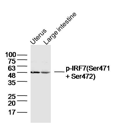 Sample:
Uterus (Mouse) Lysate at 40 ug
Large intestine (Mouse) Lysate at 40 ug
Primary: Anti-Phospho-IRF7 (Ser471 + Ser472)(SL3196R)at 1/300 dilution
Secondary: IRDye800CW Goat Anti-Rabbit IgG at 1/20000 dilution
Predicted band size: 54kD
Observed band size: 49kD
 Sample:
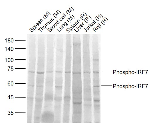
Lane 1: Mouse Spleen tissue lysates
Lane 2: Mouse Thymus tissue lysates
Lane 3: Mouse Blood cell lysates
Lane 4: Mouse Lung tissue lysates
Lane 5: Rat Spleen tissue lysates
Lane 6: Rat Liver tissue lysates
Lane 7: Human Jurkat cell lysates
Lane 8: Human Raji cell lysates
Primary: Anti-Phospho-IRF7 (Ser471 + Ser472) (SL3196R) at 1/1000 dilution
Secondary: IRDye800CW Goat Anti-Rabbit IgG at 1/20000 dilution
Predicted band size: 54 kD
Observed band size: 70,55 kD
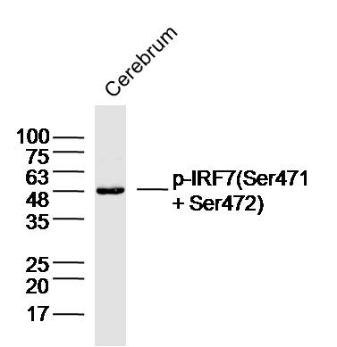 Sample:Cerebrum (Rat) Lysate at 40 ug
Primary: Anti-Phospho-IRF7 (Ser471 + Ser472)(SL3196R)at 1/300 dilution
Secondary: IRDye800CW Goat Anti-Rabbit IgG at 1/20000 dilution
Predicted band size: 54kD
Observed band size: 48kD
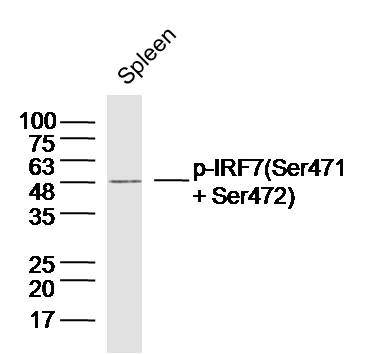 Sample:Spleen (Mouse) Lysate at 40 ug
Primary: Anti-Phospho-IRF7 (Ser471 + Ser472)(SL3196R)at 1/300 dilution
Secondary: IRDye800CW Goat Anti-Rabbit IgG at 1/20000 dilution
Predicted band size: 54kD
Observed band size: 49kD
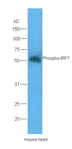 Sample: Heart(Mouse) lysate at 30ug;
Primary: Anti-Phospho-IRF7 (Ser471+Ser472) (SL3196R) at 1:200 dilution;
Secondary: HRP conjugated Goat Anti-Rabbit IgG(SL0295G-HRP) at 1: 5000 dilution;
Predicted band size : 54kD
Observed band size : 51kD
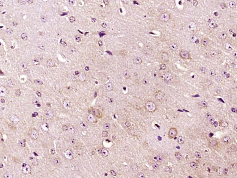 Paraformaldehyde-fixed, paraffin embedded (Mouse brain); Antigen retrieval by boiling in sodium citrate buffer (pH6.0) for 15min; Block endogenous peroxidase by 3% hydrogen peroxide for 20 minutes; Blocking buffer (normal goat serum) at 37°C for 30min; Antibody incubation with (Phospho-IRF7 (Ser471 + Ser472)) Polyclonal Antibody, Unconjugated (SL3196R) at 1:400 overnight at 4°C, followed by operating according to SP Kit(Rabbit) (sp-0023) instructionsand DAB staining.
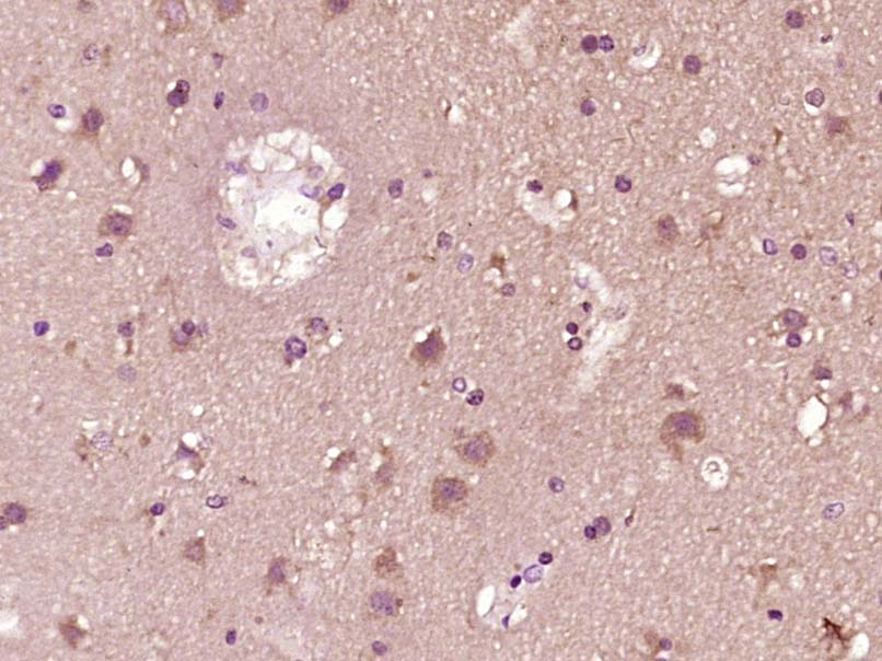 Paraformaldehyde-fixed, paraffin embedded (Human brain glioma); Antigen retrieval by boiling in sodium citrate buffer (pH6.0) for 15min; Block endogenous peroxidase by 3% hydrogen peroxide for 20 minutes; Blocking buffer (normal goat serum) at 37°C for 30min; Antibody incubation with (Phospho-IRF7 (Ser471 + Ser472)) Polyclonal Antibody, Unconjugated (SL3196R) at 1:400 overnight at 4°C, followed by operating according to SP Kit(Rabbit) (sp-0023) instructionsand DAB staining.
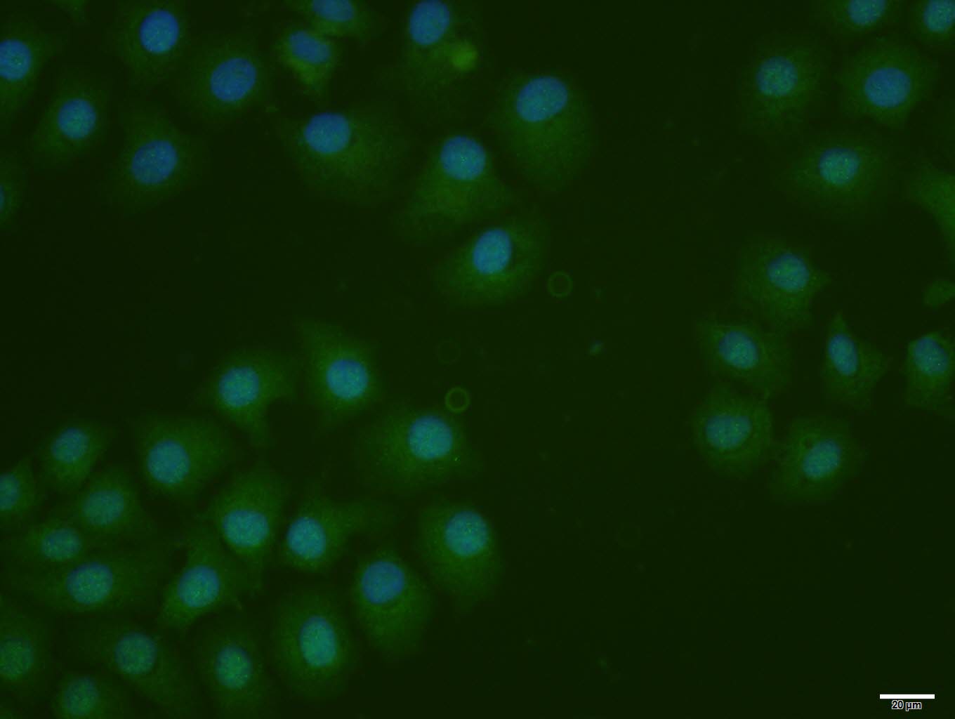 HepG2 cell; 4% Paraformaldehyde-fixed; Triton X-100 at room temperature for 20 min; Blocking buffer (normal goat serum, C-0005) at 37°C for 20 min; Antibody incubation with (Phospho-IRF7 (Ser471 + Ser472)) polyclonal Antibody, Unconjugated (SL3196R) 1:100, 90 minutes at 37°C; followed by a conjugated Goat Anti-Rabbit IgG antibody at 37°C for 90 minutes, DAPI (blue, C02-04002) was used to stain the cell nuclei.
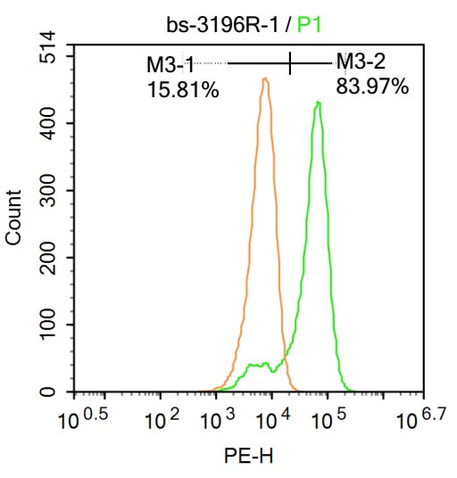 Blank control:Molt-4.
Primary Antibody (green line): Rabbit Anti-Phospho-IRF7 (Ser471 + Ser472) antibody (SL3196R)
Dilution: 1μg /10^6 cells;
Isotype Control Antibody (orange line): Rabbit IgG .
Secondary Antibody : Goat anti-rabbit IgG-AF647
Dilution: 1μg /test.
Protocol
The cells were fixed with 4% PFA (10min at room temperature)and then permeabilized with 90% ice-cold methanol for 20 min at-20℃. The cells were then incubated in 5%BSA to block non-specific protein-protein interactions for 30 min at at room temperature .Cells stained with Primary Antibody for 30 min at room temperature. The secondary antibody used for 40 min at room temperature. Acquisition of 20,000 events was performed.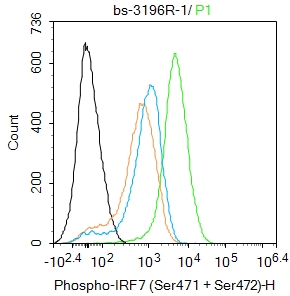 Blank control:Mouse spleen.
Primary Antibody (green line): Rabbit Anti-Phospho-IRF7 (Ser471 + Ser472) antibody (SL3196R)
Dilution: 1μg /10^6 cells;
Isotype Control Antibody (orange line): Rabbit IgG .
Secondary Antibody : Goat anti-rabbit IgG-FITC
Dilution: 1μg /test.
Protocol
The cells were fixed with 4% PFA (10min at room temperature)and then permeabilized with 90% ice-cold methanol for 20 min at-20℃. The cells were then incubated in 5%BSA to block non-specific protein-protein interactions for 30 min at room temperature .Cells stained with Primary Antibody for 30 min at room temperature. The secondary antibody used for 40 min at room temperature. Acquisition of 20,000 events was performed.
Cartpieces
Totalgoods,subtotals:¥Checkout
References (0)
No References
Bought notes(bought amounts latest0)
No one bought this product
User Comment(Total0User Comment Num)
- No comment


 +86 571 56623320
 +86 18668110335

