
Rabbit Anti-IRF3 antibody
Interferon regulatory factor 3; IRF-3; IRF 3; IRF3_HUMAN.
View History [Clear]
Details
Product Name IRF3 Chinese Name Interferon调节因子3 Alias Interferon regulatory factor 3; IRF-3; IRF 3; IRF3_HUMAN. literatures Research Area Tumour immunology Apoptosis transcriptional regulatory factor Immunogen Species Rabbit Clonality Polyclonal React Species Human, Mouse, Rat, (predicted: Dog, Pig, Cow, Horse, ) Applications WB=1:500-2000 ELISA=1:5000-10000 IHC-P=1:100-500 IHC-F=1:100-500 Flow-Cyt=2ug/Test IF=1:100-500 (Paraffin sections need antigen repair)
not yet tested in other applications.
optimal dilutions/concentrations should be determined by the end user.Theoretical molecular weight 47kDa Cellular localization The nucleus cytoplasmic Form Liquid Concentration 1mg/ml immunogen KLH conjugated synthetic peptide derived from human IRF3: 51-150/427 Lsotype IgG Purification affinity purified by Protein A Buffer Solution 0.01M TBS(pH7.4) with 1% BSA, 0.03% Proclin300 and 50% Glycerol. Storage Shipped at 4℃. Store at -20 °C for one year. Avoid repeated freeze/thaw cycles. Attention This product as supplied is intended for research use only, not for use in human, therapeutic or diagnostic applications. PubMed PubMed Product Detail This gene encodes a member of the interferon regulatory transcription factor (IRF) family. The encoded protein is found in an inactive cytoplasmic form that upon serine/threonine phosphorylation forms a complex with CREBBP. This complex translocates to the nucleus and activates the transcription of interferons alpha and beta, as well as other interferon-induced genes. Alternatively spliced transcript variants encoding multiple isoforms have been observed for this gene. [provided by RefSeq, Nov 2011].
Function:
Key transcriptional regulator of type I interferon (IFN)-dependent immune responses and plays a critical role in the innate immune response against DNA and RNA viruses. Regulates the transcription of type I IFN genes (IFN-alpha and IFN-beta) and IFN-stimulated genes (ISG) by binding to an interferon-stimulated response element (ISRE) in their promoters. Acts as a more potent activator of the IFN-beta (IFNB) gene than the IFN-alpha (IFNA) gene and plays a critical role in both the early and late phases of the IFNA/B gene induction. Found in an inactive form in the cytoplasm of uninfected cells and following viral infection, double-stranded RNA (dsRNA), or toll-like receptor (TLR) signaling, becomes phosphorylated by IKBKE and TBK1 kinases. This induces a conformational change, leading to its dimerization and nuclear localization and association with CREB binding protein (CREBBP) to form dsRNA-activated factor 1 (DRAF1), a complex which activates the transcription of the type I IFN and ISG genes. Can activate distinct gene expression programs in macrophages and can induce significant apoptosis in primary macrophages.
Subunit:
Monomer. Homodimer; phosphorylation-induced. Heterodimer with IRF7. Interacts with CREBBP. May interact with MAVS. Interacts with IKBKE and TBK1. Interacts with TICAM1 and TICAM2. Interacts with rotavirus A NSP1 (via C-terminus); this interaction leads to the proteasome-dependent degradation of IRF3. Interacts with RBCK1. Interacts with TRIM21. Interacts with HERC5.
Subcellular Location:
Cytoplasm. Nucleus. Note=Shuttles between cytoplasmic and nuclear compartments, with export being the prevailing effect. When activated, IRF3 interaction with CREBBP prevents its export to the cytoplasm.
Tissue Specificity:
Expressed constitutively in a variety of tissues.
Post-translational modifications:
Constitutively phosphorylated on many serines residues. C-terminal serine/threonine cluster is phosphorylated in response of induction by IKBKE and TBK1. Ser-385 and Ser-386 may be specifically phosphorylated in response to induction. An alternate model propose that the five serine/threonine residues between 396 and 405 are phosphorylated in response to a viral infection. Phosphorylation, and subsequent activation of IRF3 is inhibited by vaccinia virus protein E3.
Ubiquitinated; ubiquitination involves RBCK1 leading to proteasomal degradation. Polyubiquitinated; ubiquitination involves TRIM21 leading to proteasomal degradation.
ISGylated by HERC5 resulting in sustained IRF3 activation and in the inhibition of IRF3 ubiquitination by disrupting PIN1 binding. The phosphorylation state of IRF3 does not alter ISGylation.
Similarity:
Belongs to the IRF family.
Contains 1 IRF tryptophan pentad repeat DNA-binding domain.
SWISS:
Q14653
Gene ID:
3661
Database links:Entrez Gene: 3661 Human
Entrez Gene: 54131 Mouse
Omim: 603734 Human
SwissProt: Q14653 Human
SwissProt: P70671 Mouse
Unigene: 289052 Human
Unigene: 75254 Human
Unigene: 3960 Mouse
Interferon调节因子家族是一大类对Interferon起调控作用的转录因子的统称。 一般认为Interferon调节因子(IRF)通过调节Interferon的表达而行使其抗病毒、应激、免疫调节功能。近年来,研究人员发现IRF在Apoptosis、细胞周期、Cell differentiation、Tumour发生中也起着重要的调节作用。Product Picture 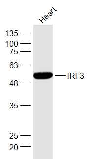 Sample:
Heart (Mouse) Lysate at 40 ug
Primary: Anti-IRF3 (SL2993R) at 1/1000 dilution
Secondary: IRDye800CW Goat Anti-Rabbit IgG at 1/20000 dilution
Predicted band size: 47 kD
Observed band size: 51 kD
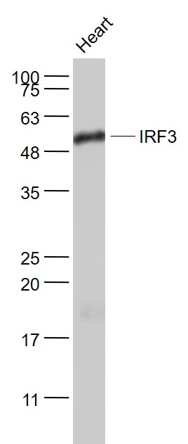 Sample:
Heart (Mouse) Lysate at 40 ug
Primary: Anti- IRF3 (SL2993R) at 1/1000 dilution
Secondary: IRDye800CW Goat Anti-Rabbit IgG at 1/20000 dilution
Predicted band size: 47 kD
Observed band size: 55 kD
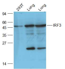 Sample:
293T(Human) Cell Lysate at 30 ug
Lung (Mouse) Lysate at 40 ug
Lung (Rat) Lysate at 40 ug
Primary: Anti-IRF3 (SL2993R) at 1/2000 dilution
Secondary: IRDye800CW Goat Anti-Rabbit IgG at 1/20000 dilution
Predicted band size: 47 kD
Observed band size: 47 kD
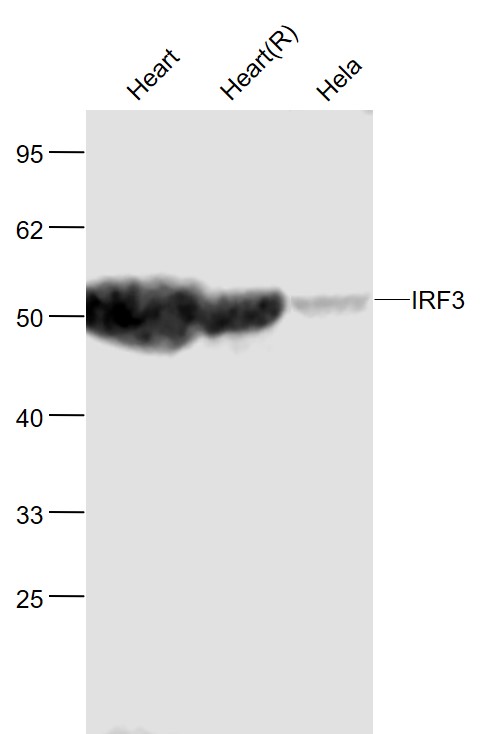 Sample:
Heart(Mouse) Lysate at 40 ug
Heart(Rat) Lysate at 40 ug
Hela(Human) Cell Lysate at 30 ug
Primary: Anti-IRF3 (SL2993R) at 1/1000 dilution
Secondary: IRDye800CW Goat Anti-Rabbit IgG at 1/20000 dilution
Predicted band size: 50 kD
Observed band size: 50 kD
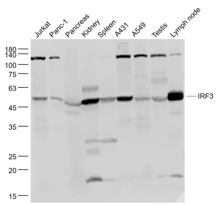 Sample:
Jurkat(Human) Cell Lysate at 30 ug
Panc-1(Human) Cell Lysate at 30 ug
Pancreas (Mouse) Lysate at 40 ug
Kidney (Mouse) Lysate at 40 ug
Spleen (Mouse) Lysate at 40 ug
A431(Human) Cell Lysate at 30 ug
A549(Human) Cell Lysate at 30 ug
Testis (Mouse) Lysate at 40 ug
Lymph node (Mouse) Lysate at 40 ug
Primary: Anti- IRF3 (SL2993R) at 1/1000 dilution
Secondary: IRDye800CW Goat Anti-Rabbit IgG at 1/20000 dilution
Predicted band size: 47 kD
Observed band size: 54 kD
 Sample:
Hepg2(Human) Lysate at 40 ug
Primary: Anti-IRF3 (SL2993R) at 1/1000 dilution
Secondary: IRDye800CW Goat Anti-Rabbit IgG at 1/20000 dilution
Predicted band size: 47 kD
Observed band size: 51 kD
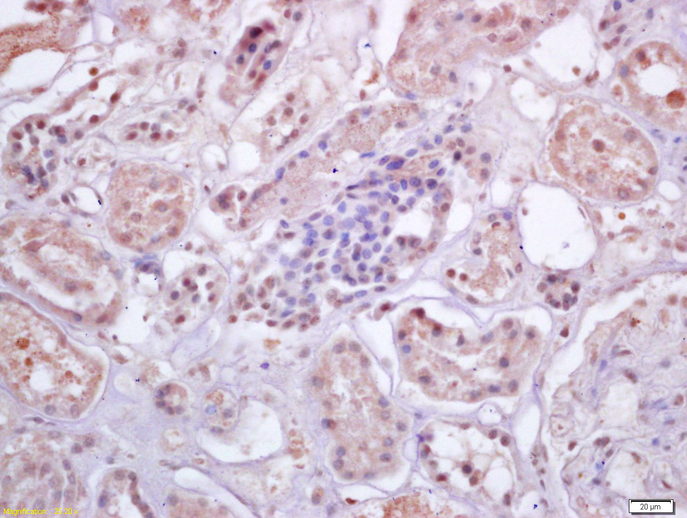 Tissue/cell: human kidney tissue; 4% Paraformaldehyde-fixed and paraffin-embedded;
Antigen retrieval: citrate buffer ( 0.01M, pH 6.0 ), Boiling bathing for 15min; Block endogenous peroxidase by 3% Hydrogen peroxide for 30min; Blocking buffer (normal goat serum,C-0005) at 37℃ for 20 min;
Incubation: Anti-IRF3 Polyclonal Antibody, Unconjugated(SL2993R) 1:200, overnight at 4°C, followed by conjugation to the secondary antibody(SP-0023) and DAB(C-0010) staining
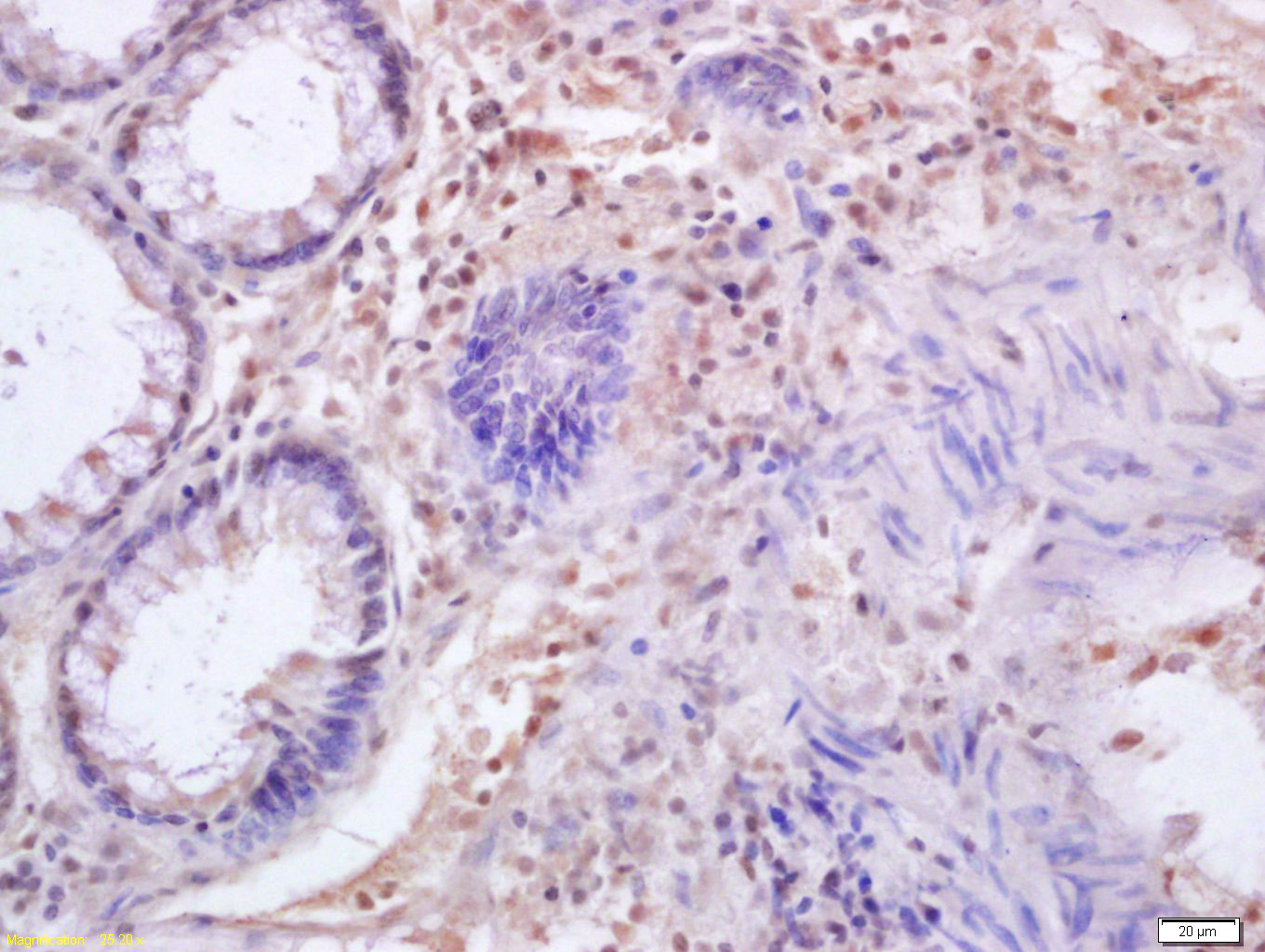 Tissue/cell: human colon carcinoma; 4% Paraformaldehyde-fixed and paraffin-embedded;
Antigen retrieval: citrate buffer ( 0.01M, pH 6.0 ), Boiling bathing for 15min; Block endogenous peroxidase by 3% Hydrogen peroxide for 30min; Blocking buffer (normal goat serum,C-0005) at 37℃ for 20 min;
Incubation: Anti-IRF3 Polyclonal Antibody, Unconjugated(SL2993R) 1:200, overnight at 4°C, followed by conjugation to the secondary antibody(SP-0023) and DAB(C-0010) staining
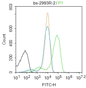 Blank control:Jurkat.
Primary Antibody (green line): Rabbit Anti-IRF3 antibody (SL2993R)
Dilution: 2μg /10^6 cells;
Isotype Control Antibody (orange line): Rabbit IgG .
Secondary Antibody : Goat anti-rabbit IgG-FITC
Dilution: 1μg /test.
Protocol
The cells were fixed with 4% PFA (10min at room temperature)and then permeabilized with 90% ice-cold methanol for 20 min at-20℃. The cells were then incubated in 5%BSA to block non-specific protein-protein interactions for 30 min at room temperature .Cells stained with Primary Antibody for 30 min at room temperature. The secondary antibody used for 40 min at room temperature. Acquisition of 20,000 events was performed.
Cartpieces
Totalgoods,subtotals:¥Checkout
Bought notes(bought amounts latest0)
No one bought this product
User Comment(Total0User Comment Num)
- No comment


 +86 571 56623320
 +86 18668110335

