
Rabbit Anti-Vitamin D Receptor antibody
Vitamin D3 receptor; 125 dihydroxyvitamin D3 receptor; 1 antibody 1,25-@dihydroxyvitamin D3 receptor; 125 dihydroxyvitamin D3 receptor; 25-dihydroxyvitamin D3 receptor; NR1I1; Nuclear receptor subfamily 1 group I member 1; VDR; VDR_HUMAN; Vitamin D (1,25-
View History [Clear]
Details
Product Name Vitamin D Receptor Chinese Name 维生素D3受体抗体 Alias Vitamin D3 receptor; 125 dihydroxyvitamin D3 receptor; 1 antibody 1,25-@dihydroxyvitamin D3 receptor; 125 dihydroxyvitamin D3 receptor; 25-dihydroxyvitamin D3 receptor; NR1I1; Nuclear receptor subfamily 1 group I member 1; VDR; VDR_HUMAN; Vitamin D (1,25- dihydroxyvitamin D3) receptor; Vitamin D hormone receptor; Vitamin D receptor; Vitamin D3 receptor, literatures Research Area Cell biology immunology Chromatin and nuclear signals Immunogen Species Rabbit Clonality Polyclonal React Species Human, Rat, (predicted: Mouse, Chicken, Pig, Cow, Horse, Rabbit, ) Applications IHC-P=1:100-500 IHC-F=1:100-500 ICC=1:100 IF=1:100-500 (Paraffin sections need antigen repair)
not yet tested in other applications.
optimal dilutions/concentrations should be determined by the end user.Theoretical molecular weight 47kDa Cellular localization The nucleus Form Liquid Concentration 1mg/ml immunogen KLH conjugated synthetic peptide derived from human Vitamin D Receptor: 65-180/427 Lsotype IgG Purification affinity purified by Protein A Buffer Solution 0.01M TBS(pH7.4) with 1% BSA, 0.03% Proclin300 and 50% Glycerol. Storage Shipped at 4℃. Store at -20 °C for one year. Avoid repeated freeze/thaw cycles. Attention This product as supplied is intended for research use only, not for use in human, therapeutic or diagnostic applications. PubMed PubMed Product Detail Nuclear hormone receptor. Transcription factor that mediates the action of vitamin D3 by controlling the expression of hormone sensitive genes. Regulates transcription of hormone sensitive genes via its association with the WINAC complex, a chromatin-remodeling complex. Recruited to promoters via its interaction with the WINAC complex subunit BAZ1B/WSTF, which mediates the interaction with acetylated histones, an essential step for VDR-promoter association. Plays a central role in calcium homeostasis.
Function:
Nuclear hormone receptor. Transcription factor that mediates the action of vitamin D3 by controlling the expression of hormone sensitive genes. Regulates transcription of hormone ensitive genes via its association with the WINAC complex, a chromatin-remodeling complex. Recruited to promoters via its interaction with the WINAC complex subunit BAZ1B/WSTF, which mediates the interaction with acetylated histones, an essential step for VDR-promoter association. Plays a central role in calcium homeostasis.
Subunit:
Homodimer in the absence of bound vitamin D3. Heterodimer with RXRA after vitamin D3 binding. Interacts with SMAD3. Interacts with MED1, NCOA1, NCOA2, NCOA3 and NCOA6 coactivators, leading to a strong increase of transcription of target genes. Interacts (in a ligand-dependent manner) with BAZ1B/WSTF.
Subcellular Location:
Nucleus.
DISEASE:
Defects in VDR are the cause of rickets vitamin D-dependent type 2A (VDDR2A) [MIM:277440]. A disorder of vitamin D metabolism resulting in severe rickets, hypocalcemia and secondary hyperparathyroidism. Most patients have total alopecia in addition to rickets.
Similarity:
Belongs to the nuclear hormone receptor family. NR1 subfamily.
Contains 1 nuclear receptor DNA-binding domain.
SWISS:
P11473
Gene ID:
7421
Database links:Entrez Gene: 7421 Human
Entrez Gene: 22337 Mouse
Omim: 601769 Human
SwissProt: P11473 Human
SwissProt: P48281 Mouse
Unigene: 524368 Human
Unigene: 245084 Mouse
Unigene: 10911 Rat
Product Picture 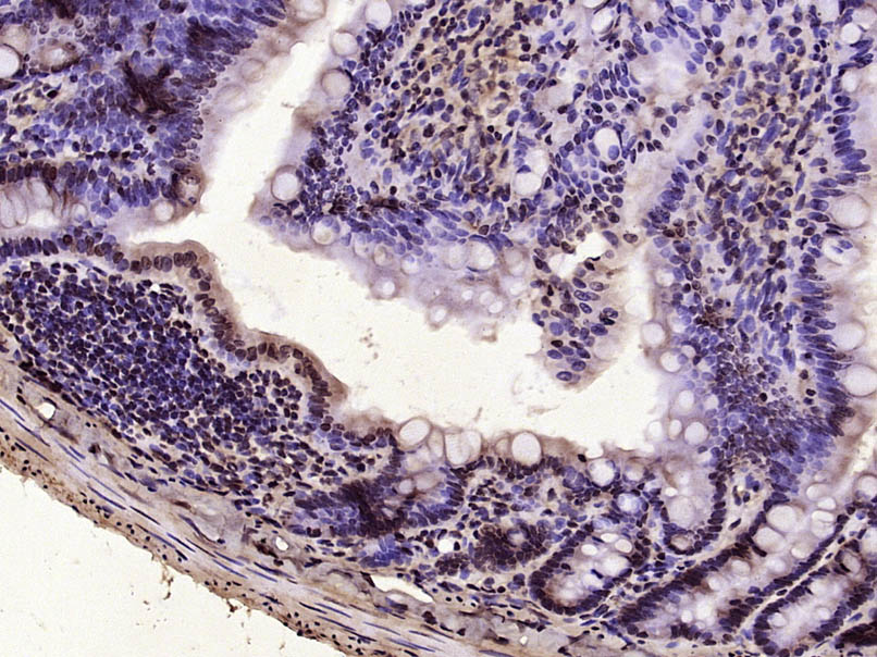 Paraformaldehyde-fixed, paraffin embedded (Rat small intestine); Antigen retrieval by microwave in sodium citrate buffer (pH6.0) ; Block endogenous peroxidase by 3% hydrogen peroxide for 30 minutes; Blocking buffer (3% BSA) at RT for 30min; Antibody incubation with (Vitamin D Receptor) Polyclonal Antibody, Unconjugated (SL2987R) at 1:400 overnight at 4℃, followed by conjugation to the secondary antibody (labeled with HRP)and DAB staining.
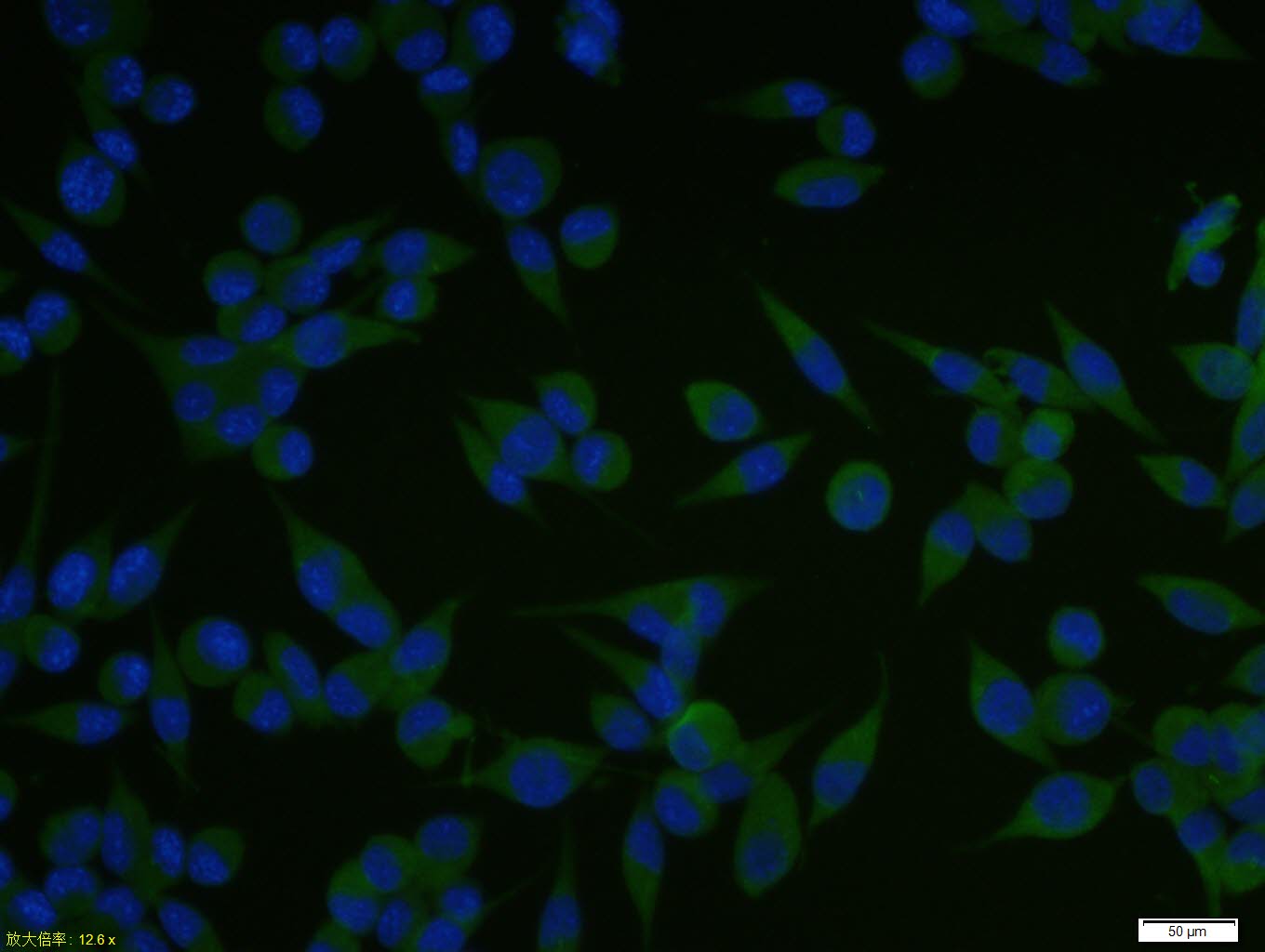 A431 cell; 4% Paraformaldehyde-fixed; Triton X-100 at room temperature for 20 min; Blocking buffer (normal goat serum, C-0005) at 37°C for 20 min; Antibody incubation with (Vitamin D Receptor) polyclonal Antibody, Unconjugated (SL2987R) 1:100, 90 minutes at 37°C; followed by a conjugated Goat Anti-Rabbit IgG antibody at 37°C for 90 minutes, DAPI (blue, C02-04002) was used to stain the cell nuclei.A431 cell; 4% Paraformaldehyde-fixed; Triton X-100 at room temperature for 20 min; Blocking buffer (normal goat serum, C-0005) at 37°C for 20 min; Antibody incubation with (Vitamin D Receptor) polyclonal Antibody, Unconjugated (SL2987R) 1:100, 90 minutes at 37°C; followed by a conjugated Goat Anti-Rabbit IgG antibody at 37°C for 90 minutes, DAPI (blue, C02-04002) was used to stain the cell nuclei.
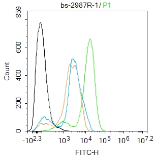 Blank control:THP-1.
Primary Antibody (green line): Rabbit Anti-Vitamin D Receptor antibody (SL2987R)
Dilution: 1ug/Test;
Secondary Antibody : Goat anti-rabbit IgG-FITC
Dilution: 0.5ug/Test.
Protocol
The cells were fixed with 4% PFA (10min at room temperature)and then permeabilized with 90% ice-cold methanol for 20 min at -20℃.The cells were then incubated in 5%BSA to block non-specific protein-protein interactions for 30 min at room temperature .Cells stained with Primary Antibody for 30 min at room temperature. The secondary antibody used for 40 min at room temperature. Acquisition of 20,000 events was performed.
Cartpieces
Totalgoods,subtotals:¥Checkout
Bought notes(bought amounts latest0)
No one bought this product
User Comment(Total0User Comment Num)
- No comment


 +86 571 56623320
 +86 18668110335

