
Rabbit Anti-CACNA1G antibody
calcium channel voltage dependent alpha 1G subunit; calcium channel voltage dependent T type alpha 1G subunit; calcium channel voltage dependent T type alpha1G subunit; CaV T1; Cav3 1; cav3 1c; NBR13; voltage dependent calcium channel alpha 1G subunit iso
View History [Clear]
Details
Product Name CACNA1G Chinese Name 电压依赖性钙通道Cav3.1抗体 Alias calcium channel voltage dependent alpha 1G subunit; calcium channel voltage dependent T type alpha 1G subunit; calcium channel voltage dependent T type alpha1G subunit; CaV T1; Cav3 1; cav3 1c; NBR13; voltage dependent calcium channel alpha 1G subunit isoform 11; voltage dependent T type calcium channel subunit alpha 1G; CAC1G_HUMAN; Cav3.1 literatures Research Area Channel protein Immunogen Species Rabbit Clonality Polyclonal React Species Human, Mouse, Rat, (predicted: Dog, Pig, Cow, Guinea Pig, ) Applications ELISA=1:1000-10000 IHC-P=1:100-500 IHC-F=1:100-500 Flow-Cyt=1μg /Test IF=1:100-500 (Paraffin sections need antigen repair)
not yet tested in other applications.
optimal dilutions/concentrations should be determined by the end user.Theoretical molecular weight 262kDa Cellular localization The cell membrane Form Liquid Concentration 1mg/ml immunogen KLH conjugated synthetic peptide derived from human CACNA1G/Cav31: 901-1000/2377 <Extracellular> Lsotype IgG Purification affinity purified by Protein A Buffer Solution 0.01M TBS(pH7.4) with 1% BSA, 0.03% Proclin300 and 50% Glycerol. Storage Shipped at 4℃. Store at -20 °C for one year. Avoid repeated freeze/thaw cycles. Attention This product as supplied is intended for research use only, not for use in human, therapeutic or diagnostic applications. PubMed PubMed Product Detail Voltage-sensitive calcium channels mediate the entry of calcium ions into excitable cells, and are also involved in a variety of calcium-dependent processes, including muscle contraction, hormone or neurotransmitter release, gene expression, cell motility, cell division, and cell death. This gene encodes a T-type, low-voltage activated calcium channel. The T-type channels generate currents that are both transient, owing to fast inactivation, and tiny, owing to small conductance. T-type channels are thought to be involved in pacemaker activity, low-threshold calcium spikes, neuronal oscillations and resonance, and rebound burst firing. Many alternatively spliced transcript variants encoding different isoforms have been described for this gene. [provided by RefSeq, Sep 2011]
Function:
Voltage-sensitive calcium channels (VSCC) mediate the entry of calcium ions into excitable cells and are also involved in a variety of calcium-dependent processes, including muscle contraction, hormone or neurotransmitter release, gene expression, cell motility, cell division and cell death. The isoform alpha-1G gives rise to T-type calcium currents. T-type calcium channels belong to the 'low-voltage activated (LVA)' group and are strongly blocked by mibefradil. A particularity of this type of channels is an opening at quite negative potentials and a voltage-dependent inactivation. T-type channels serve pacemaking functions in both central neurons and cardiac nodal cells and support calcium signaling in secretory cells and vascular smooth muscle. They may also be involved in the modulation of firing patterns of neurons which is important for information processing as well as in cell growth processes.
Subcellular Location:
Membrane; Multi-pass membrane protein.
Tissue Specificity:
Highly expressed in brain, in particular in the amygdala, subthalamic nuclei, cerebellum and thalamus. Moderate expression in heart; low expression in placenta, kidney and lung. Also expressed in colon and bone marrow and in tumoral cells to a lesser extent. Highly expressed in fetal brain, but also in peripheral fetal tissues as heart, kidney and lung, suggesting a developmentally regulated expression.
Similarity:
Belongs to the calcium channel alpha-1 subunit (TC 1.A.1.11) family. CACNA1G subfamily.
SWISS:
O43497
Gene ID:
8913
Database links:Entrez Gene: 8913 Human
Entrez Gene: 12291 Mouse
Omim: 604065 Human
SwissProt: O43497 Human
Unigene: 29585 Mouse
Unigene: 86960 Rat
Product Picture 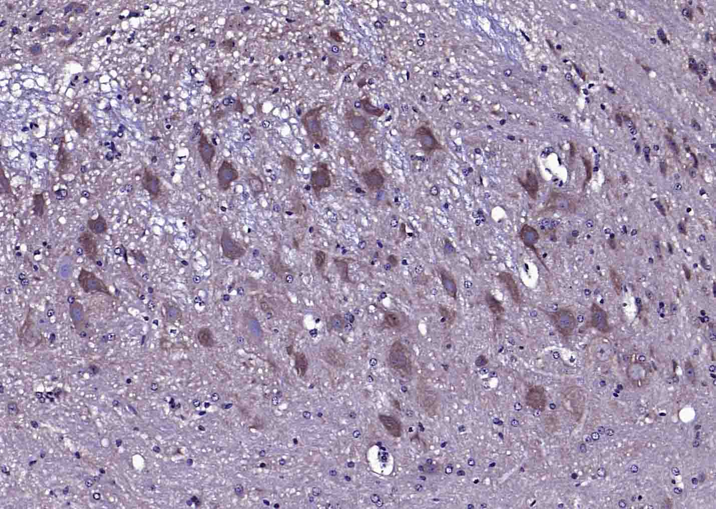 Paraformaldehyde-fixed, paraffin embedded (Rat cerebellum); Antigen retrieval by boiling in sodium citrate buffer (pH6.0) for 15min; Block endogenous peroxidase by 3% hydrogen peroxide for 20 minutes; Blocking buffer (normal goat serum) at 37°C for 30min; Antibody incubation with (CACNA1G) Polyclonal Antibody, Unconjugated (SL2781R) at 1:200 overnight at 4°C, followed by operating according to SP Kit(Rabbit) (sp-0023) instructionsand DAB staining
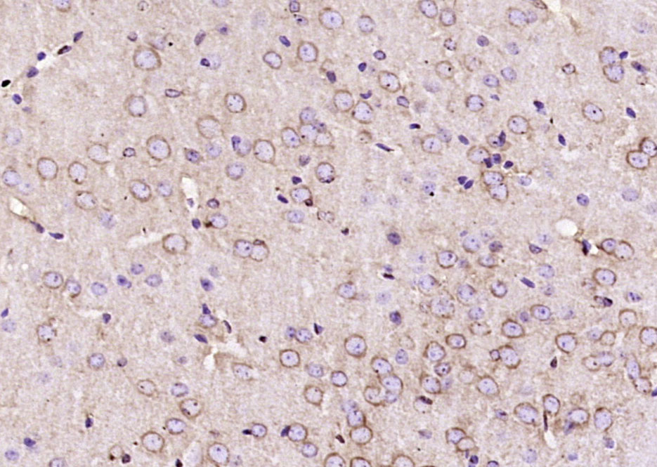 Paraformaldehyde-fixed, paraffin embedded (mouse brain); Antigen retrieval by boiling in sodium citrate buffer (pH6.0) for 15min; Block endogenous peroxidase by 3% hydrogen peroxide for 20 minutes; Blocking buffer (normal goat serum) at 37°C for 30min; Antibody incubation with (CACNA1G) Polyclonal Antibody, Unconjugated (SL2781R) at 1:200 overnight at 4°C, followed by operating according to SP Kit(Rabbit) (sp-0023) instructionsand DAB staining.
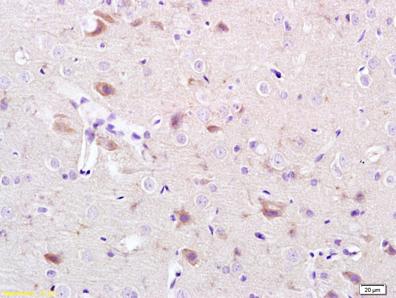 Tissue/cell: rat brain tissue; 4% Paraformaldehyde-fixed and paraffin-embedded;
Antigen retrieval: citrate buffer ( 0.01M, pH 6.0 ), Boiling bathing for 15min; Block endogenous peroxidase by 3% Hydrogen peroxide for 30min; Blocking buffer (normal goat serum,C-0005) at 37℃ for 20 min;
Incubation: Anti-CACNA1G/Cav3.1 Polyclonal Antibody, Unconjugated(SL2781R) 1:200, overnight at 4°C, followed by conjugation to the secondary antibody(SP-0023) and DAB(C-0010) staining
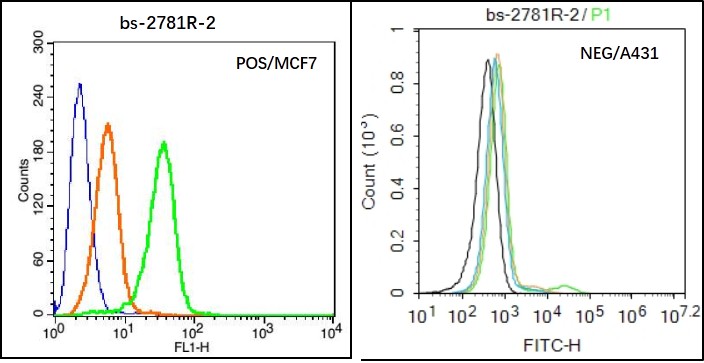 Black line : Positive blank control (MCF7); Negative blank control (431)
Green line : Primary Antibody (Rabbit Anti-CACNA1G antibody (SL2781R) )
Orange line:Isotype Control Antibody (Rabbit IgG) .
Blue line : Secondary Antibody (Goat anti-rabbit IgG-AF488)
MCF7(Positive)and A431(Negative control)cells (black) were incubated in 5% BSA blocking buffer for 30 min at room temperature. Cells were then stained with CACNA1G Antibody(SL2781R)at 1:50 dilution in blocking buffer and incubated for 30 min at room temperature, washed twice with 2% BSA in PBS, followed by secondary antibody(blue) incubation for 40 min at room temperature. Acquisitions of 20,000 events were performed. Cells stained with primary antibody (green), and isotype control (orange).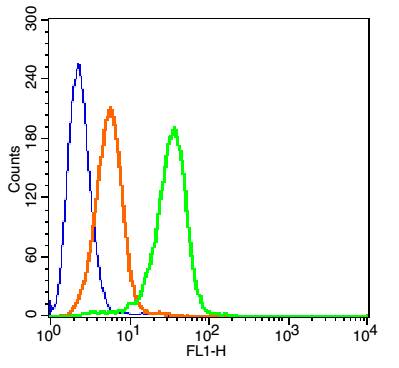 Blank control(blue): MCF7 Cells(fixed with 2% paraformaldehyde (10 min). The cells were washed twice with 1 X PBS).
Primary Antibody: Rabbit Anti- CACNA1G/AF488 Conjugated antibody (SL2781R-AF488), Dilution: 1μg in 100 μL 1X PBS containing 0.5% BSA;
Isotype Control Antibody: Rabbit IgG/AF488(orange) ,used under the same conditions.
Cartpieces
Totalgoods,subtotals:¥Checkout
Bought notes(bought amounts latest0)
No one bought this product
User Comment(Total0User Comment Num)
- No comment


 +86 571 56623320


