
Rabbit Anti-Cofilin antibody
Cofilin-1; 18 kDa phosphoprotein; CFL 1; CFL; CFL1; Cofilin 1; Cofilin 1 non muscle; Cofilin non muscle isoform; p18; COF1_HUMAN.
View History [Clear]
Details
Product Name Cofilin Chinese Name 丝切蛋白抗体 Alias Cofilin-1; 18 kDa phosphoprotein; CFL 1; CFL; CFL1; Cofilin 1; Cofilin 1 non muscle; Cofilin non muscle isoform; p18; COF1_HUMAN. literatures Research Area Tumour immunology Growth factors and hormones transcriptional regulatory factor Immunogen Species Rabbit Clonality Polyclonal React Species Human, Mouse, Rat, (predicted: Chicken, Dog, Pig, Cow, Rabbit, ) Applications ELISA=1:5000-10000 IHC-P=1:100-500 IHC-F=1:100-500 Flow-Cyt=1ug/Test IF=1:100-500 (Paraffin sections need antigen repair)
not yet tested in other applications.
optimal dilutions/concentrations should be determined by the end user.Theoretical molecular weight 18kDa Cellular localization The nucleus cytoplasmic Form Liquid Concentration 1mg/ml immunogen Synthetic peptide from the human cofilin conjugated to KLH: 2-100/166 Lsotype IgG Purification affinity purified by Protein A Buffer Solution 0.01M TBS(pH7.4) with 1% BSA, 0.03% Proclin300 and 50% Glycerol. Storage Shipped at 4℃. Store at -20 °C for one year. Avoid repeated freeze/thaw cycles. Attention This product as supplied is intended for research use only, not for use in human, therapeutic or diagnostic applications. PubMed PubMed Product Detail The protein encoded by this gene can polymerize and depolymerize F-actin and G-actin in a pH-dependent manner. Increased phosphorylation of this protein by LIM kinase aids in Rho-induced reorganization of the actin cytoskeleton. Cofilin is a widely distributed intracellular actin-modulating protein that binds and depolymerizes filamentous F-actin and inhibits the polymerization of monomeric G-actin in a pH-dependent manner. It is involved in the translocation of actin-cofilin complex from cytoplasm to nucleus.[supplied by OMIM, Apr 2004].
Function:
Controls reversibly actin polymerization and depolymerization in a pH-sensitive manner. It has the ability to bind G- and F-actin in a 1:1 ratio of cofilin to actin. It is the major component of intranuclear and cytoplasmic actin rods.
Subunit:
Can bind G- and F-actin in a 1:1 ratio of cofilin to actin. It is a major component of intranuclear and cytoplasmic actin rods.
Subcellular Location:
Nucleus matrix. Cytoplasm, cytoskeleton. Almost completely in nucleus in cells exposed to heat shock or 10% dimethyl sulfoxide.
Tissue Specificity:
Widely distributed in various tissues.
Post-translational modifications:
Inactivated by phosphorylation on Ser-3. Phosphorylated on Ser-3 in resting cells. Dephosphorylated by PDXP/chronophin; this restores its activity in promoting actin filament depolymerization. The phosphorylation of Ser-24 may prevent recognition of the nuclear localization signal.
Similarity:
Belongs to the actin-binding proteins ADF family.
Contains 1 ADF-H domain.
SWISS:
P23528
Gene ID:
1072
Database links:Entrez Gene: 1072 Human
Entrez Gene: 12631 Mouse
Omim: 601442 Human
SwissProt: P23528 Human
SwissProt: P18760 Mouse
Unigene: 170622 Human
Unigene: 329655 Mouse
Unigene: 11675 Rat
丝切蛋白(cofilin)是Cytoskeleton塑形的重要调节因素,其主要功能是分解肌动蛋白微丝和增加肌动蛋白单体从肌动蛋白微丝的末端解离的速度,从而促进肌动蛋白微丝的循环,是目前为止已发现的肌动蛋白Binding protein家族中唯一能够改变肌动蛋白微丝扭曲结构的蛋白.Product Picture 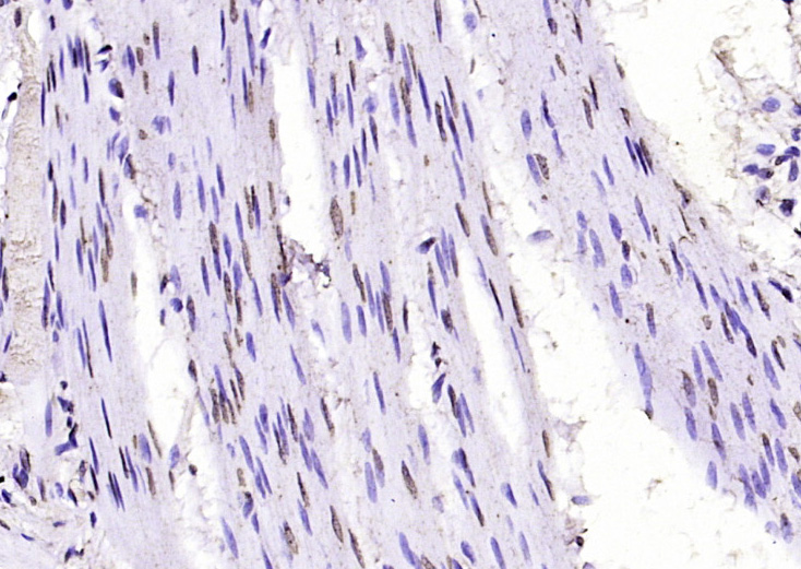 Paraformaldehyde-fixed, paraffin embedded (rat stomach tissue); Antigen retrieval by boiling in sodium citrate buffer (pH6.0) for 15min; Block endogenous peroxidase by 3% hydrogen peroxide for 20 minutes; Blocking buffer (normal goat serum) at 37°C for 30min; Antibody incubation with (cofilin) Polyclonal Antibody, Unconjugated (SL2759R) at 1:200 overnight at 4°C, followed by operating according to SP Kit(Rabbit) (sp-0023) instructionsand DAB staining.
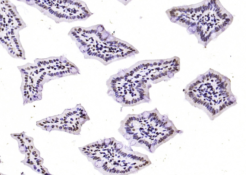 Paraformaldehyde-fixed, paraffin embedded (mouse intestine tissue); Antigen retrieval by boiling in sodium citrate buffer (pH6.0) for 15min; Block endogenous peroxidase by 3% hydrogen peroxide for 20 minutes; Blocking buffer (normal goat serum) at 37°C for 30min; Antibody incubation with (cofilin) Polyclonal Antibody, Unconjugated (SL2759R) at 1:200 overnight at 4°C, followed by operating according to SP Kit(Rabbit) (sp-0023) instructionsand DAB staining.
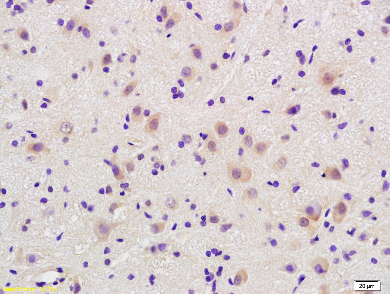 Tissue/cell: rat brain tissue; 4% Paraformaldehyde-fixed and paraffin-embedded;
Antigen retrieval: citrate buffer ( 0.01M, pH 6.0 ), Boiling bathing for 15min; Block endogenous peroxidase by 3% Hydrogen peroxide for 30min; Blocking buffer (normal goat serum,C-0005) at 37℃ for 20 min;
Incubation: Anti-cofilin Polyclonal Antibody, Unconjugated(SL2759R) 1:200, overnight at 4°C, followed by conjugation to the secondary antibody(SP-0023) and DAB(C-0010) staining
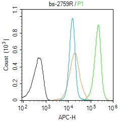 Blank control (Black line): Molt4 (Black).
Primary Antibody (green line): Rabbit Anti-cofilin antibody (SL2759R)
Dilution: 1μg /10^6 cells;
Isotype Control Antibody (orange line): Rabbit IgG .
Secondary Antibody (white blue line): Goat anti-rabbit IgG-AF647
Dilution: 1μg /test.
Protocol
The cells were fixed with 4% PFA (10min at room temperature)and then permeabilized with 90% ice-cold methanol for 20 min at room temperature. The cells were then incubated in 5%BSA to block non-specific protein-protein interactions for 30 min at room temperature .Cells stained with Primary Antibody for 30 min at room temperature. The secondary antibody used for 40 min at room temperature. Acquisition of 20,000 events was performed.
Cartpieces
Totalgoods,subtotals:¥Checkout
Bought notes(bought amounts latest0)
No one bought this product
User Comment(Total0User Comment Num)
- No comment


 +86 571 56623320
 +86 18668110335

