
Rabbit Anti-phospho-Akt (Thr308)antibody
AKT (phospho T308); p-AKT (phospho T308); Akt(Phospho-Thr308); Akt(Phospho T308); C AKT; MGC9965; Oncogene AKT1; PKB; PRKBA; Protein Kinase B Alpha; RAC Alpha; RAC; RAC PK Alpha; RAC Serine/Threonine Protein Kinase; vAKT Murine Thymoma Viral Oncogene Homo
View History [Clear]
Details
Product Name phospho-Akt (Thr308) Chinese Name 磷酸化蛋白激酶B抗体 Alias AKT (phospho T308); p-AKT (phospho T308); Akt(Phospho-Thr308); Akt(Phospho T308); C AKT; MGC9965; Oncogene AKT1; PKB; PRKBA; Protein Kinase B Alpha; RAC Alpha; RAC; RAC PK Alpha; RAC Serine/Threonine Protein Kinase; vAKT Murine Thymoma Viral Oncogene Homolog 1; AKT1_HUMAN. literatures Product Type Phosphorylated anti Research Area Cell biology Neurobiology Signal transduction Apoptosis Cyclin Kinases and Phosphatases Immunogen Species Rabbit Clonality Polyclonal React Species Human, Mouse, Rat, (predicted: Chicken, Dog, Pig, Cow, Rabbit, Sheep, ) Applications WB=1:500-2000 ELISA=1:5000-10000 IHC-P=1:100-500 IHC-F=1:100-500 Flow-Cyt=1μg /test IF=1:100-500 (Paraffin sections need antigen repair)
not yet tested in other applications.
optimal dilutions/concentrations should be determined by the end user.Theoretical molecular weight 56kDa Cellular localization The nucleus cytoplasmic The cell membrane Form Liquid Concentration 1mg/ml immunogen KLH conjugated Synthesised phosphopeptide derived from human Akt around the phosphorylation site of Thr308: MK(p-T)FC Lsotype IgG Purification affinity purified by Protein A Buffer Solution 0.01M TBS(pH7.4) with 1% BSA, 0.03% Proclin300 and 50% Glycerol. Storage Shipped at 4℃. Store at -20 °C for one year. Avoid repeated freeze/thaw cycles. Attention This product as supplied is intended for research use only, not for use in human, therapeutic or diagnostic applications. PubMed PubMed Product Detail This gene encodes one of the three members of the human AKT serine-threonine protein kinase family which are often referred to as protein kinase B alpha, beta, and gamma. These highly similar AKT proteins all have an N-terminal pleckstrin homology domain, a serine/threonine-specific kinase domain and a C-terminal regulatory domain. These proteins are phosphorylated by phosphoinositide 3-kinase (PI3K). AKT/PI3K forms a key component of many signalling pathways that involve the binding of membrane-bound ligands such as receptor tyrosine kinases, G-protein coupled receptors, and integrin-linked kinase. These AKT proteins therefore regulate a wide variety of cellular functions including cell proliferation, survival, metabolism, and angiogenesis in both normal and malignant cells. AKT proteins are recruited to the cell membrane by phosphatidylinositol 3,4,5-trisphosphate (PIP3) after phosphorylation of phosphatidylinositol 4,5-bisphosphate (PIP2) by PI3K. Subsequent phosphorylation of both threonine residue 308 and serine residue 473 is required for full activation of the AKT1 protein encoded by this gene. Phosphorylation of additional residues also occurs, for example, in response to insulin growth factor-1 and epidermal growth factor. Protein phosphatases act as negative regulators of AKT proteins by dephosphorylating AKT or PIP3. The PI3K/AKT signalling pathway is crucial for tumor cell survival. Survival factors can suppress apoptosis in a transcription-independent manner by activating AKT1 which then phosphorylates and inactivates components of the apoptotic machinery. AKT proteins also participate in the mammalian target of rapamycin (mTOR) signalling pathway which controls the assembly of the eukaryotic translation initiation factor 4F (eIF4E) complex and this pathway, in addition to responding to extracellular signals from growth factors and cytokines, is disregulated in many cancers. Mutations in this gene are associated with multiple types of cancer and excessive tissue growth including Proteus syndrome and Cowden syndrome 6, and breast, colorectal, and ovarian cancers. Multiple alternatively spliced transcript variants have been found for this gene. [provided by RefSeq, Jul 2020]
Subunit:
Interacts (via PH domain) with TCL1A; this enhances AKT3 phosphorylation and activation. Interacts with TRAF6.
Subcellular Location:
Cytoplasm. Nucleus. Cell membrane. Note=Nucleus after activation by integrin-linked protein kinase 1 (ILK1). Nuclear translocation is enhanced by interaction with TCL1A. Phosphorylation on Tyr-176 by TNK2 results in its localization to the cell membrane where it is targeted for further phosphorylations on Thr-308 and Ser-473 leading to its activation and the activated form translocates to the nucleus.
Tissue Specificity:
Widely expressed. Low levels found in liver with slightly higher levels present in thymus and testis.
Similarity:
Belongs to the protein kinase superfamily. AGC Ser/Thr protein kinase family. RAC subfamily.
Contains 1 AGC-kinase C-terminal domain.
Contains 1 PH domain.
Contains 1 protein kinase domain.
SWISS:
P31749
Gene ID:
207
Database links:Entrez Gene: 207 Human
Entrez Gene: 11651 Mouse
Omim: 164730 Human
SwissProt: O57513 Chicken
SwissProt: P31749 Human
SwissProt: P31750 Mouse
Unigene: 525622 Human
Unigene: 6645 Mouse
Unigene: 11422 Rat
Product Picture 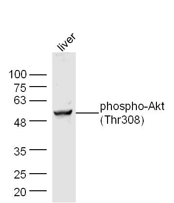 Sample: Liver (Mouse) Lysate at 30 ug
Primary: Anti- phospho-Akt(Thr308)(SL2720R) at 1/300 dilution
Secondary: IRDye800CW Goat Anti-Mouse IgG at 1/10000 dilution
Predicted band size: 56 kD
Observed band size: 56 kD
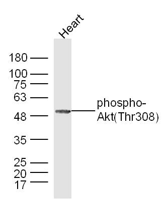 Western blot analysis of extracts from Heart,using phospho-Akt(thr308) Antibody.
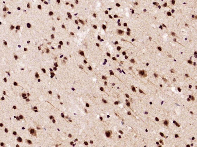 Paraformaldehyde-fixed, paraffin embedded (Mouse brain); Antigen retrieval by boiling in sodium citrate buffer (pH6.0) for 15min; Block endogenous peroxidase by 3% hydrogen peroxide for 20 minutes; Blocking buffer (normal goat serum) at 37°C for 30min; Antibody incubation with (phospho-Akt (Thr308)) Polyclonal Antibody, Unconjugated (SL2720R) at 1:400 overnight at 4°C, followed by operating according to SP Kit(Rabbit) (sp-0023) instructions and DAB staining.
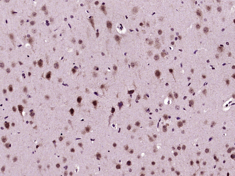 Paraformaldehyde-fixed, paraffin embedded (Rat brain); Antigen retrieval by boiling in sodium citrate buffer (pH6.0) for 15min; Block endogenous peroxidase by 3% hydrogen peroxide for 20 minutes; Blocking buffer (normal goat serum) at 37°C for 30min; Antibody incubation with (phospho-Akt (Thr308)) Polyclonal Antibody, Unconjugated (SL2720R) at 1:400 overnight at 4°C, followed by operating according to SP Kit(Rabbit) (sp-0023) instructions and DAB staining.
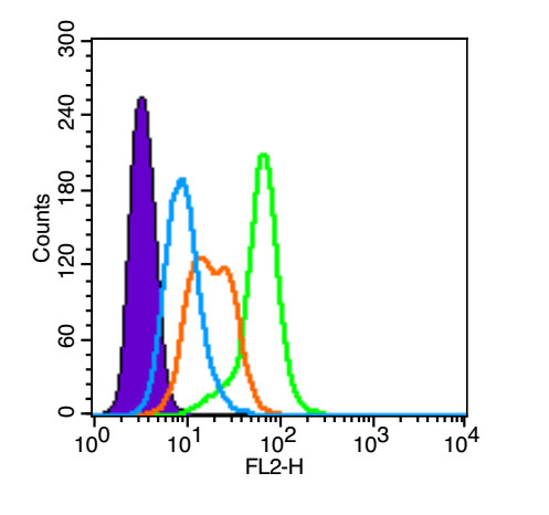 Blank control (blue line): A431(Black).
Primary Antibody (green line): Rabbit Anti-phospho-Akt (Thr308) antibody (SL2720R)
Dilution: 1μg /10^6 cells;
Isotype Control Antibody (orange line): Rabbit IgG .
Secondary Antibody (white blue line): Goat anti-rabbit IgG-PE(Jackson lab)
Dilution: 1μg /test.
Protocol
The cells were fixed with 4% paraformaldehyde (10 min) , then permeabilized with 90% ice-cold methanol for 20 min on ice. Cells stained with Primary Antibody for 30 min at room temperature. The cells were then incubated in 5%BSA to block non-specific protein-protein interactions followed by the antibody for 15 min at room temperature. The secondary antibody used for 40 min at room temperature. Acquisition of 20,000 events was performed.
Cartpieces
Totalgoods,subtotals:¥Checkout
Bought notes(bought amounts latest0)
No one bought this product
User Comment(Total0User Comment Num)
- No comment


 +86 571 56623320
 +86 18668110335

