
Rabbit Anti-ACHE antibody
Acetylcholinesterase; Acetylcholine acetylhydrolase; Acetylcholinesterase YT blood group; ACHE protein; Apoptosis related acetylcholinesterase; ARACHE; N ACHE; N-ACHE; YT; Yt blood group; ACES_HUMAN.
View History [Clear]
Details
Product Name ACHE Chinese Name 乙酰胆碱酯酶抗体 Alias Acetylcholinesterase; Acetylcholine acetylhydrolase; Acetylcholinesterase YT blood group; ACHE protein; Apoptosis related acetylcholinesterase; ARACHE; N ACHE; N-ACHE; YT; Yt blood group; ACES_HUMAN. literatures Research Area Neurobiology Signal transduction Immunogen Species Rabbit Clonality Polyclonal React Species Human, Mouse, (predicted: Rat, Dog, Cow, Horse, ) Applications WB=1:500-2000 ELISA=1:5000-10000 IHC-P=1:100-500 IHC-F=1:100-500 IF=1:100-500 (Paraffin sections need antigen repair)
not yet tested in other applications.
optimal dilutions/concentrations should be determined by the end user.Theoretical molecular weight 68kDa Cellular localization The nucleus cytoplasmic The cell membrane Extracellular matrix Secretory protein Form Liquid Concentration 1mg/ml immunogen KLH conjugated synthetic peptide derived from human AchE: 521-614/614 Lsotype IgG Purification affinity purified by Protein A Buffer Solution 0.01M TBS(pH7.4) with 1% BSA, 0.03% Proclin300 and 50% Glycerol. Storage Shipped at 4℃. Store at -20 °C for one year. Avoid repeated freeze/thaw cycles. Attention This product as supplied is intended for research use only, not for use in human, therapeutic or diagnostic applications. PubMed PubMed Product Detail Acetylcholinesterase hydrolyzes the neurotransmitter, acetylcholine at neuromuscular junctions and brain cholinergic synapses, and thus terminates signal transmission. It is also found on the red blood cell membranes, where it constitutes the Yt blood group antigen. Acetylcholinesterase exists in multiple molecular forms which possess similar catalytic properties, but differ in their oligomeric assembly and mode of cell attachment to the cell surface. It is encoded by the single ACHE gene, and the structural diversity in the gene products arises from alternative mRNA splicing, and post-translational associations of catalytic and structural subunits. The major form of acetylcholinesterase found in brain, muscle and other tissues is the hydrophilic species, which forms disulfide-linked oligomers with collagenous, or lipid-containing structural subunits. The other, alternatively spliced form, expressed primarily in the erythroid tissues, differs at the C-terminal end, and contains a cleavable hydrophobic peptide with a GPI-anchor site. It associates with the membranes through the phosphoinositide (PI) moieties added post-translationally.
Function:
Terminates signal transduction at the neuromuscular junction by rapid hydrolysis of the acetylcholine released into the synaptic cleft. Role in neuronal apoptosis.
Subunit:
Interacts with PRIMA1. The interaction with PRIMA1 is required to anchor it to the basal lamina of cells and organize into tetramers. Isoform H generates GPI-anchored dimers; disulfide linked. Isoform T generates multiple structures, ranging from monomers and dimers to collagen-tailed and hydrophobic-tailed forms, in which catalytic tetramers are associated with anchoring proteins that attach them to the basal lamina or to cell membranes. In the collagen-tailed forms, isoform T subunits are associated with a specific collagen, COLQ, which triggers the formation of isoform T tetramers, from monomers and dimers. Isoform R may be monomeric.
Subcellular Location:
Cell junction, synapse. Secreted. Cell membrane; Peripheral membrane protein.
Isoform T: Nucleus. Note=Only observed in apoptotic nuclei.
Isoform H: Cell membrane; Lipid-anchor, GPI-anchor; Extracellular side.
Tissue Specificity:
Isoform H is highly expressed in erythrocytes.
Similarity:
Belongs to the type-B carboxylesterase/lipase family.
SWISS:
P22303
Gene ID:
43
Database links:
Entrez Gene: 43 Human
Entrez Gene: 11423 Mouse
Omim: 100740 Human
SwissProt: P22303 Human
SwissProt: P21836 Mouse
Unigene: 154495 Human
Unigene: 255464 Mouse
Unigene: 105879 Rat
Product Picture 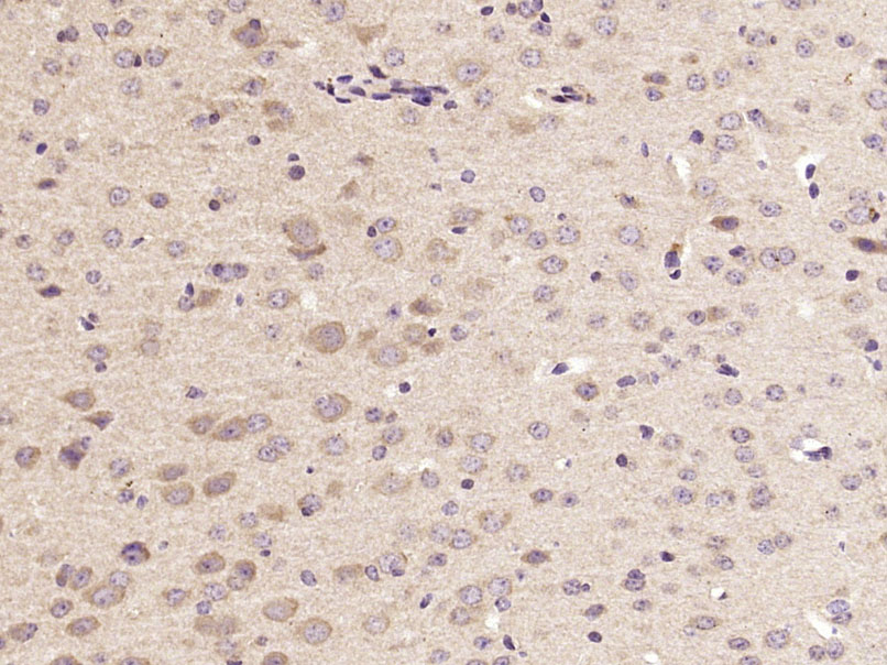 Paraformaldehyde-fixed, paraffin embedded (Mouse brain); Antigen retrieval by microwave in sodium citrate buffer (pH6.0) ; Block endogenous peroxidase by 3% hydrogen peroxide for 30 minutes; Blocking buffer (3% BSA) at RT for 30min; Antibody incubation with (ACHE) Polyclonal Antibody, Unconjugated (SL2511R) at 1:400 overnight at 4℃, followed by conjugation to the secondary antibody (labeled with HRP)and DAB staining.
Cartpieces
Totalgoods,subtotals:¥Checkout
References (0)
No References
Bought notes(bought amounts latest0)
No one bought this product
User Comment(Total0User Comment Num)
- No comment


 +86 571 56623320
 +86 18668110335

