
Rabbit Anti-CD1E antibody
CD_antigen=CD1e;CD1e;CD1e antigen;CD1E antigen; CD1e molecule;CD1E_HUMAN;Differentiation antigen CD1-alpha-3;hCD1e;Leukocyte differentiation antigen;R2;R2G1;sCD1e;soluble;T-cell surface glycoprotein CD1e.
View History [Clear]
Details
Product Name CD1E Chinese Name T细胞表面glycoproteinCD1E抗体 Alias CD_antigen=CD1e; CD1e; CD1e antigen; CD1E antigen; CD1e molecule; CD1E_HUMAN; Differentiation antigen CD1-alpha-3; hCD1e; Leukocyte differentiation antigen; R2; R2G1; sCD1e; soluble; T-cell surface glycoprotein CD1e. Research Area immunology Immunogen Species Rabbit Clonality Polyclonal React Species Human, Applications WB=1:500-2000 Flow-Cyt=1ug/Test
not yet tested in other applications.
optimal dilutions/concentrations should be determined by the end user.Theoretical molecular weight 42kDa Cellular localization cytoplasmic Form Liquid Concentration 1mg/ml immunogen KLH conjugated synthetic peptide derived from human CD1E: 81-180/388 Lsotype IgG Purification affinity purified by Protein A Buffer Solution 0.01M TBS(pH7.4) with 1% BSA, 0.03% Proclin300 and 50% Glycerol. Storage Shipped at 4℃. Store at -20 °C for one year. Avoid repeated freeze/thaw cycles. Attention This product as supplied is intended for research use only, not for use in human, therapeutic or diagnostic applications. PubMed PubMed Product Detail This gene encodes a member of the CD1 family of transmembrane glycoproteins, which are structurally related to the major histocompatibility complex (MHC) proteins and form heterodimers with beta-2-microglobulin. The CD1 proteins mediate the presentation of primarily lipid and glycolipid antigens of self or microbial origin to T cells. The human genome contains five CD1 family genes organized in a cluster on chromosome 1. The CD1 family members are thought to differ in their cellular localization and specificity for particular lipid ligands. The protein encoded by this gene localizes within Golgi compartments, endosomes, and lysosomes, and is cleaved into a stable soluble form. The soluble form is required for the intracellular processing of some glycolipids into a form that can be presented by other CD1 family members. Many alternatively spliced transcript variants encoding different isoforms have been described. Additional transcript variants have been found; however, their biological validity has not been determined. [provided by RefSeq, Jun 2010]
Function:
T-cell surface glycoprotein CD1e, soluble binds diacetylated lipids, including phosphatidyl inositides and diacylated sulfoglycolipids, and is required for the presentation of glycolipid antigens on the cell surface. The membrane-associated form is not active.
Subcellular Location:
Lysosome lumen and Golgi apparatus membrane. Early endosome. Late endosome. Predominantly localized in the trans-Golgi network in immature dendritic cells, and as a cleaved, soluble protein in the lysosome lumen of mature dendritic cells.
Tissue Specificity:
Expressed on cortical thymocytes, dendritic cells, Langerhans cells, on certain T-cell leukemias, and in various other tissues.
Post-translational modifications:
Mono-ubiquitinated. Proteolytically cleaved in late endosomes to yield a soluble form.
Similarity:
Contains 1 Ig-like (immunoglobulin-like) domain.
SWISS:
P15812
Gene ID:
913
Database links:Entrez Gene: 913 Human
Omim: 188411 Human
SwissProt: P15812 Human
Unigene: 249217 Human
Product Picture 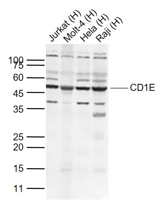 Sample:
Lane 1: Jurkat (Human) Cell Lysate at 30 ug
Lane 2: Molt-4 (Human) Cell Lysate at 30 ug
Lane 3: Hela (Human) Cell Lysate at 30 ug
Lane 4: Raji (Human) Cell Lysate at 30 ug
Primary:
Anti-CD1E (SL23497R) at 1/500 dilution
Secondary: IRDye800CW Goat Anti-Rabbit IgG at 1/20000 dilution
Predicted band size: 42 kD
Observed band size: 50 kD
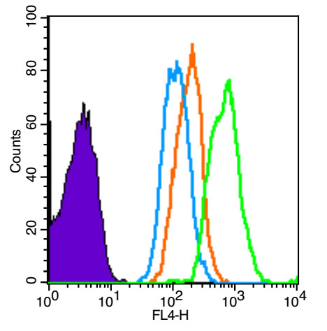 Blank control (Black line): Molt-4 (Black).
Primary Antibody (green line): Rabbit Anti-CD1E antibody (SL23497R)
Dilution: 1μg /10^6 cells;
Isotype Control Antibody (orange line): Rabbit IgG .
Secondary Antibody (white blue line): Goat anti-rabbit IgG-AF647
Dilution: 1μg /test.
Protocol
The cells were fixed with 4% PFA (10min at room temperature)and then permeabilized with PBST for 20 min at room temperature. The cells were then incubated in 5% BSA to block non-specific protein-protein interactions for 30 min at room temperature .Cells stained with Primary Antibody for 30 min at room temperature. The secondary antibody used for 40 min at room temperature. Acquisition of 20,000 events was performed.
Cartpieces
Totalgoods,subtotals:¥Checkout
References (0)
No References
Bought notes(bought amounts latest0)
No one bought this product
User Comment(Total0User Comment Num)
- No comment


 +86 571 56623320
 +86 18668110335

