
Rabbit Anti-CD1B antibody
CD1b; CD1b antigen; CD1B antigen b polypeptide; CD1b molecule; CD1B_HUMAN; Cortical thymocyte antigen CD1B ; R1; T-cell surface glycoprotein CD1b; T6/Leu6; Thymocite antigen CD1B.
View History [Clear]
Details
Product Name CD1B Chinese Name TlymphocyteCD1B抗体 Alias CD1b; CD1b antigen; CD1B antigen b polypeptide; CD1b molecule; CD1B_HUMAN; Cortical thymocyte antigen CD1B ; R1; T-cell surface glycoprotein CD1b; T6/Leu6; Thymocite antigen CD1B. Research Area immunology Cell type markers b-lymphocyte Immunogen Species Rabbit Clonality Polyclonal React Species Human, Applications ELISA=1:5000-10000 IHC-P=1:100-500 IHC-F=1:100-500 Flow-Cyt=1ug/Test ICC=1:100-500 IF=1:100-500 (Paraffin sections need antigen repair)
not yet tested in other applications.
optimal dilutions/concentrations should be determined by the end user.Theoretical molecular weight 35kDa Cellular localization cytoplasmic Form Liquid Concentration 1mg/ml immunogen KLH conjugated synthetic peptide derived from human CD1B : 51/150/333 <Extracellular> Lsotype IgG Purification affinity purified by Protein A Buffer Solution 0.01M TBS(pH7.4) with 1% BSA, 0.03% Proclin300 and 50% Glycerol. Storage Shipped at 4℃. Store at -20 °C for one year. Avoid repeated freeze/thaw cycles. Attention This product as supplied is intended for research use only, not for use in human, therapeutic or diagnostic applications. PubMed PubMed Product Detail This gene encodes a member of the CD1 family of transmembrane glycoproteins, which are structurally related to the major histocompatibility complex (MHC) proteins and form heterodimers with beta-2-microglobulin. The CD1 proteins mediate the presentation of primarily lipid and glycolipid antigens of self or microbial origin to T cells. The human genome contains five CD1 family genes organized in a cluster on chromosome 1. The CD1 family members are thought to differ in their cellular localization and specificity for particular lipid ligands. The protein encoded by this gene localizes to late endosomes and lysosomes via a tyrosine-based motif in the cytoplasmic tail, and requires vesicular acidification to bind lipid antigens. [provided by RefSeq, Jul 2008]
Function:
Antigen-presenting protein that binds self and non-self lipid and glycolipid antigens and presents them to T-cell receptors on natural killer T-cells.
Subcellular Location:
Cell membrane. Endosome membrane. Lysosome membrane. Subject to intracellular trafficking between the cell membrane, endosomes and lysosomes. Localizes to cell surface lipid rafts.
Tissue Specificity:
Cell membrane. Endosome membrane. Lysosome membrane. Subject to intracellular trafficking between the cell membrane, endosomes and lysosomes. Localizes to cell surface lipid rafts.
Similarity:
Contains 1 Ig-like (immunoglobulin-like) domain.
SWISS:
P29016
Gene ID:
910
Database links:Entrez Gene: 910 Human
Omim: 188360 Human
SwissProt: P29016 Human
Unigene: 1310 Human
Product Picture 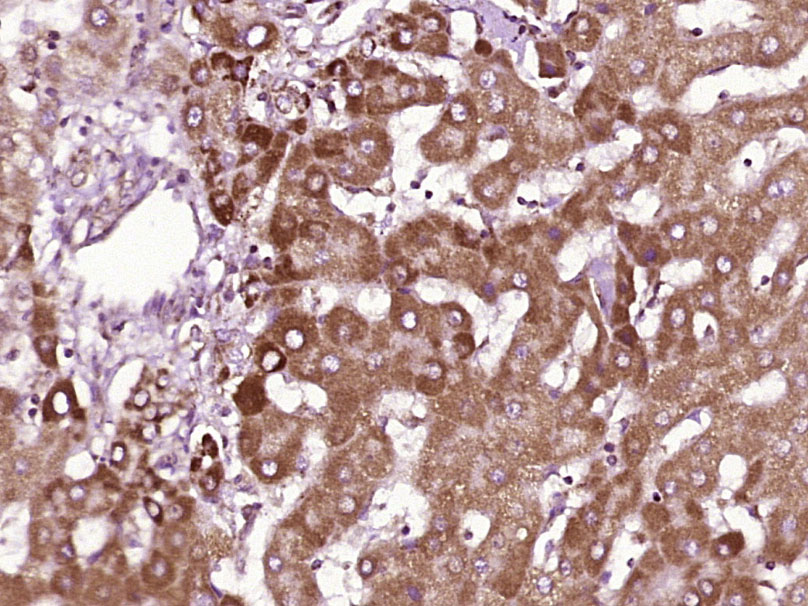 Paraformaldehyde-fixed, paraffin embedded (human liver carcinoma); Antigen retrieval by boiling in sodium citrate buffer (pH6.0) for 15min; Block endogenous peroxidase by 3% hydrogen peroxide for 20 minutes; Blocking buffer (normal goat serum) at 37°C for 30min; Antibody incubation with (CD1B) Polyclonal Antibody, Unconjugated (SL23494R) at 1:400 overnight at 4°C, followed by operating according to SP Kit(Rabbit) (sp-0023) instructionsand DAB staining.
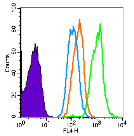 Blank control (Black line): Molt-4 (Black).
Primary Antibody (green line): Rabbit Anti-CD1B antibody (SL23494R)
Dilution: 1μg /10^6 cells;
Isotype Control Antibody (orange line): Rabbit IgG .
Secondary Antibody (white blue line): Goat anti-rabbit IgG-AF647
Dilution: 1μg /test.
Protocol
The cells were fixed with 4% PFA (10min at room temperature)and then permeabilized with PBST for 20 min at room temperature. The cells were then incubated in 5% BSA to block non-specific protein-protein interactions for 30 min at room temperature .Cells stained with Primary Antibody for 30 min at room temperature. The secondary antibody used for 40 min at room temperature. Acquisition of 20,000 events was performed.
Cartpieces
Totalgoods,subtotals:¥Checkout
References (0)
No References
Bought notes(bought amounts latest0)
No one bought this product
User Comment(Total0User Comment Num)
- No comment


 +86 571 56623320
 +86 18668110335

