
Rabbit Anti-Progesterone Receptor antibody
NR3C3; Nuclear receptor subfamily 3 group C member 3; PGR; PR; PRA; PRB; Progesterone receptor; Progestin receptor form A; Progestin receptor form B; PRGR_HUMAN; Progestin receptor form A; Progestin receptor form B.
View History [Clear]
Details
Product Name Progesterone Receptor Chinese Name 孕激素受体抗体 Alias NR3C3; Nuclear receptor subfamily 3 group C member 3; PGR; PR; PRA; PRB; Progesterone receptor; Progestin receptor form A; Progestin receptor form B; PRGR_HUMAN; Progestin receptor form A; Progestin receptor form B. literatures Research Area Tumour immunology Signal transduction Growth factors and hormones TumourCell biologyMaker Immunogen Species Rabbit Clonality Polyclonal React Species Human, Mouse, Rat, (predicted: Rabbit, ) Applications WB=1:500-2000 IHC-P=1:100-500 IHC-F=1:100-500 Flow-Cyt=1ug/Test ICC=1:100-500 IF=1:100-500 (Paraffin sections need antigen repair)
not yet tested in other applications.
optimal dilutions/concentrations should be determined by the end user.Theoretical molecular weight 103kDa Cellular localization The nucleus cytoplasmic Form Liquid Concentration 1mg/ml immunogen KLH conjugated synthetic peptide derived from human Progesterone Receptor: 221-320/933 Lsotype IgG Purification affinity purified by Protein A Buffer Solution 0.01M TBS(pH7.4) with 1% BSA, 0.03% Proclin300 and 50% Glycerol. Storage Shipped at 4℃. Store at -20 °C for one year. Avoid repeated freeze/thaw cycles. Attention This product as supplied is intended for research use only, not for use in human, therapeutic or diagnostic applications. PubMed PubMed Product Detail Estrogen and progesterone receptor are members of a family of transcription factors that are regulated by the binding of their cognate ligands. The interaction of hormone-bound estrogen receptors with estrogen responsive elements(EREs) alters transcription of ERE-containing genes. The carboxy terminal region of the estrgen receptor contains the ligand binding domain, the amino terminus serves as the transactivation domain, and the DNA binding domain is centrally located. Two forms of estrogen receptor have been identified, ER alpha and ER beta. ER alpha and ER beta have been shown to be differentially activated by various ligands. The biological response to progesterone is mediated by two distinct forms of the human progesterone receptor (hPR-Aand hPR-B), which arise from alternative splicing. In most cells, hPR-B functions as a transcriptional activator of progesterone-responsive gene, whereas hPR-A function as a transcriptional inhibitor of all steroid hormone receptors.
Function:
The steroid hormones and their receptors are involved in the regulation of eukaryotic gene expression and affect cellular proliferation and differentiation in target tissues. Progesterone receptor isoform B (PRB) is involved activation of c-SRC/MAPK signaling on hormone stimulation.
Isoform A is inactive in stimulating c-Src/MAPK signaling on hormone stimulation.
Subunit:
Interacts with SMARD1 and UNC45A. Interacts with CUEDC2; the interaction promotes ubiquitination, decreases sumoylation, and repesses transcriptional activity. Interacts with PIAS3; the interaction promotes sumoylation of PR in a hormone-dependent manner, inhibits DNA-binding, and alters nuclear export. Interacts with SP1; the interaction requires ligand-induced phosphorylation on Ser-345 by ERK1/2 MAPK. Interacts with PRMT2.
Subcellular Location:
Nucleus. Cytoplasm. Note=Nucleoplasmic shuttling is both homone- and cell cycle-dependent. On hormone stimulation, retained in the cytoplasm in the G(1) and G(2)/M phases. Isoform A: Nucleus. Cytoplasm. Note=Mainly nuclear.
Post-translational modifications:
Phosphorylated on multiple serine sites. Several of these sites are hormone-dependent. Phosphorylation on Ser-294 occurs preferentially on isoform B, is highly hormone-dependent and modulates ubiquitination and sumoylation on Lys-388. Phosphorylation on Ser-102 and Ser-345 also requires induction by hormone. Basal phosphorylation on Ser-81, Ser-162, Ser-190 and Ser-400 is increased in response to progesterone and can be phosphorylated in vitro by the CDK2-A1 complex. Increased levels of phosphorylation on Ser-400 also in the presence of EGF, heregulin, IGF, PMA and FBS. Phosphorylation at this site by CDK2 is ligand-independent, and increases nuclear translocation and transcriptional activity. Phosphorylation at Ser-162 and Ser-294, but not at Ser-190, is impaired during the G(2)/M phase of the cell cycle. Phosphorylation on Ser-345 by ERK1/2 MAPK is required for interaction with SP1.
Sumoylation is hormone-dependent and represses transcriptional activity. Sumoylation on all three sites is enhanced by PIAS3. Desumoylated by SENP1. Sumoylation on Lys-388, the main site of sumoylation, is repressed by ubiquitination on the same site, and modulated by phosphorylation at Ser-294.
Similarity:
Belongs to the nuclear hormone receptor family. NR3 subfamily.
Contains 1 nuclear receptor DNA-binding domain.
SWISS:
P06401
Gene ID:
5241
Database links:Entrez Gene: 5241 Human
Entrez Gene: 18667 Mouse
Entrez Gene: 100009094 Rabbit
Omim: 607311 Human
SwissProt: P06401 Human
SwissProt: Q00175 Mouse
SwissProt: P06186 Rabbit
Unigene: 2905 Human
Unigene: 32405 Human
Unigene: 742403 Human
Unigene: 12798 Mouse
Unigene: 437703 Mouse
Unigene: 1947 Rabbit
Unigene: 10303 Rat
Product Picture
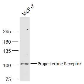
MCF-7(Human) Cell Lysate at 30 ug
Primary: Anti-Progesterone Receptor (SL23376R) at 1/1000 dilution
Secondary: IRDye800CW Goat Anti-Rabbit IgG at 1/20000 dilution
Predicted band size: 103 kD
Observed band size: 103 kD
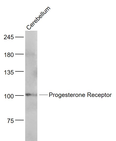
Sample:
Cerebellum (Mouse) Lysate at 40 ug
Primary: Anti- Progesterone Receptor (SL23376R) at 1/1000 dilution
Secondary: IRDye800CW Goat Anti-Rabbit IgG at 1/20000 dilution
Predicted band size: 103 kD
Observed band size: 103 kD
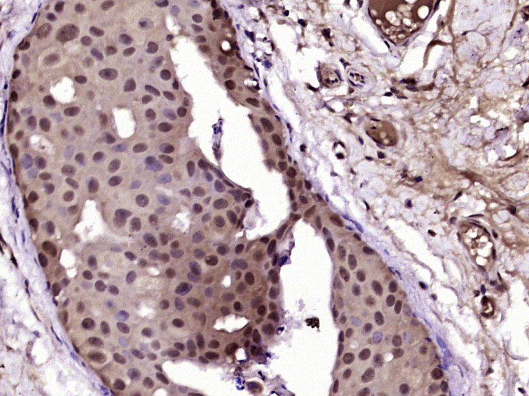
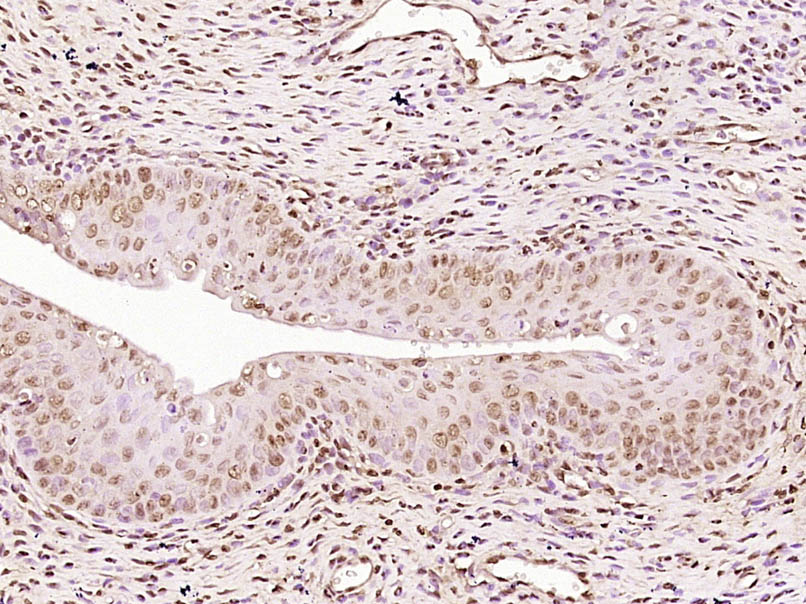
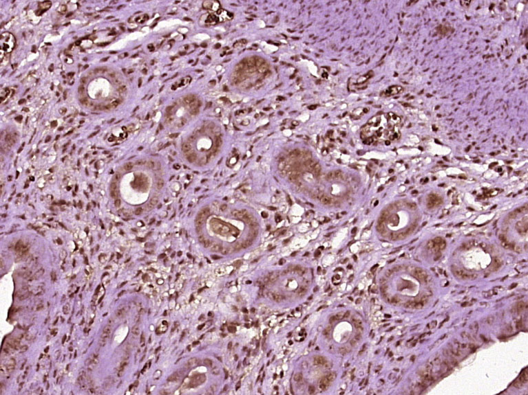
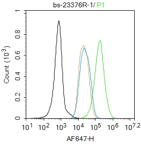
Paraformaldehyde-fixed, paraffin embedded (Human breast cancer); Antigen retrieval by boiling in sodium citrate buffer (pH6.0) for 15min; Block endogenous peroxidase by 3% hydrogen peroxide for 20 minutes; Blocking buffer (normal goat serum) at 37°C for 30min; Antibody incubation with (Progesterone Receptor) Polyclonal Antibody, Unconjugated (SL23376R) at 1:400 overnight at 4°C, followed by operating according to SP Kit(Rabbit) (sp-0023) instructions and DAB staining.Paraformaldehyde-fixed, paraffin embedded (Rat uterus); Antigen retrieval by boiling in sodium citrate buffer (pH6.0) for 15min; Block endogenous peroxidase by 3% hydrogen peroxide for 20 minutes; Blocking buffer (normal goat serum) at 37°C for 30min; Antibody incubation with (Progesterone Receptor) Polyclonal Antibody, Unconjugated (SL23376R) at 1:400 overnight at 4°C, followed by operating according to SP Kit(Rabbit) (sp-0023) instructionsand DAB staining.Paraformaldehyde-fixed, paraffin embedded (mouse uterus tissue); Antigen retrieval by boiling in sodium citrate buffer (pH6.0) for 15min; Block endogenous peroxidase by 3% hydrogen peroxide for 20 minutes; Blocking buffer (normal goat serum) at 37°C for 30min; Antibody incubation with (PRGR) Polyclonal Antibody, Unconjugated (SL23376R) at 1:400 overnight at 4°C, followed by operating according to SP Kit(Rabbit) (sp-0023) instructionsand DAB staining.Blank control: MCF7.
Primary Antibody (green line): Rabbit Anti-Progesterone Receptor antibody (SL23376R)
Dilution: 1μg /10^6 cells;
Isotype Control Antibody (orange line): Rabbit IgG .
Secondary Antibody : Goat anti-rabbit IgG-AF647
Dilution: 1μg /test.
Protocol
The cells were fixed with 4% PFA (10min at room temperature)and then permeabilized with 90% ice-cold methanol for 20 min at -20℃. The cells were then incubated in 5%BSA to block non-specific protein-protein interactions for 30 min at room temperature .Cells stained with Primary Antibody for 30 min at room temperature. The secondary antibody used for 40 min at room temperature. Acquisition of 20,000 events was performed.
Partial purchase records(bought amounts latest0)
No one bought this product
User Comment(Total0User Comment Num)
- No comment


 +86 571 56623320


