
Rabbit Anti-MCL1 antibody
myeloid cell leukemia 1; myeloid cell leukemia sequence 1; MCL-1; MCL1L; MCL 1; mcl1/EAT; MGC104264; MGC1839; TM; MCL1S; EAT) Bcl 2 related protein EAT/mcl1; BCL2 related; BCL2L3; EAT; Induced myeloid leukemia cell differentiation protein Mcl 1; myeloid c
View History [Clear]
Details
Product Name MCL1 Chinese Name 髓样细胞白血病-1抗体 Alias myeloid cell leukemia 1; myeloid cell leukemia sequence 1; MCL-1; MCL1L; MCL 1; mcl1/EAT; MGC104264; MGC1839; TM; MCL1S; EAT) Bcl 2 related protein EAT/mcl1; BCL2 related; BCL2L3; EAT; Induced myeloid leukemia cell differentiation protein Mcl 1; myeloid cell leukemia sequence 1; Myeloid cell leukemia sequence 1 BCL2 related; Myeloid cell leukemia sequence 1 isoform 1; OTTHUMP00000032794; OTTHUMP00000032795; TM; bcl2-L-3; BCL2L3; EAT; Mcl-1; MCL1-ES; mcl1/EAT. literatures Research Area Tumour immunology Signal transduction Apoptosis transcriptional regulatory factor Mitochondrion Immunogen Species Rabbit Clonality Polyclonal React Species Human, Mouse, Rat, (predicted: Dog, Pig, Horse, Rabbit, ) Applications WB=1:500-2000 IHC-P=1:100-500 IHC-F=1:100-500 Flow-Cyt=1ug/Test ICC=1:100-500 IF=1:100-500 (Paraffin sections need antigen repair)
not yet tested in other applications.
optimal dilutions/concentrations should be determined by the end user.Theoretical molecular weight 39kDa Cellular localization The nucleus cytoplasmic The cell membrane Mitochondrion Form Liquid Concentration 1mg/ml immunogen KLH conjugated synthetic peptide derived from human MCL1: 101-200/350 Lsotype IgG Purification affinity purified by Protein A Buffer Solution 0.01M TBS(pH7.4) with 1% BSA, 0.03% Proclin300 and 50% Glycerol. Storage Shipped at 4℃. Store at -20 °C for one year. Avoid repeated freeze/thaw cycles. Attention This product as supplied is intended for research use only, not for use in human, therapeutic or diagnostic applications. PubMed PubMed Product Detail Mcl1 is an anti-apoptotic member of Bcl2 family originally isolated from the ML1 human myeloid leukemia cell line during phorbol ester-induced differentiation along the monocyte/macrophage pathway. Mcl1 localizes to the mitochondria, interacts with and antagonizes pro-apoptotic Bcl2 family members, and inhibits apoptosis by a number of cytotoxic stimuli. It is involved in programing of differentiation and concomitant maintenance of viability but not of proliferation. Isoform 1 inhibits apoptosis while isoform 2 promotes it. Expression increases early during phorbol-ester induced differentiation along the monocyte/macrophage pathway in myeloid leukemia cell lines ML1.
Function:
Involved in the regulation of apoptosis versus cell survival, and in the maintenance of viability but not of proliferation. Mediates its effects by interactions with a number of other regulators of apoptosis. Isoform 1 inhibits apoptosis. Isoform 2 promotes apoptosis.
Subunit:
Interacts with BAD, BOK, BIK and BFM (By similarity). Interacts with PMAIP1. Isoform 1 interacts with BAX, BAK1, TPT1 and BCL2L11. Heterodimer of isoform 1 and isoform 2. Homodimers of isoform 1 or isoform 2 are not detected. Isoform 2 does not interact with pro-apoptotic BCL2-related proteins.
Subcellular Location:
Membrane; Single-pass membrane protein (Potential). Cytoplasm. Mitochondrion. Nucleus, nucleoplasm. Note=Cytoplasmic, associated with mitochondria.
Post-translational modifications:
Cleaved by CASP3 during apoptosis. In intact cells cleavage occurs preferentially after Asp-127, yielding a pro-apoptotic 28 kDa C-terminal fragment.
Rapidly degraded in the absence of phosphorylation on Thr-163 in the PEST region.
Phosphorylated on Thr-163. Treatment with taxol or okadaic acid induces phosphorylation on additional sites.
Similarity:
Belongs to the Bcl-2 family.
SWISS:
Q07820
Gene ID:
4170
Database links:Entrez Gene: 4170 Human
Entrez Gene: 17210 Mouse
Omim: 159552 Human
SwissProt: Q07820 Human
SwissProt: P97287 Mouse
Unigene: 632486 Human
Product Picture 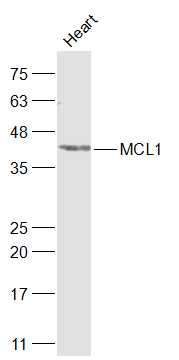 Sample:
Heart (Rat) Lysate at 40 ug
Primary: Anti-MCL1 (SL23315R) at 1/1000 dilution
Secondary: IRDye800CW Goat Anti-Rabbit IgG at 1/20000 dilution
Predicted band size: 39 kD
Observed band size: 41 kD
 Sample:
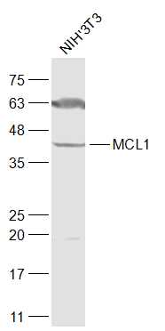
NIH/3T3(Mouse) Cell Lysate at 30 ug
Primary: Anti-MCL1 (SL23315R) at 1/1000 dilution
Secondary: IRDye800CW Goat Anti-Rabbit IgG at 1/20000 dilution
Predicted band size: 39 kD
Observed band size: 41 kD
 Sample:
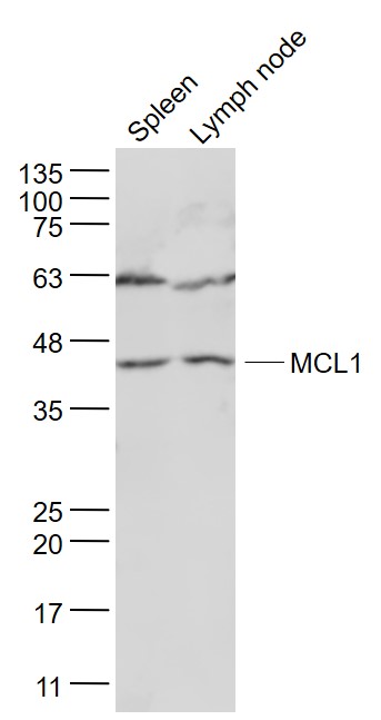
Spleen (Mouse) Lysate at 40 ug
Lymph node (Mouse) Lysate at 40 ug
Primary: Anti- MCL1 (SL23315R) at 1/1000 dilution
Secondary: IRDye800CW Goat Anti-Rabbit IgG at 1/20000 dilution
Predicted band size: 39 kD
Observed band size: 39 kD
 Sample:
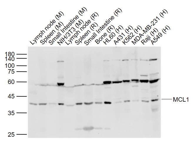
Lane 1: Lymph node (Mouse) Lysate at 40 ug
Lane 2: Spleen (Mouse) Lysate at 40 ug
Lane 3: Small intestine (Mouse) Lysate at 40 ug
Lane 4: NIH/3T3 (Mouse) Cell Lysate at 30 ug
Lane 5: Lymph node (Rat) Lysate at 40 ug
Lane 6: Spleen (Rat) Lysate at 40 ug
Lane 7: Small intestine (Rat) Lysate at 40 ug
Lane 8: Bone (Rat) Lysate at 40 ug
Lane 9: HL60 (Human) Cell Lysate at 30 ug
Lane 10: A431 (Human) Cell Lysate at 30 ug
Lane 11: K562 (Human) Cell Lysate at 30 ug
Lane 12: MDA-MB-231 (Human) Cell Lysate at 30 ug
Lane 13: Raji (Human) Cell Lysate at 30 ug
Lane 14: A549 (Human) Cell Lysate at 30 ug
Primary: Anti-MCL1 (SL23315R) at 1/1000 dilution
Secondary: IRDye800CW Goat Anti-Rabbit IgG at 1/20000 dilution
Predicted band size: 39 kD
Observed band size: 40 kD
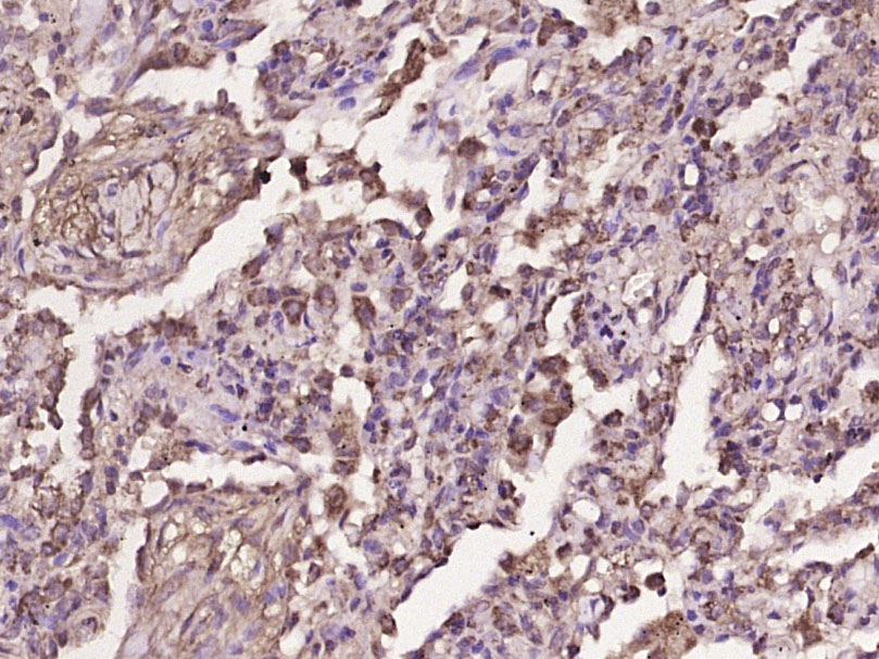 Paraformaldehyde-fixed, paraffin embedded (human lung carcinoma); Antigen retrieval by boiling in sodium citrate buffer (pH6.0) for 15min; Block endogenous peroxidase by 3% hydrogen peroxide for 20 minutes; Blocking buffer (normal goat serum) at 37°C for 30min; Antibody incubation with (MCL1) Polyclonal Antibody, Unconjugated (SL23315R) at 1:400 overnight at 4°C, followed by operating according to SP Kit(Rabbit) (sp-0023) instructionsand DAB staining.
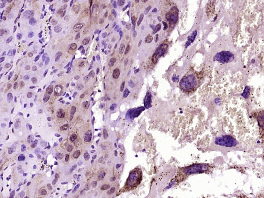 Paraformaldehyde-fixed, paraffin embedded (Mouse embryo); Antigen retrieval by boiling in sodium citrate buffer (pH6.0) for 15min; Block endogenous peroxidase by 3% hydrogen peroxide for 20 minutes; Blocking buffer (normal goat serum) at 37°C for 30min; Antibody incubation with (MCL1) Polyclonal Antibody, Unconjugated (SL23315R) at 1:400 overnight at 4°C, followed by operating according to SP Kit(Rabbit) (sp-0023) instructions and DAB staining.
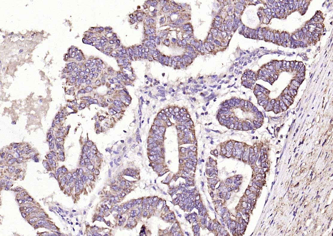 Paraformaldehyde-fixed, paraffin embedded (human colon carcinoma); Antigen retrieval by boiling in sodium citrate buffer (pH6.0) for 15min; Block endogenous peroxidase by 3% hydrogen peroxide for 20 minutes; Blocking buffer (normal goat serum) at 37°C for 30min; Antibody incubation with (MCL1) Polyclonal Antibody, Unconjugated (SL23315R) at 1:200 overnight at 4°C, followed by operating according to SP Kit(Rabbit) (sp-0023) instructionsand DAB staining.
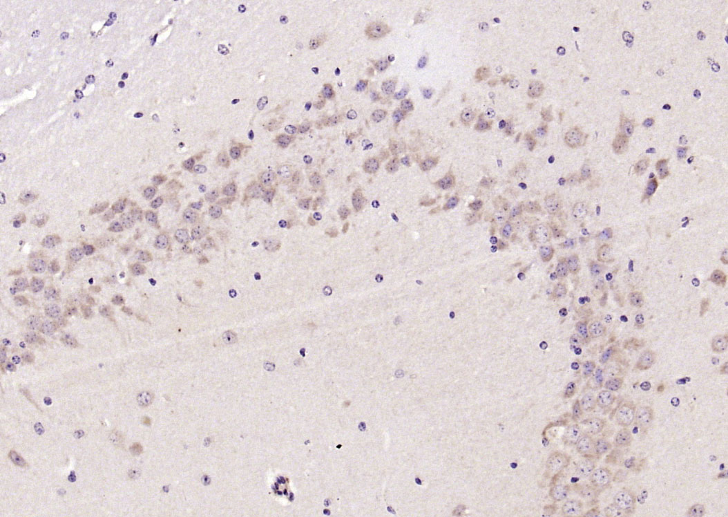 Paraformaldehyde-fixed, paraffin embedded (mouse brain); Antigen retrieval by boiling in sodium citrate buffer (pH6.0) for 15min; Block endogenous peroxidase by 3% hydrogen peroxide for 20 minutes; Blocking buffer (normal goat serum) at 37°C for 30min; Antibody incubation with (MCL1) Polyclonal Antibody, Unconjugated (SL23315R) at 1:200 overnight at 4°C, followed by operating according to SP Kit(Rabbit) (sp-0023) instructionsand DAB staining.
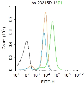 Blank control: K562.
Primary Antibody (green line): Rabbit Anti-MCL1 antibody (SL23315R)
Dilution: 1μg /10^6 cells;
Isotype Control Antibody (orange line): Rabbit IgG .
Secondary Antibody : Goat anti-rabbit IgG-FITC
Dilution: 1μg /test.
Protocol
The cells were fixed with 4% PFA (10min at room temperature)and then permeabilized with 90% ice-cold methanol for 20 min at-20℃. The cells were then incubated in 5%BSA to block non-specific protein-protein interactions for 30 min at room temperature .Cells stained with Primary Antibody for 30 min at room temperature. The secondary antibody used for 40 min at room temperature. Acquisition of 20,000 events was performed.
Cartpieces
Totalgoods,subtotals:¥Checkout
References (0)
No References
Bought notes(bought amounts latest0)
No one bought this product
User Comment(Total0User Comment Num)
- No comment


 +86 571 56623320
 +86 18668110335

