
Rabbit Anti-ITGAV antibody
Integrin alpha-V light chain; Integrin Alpha V; CD 51; CD51; CD51 antigen; HGNC; Msk 8; Msk8; Vitronectin receptor alpha polypeptide; Vitronectin receptor alpha polypeptide antigen CD51; Vitronectin receptor subunit alpha; VNRA; VTNR; ITAV_HUMAN; Integrin
View History [Clear]
Details
Product Name ITGAV Chinese Name 整合素αV(CD51)抗体 Alias Integrin alpha-V light chain; Integrin Alpha V; CD 51; CD51; CD51 antigen; HGNC; Msk 8; Msk8; Vitronectin receptor alpha polypeptide; Vitronectin receptor alpha polypeptide antigen CD51; Vitronectin receptor subunit alpha; VNRA; VTNR; ITAV_HUMAN; Integrin alpha-V Precursor. literatures Research Area Cell biology Stem cells Immunogen Species Rabbit Clonality Polyclonal React Species Human, Rat, (predicted: Mouse, ) Applications ELISA=1:5000-10000 IHC-P=1:500-2000 IHC-F=1:500-2000 (Paraffin sections need antigen repair)
not yet tested in other applications.
optimal dilutions/concentrations should be determined by the end user.Theoretical molecular weight 18/113kDa Detection molecular weight 24/125kD Cellular localization The cell membrane Form Liquid Concentration 1mg/ml immunogen KLH conjugated synthetic peptide derived from mouse Integrin alpha-V light chain Lsotype IgG Purification affinity purified by Protein A Buffer Solution 0.01M TBS(pH7.4) with 1% BSA, 0.03% Proclin300 and 50% Glycerol. Storage Shipped at 4℃. Store at -20 °C for one year. Avoid repeated freeze/thaw cycles. Attention This product as supplied is intended for research use only, not for use in human, therapeutic or diagnostic applications. PubMed PubMed Product Detail Integrins are heterodimeric proteins made up of alpha and beta subunits. At least 18 alpha and 8 beta subunits have been described in mammals. Integrin family members are membrane receptors involved in cell adhesion and recognition in a variety of processes including embryogenesis, hemostasis, tissue repair, immune response and metastatic diffusion of tumor cells.
Integrin alpha V chain interacts with the integrin beta 3 subunit/CD61 to form the alpha-V-beta-3 heterodimer/vitronectin receptor. It is expressed on endothelial cells, some activated leukocytes, NK cells, macrophages, neutrophils, and platelets. Integrin alpha V also forms heterodimers with the integrin beta 1, beta 5, beta 6, and beta 8 subunits. Alpha-V-beta-3 is an activation dependent receptor for platelet attachment and spreading on vitronectin and other matrix components. In case of HIV-1 infection, the interaction with extracellular viral Tat protein seems to enhance angiogenesis in Kaposi's sarcoma lesions. Alpha-V/beta-6 binds to foot-and-mouth disease virus (FMDV) VP1 protein and acts as a receptor for this virus By similarity. Alpha-V/beta-6 binds to coxsackievirus A9 and coxsackievirus B1 capsid proteins and acts as a receptor for these viruses.
Function:
The alpha-V integrins are receptors for vitronectin, cytotactin, fibronectin, fibrinogen, laminin, matrix metalloproteinase-2, osteopontin, osteomodulin, prothrombin, thrombospondin and vWF. They recognize the sequence R-G-D in a wide array of ligands. In case of HIV-1 infection, the interaction with extracellular viral Tat protein seems to enhance angiogenesis in Kaposi's sarcoma lesions.
Subunit:
Heterodimer of an alpha and a beta subunit. The alpha subunit is composed of an heavy and a light chain linked by a disulfide bond. Alpha-V associates with either beta-1, beta-3, beta-5, beta-6 or beta-8 subunit. Interacts with HIV-1 Tat. Alpha-V/beta-6 binds to foot-and-mouth disease virus (FMDV) VP1 protein and acts as a receptor for this viru. Alpha-V/beta-6 binds to coxsackievirus A9 and coxsackievirus B1 capsid proteins and acts as a receptor for these viruses. Interacts with RAB25.
Subcellular Location:
Membrane; Single-pass type I membrane protein.
Similarity:
Belongs to the integrin alpha chain family.
Contains 7 FG-GAP repeats.
SWISS:
P43406
Gene ID:
16410
Database links:Entrez Gene: 3685 Human
SwissProt: P06756 Human
Product Picture 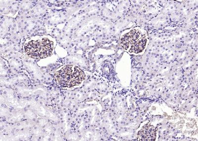 Paraformaldehyde-fixed, paraffin embedded (rat kidney); Antigen retrieval by boiling in sodium citrate buffer (pH6.0) for 15min; Block endogenous peroxidase by 3% hydrogen peroxide for 20 minutes; Blocking buffer (normal goat serum) at 37°C for 30min; Antibody incubation with (ITGAV) Polyclonal Antibody, Unconjugated (SL2203R) at 1:500 overnight at 4°C, followed by operating according to SP Kit(Rabbit) (sp-0023) instructionsand DAB staining.
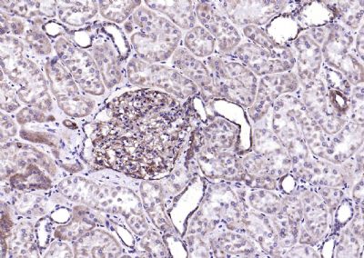 Paraformaldehyde-fixed, paraffin embedded (human kidney); Antigen retrieval by boiling in sodium citrate buffer (pH6.0) for 15min; Block endogenous peroxidase by 3% hydrogen peroxide for 20 minutes; Blocking buffer (normal goat serum) at 37°C for 30min; Antibody incubation with (ITGAV) Polyclonal Antibody, Unconjugated (SL2203R) at 1:500 overnight at 4°C, followed by operating according to SP Kit(Rabbit) (sp-0023) instructionsand DAB staining.
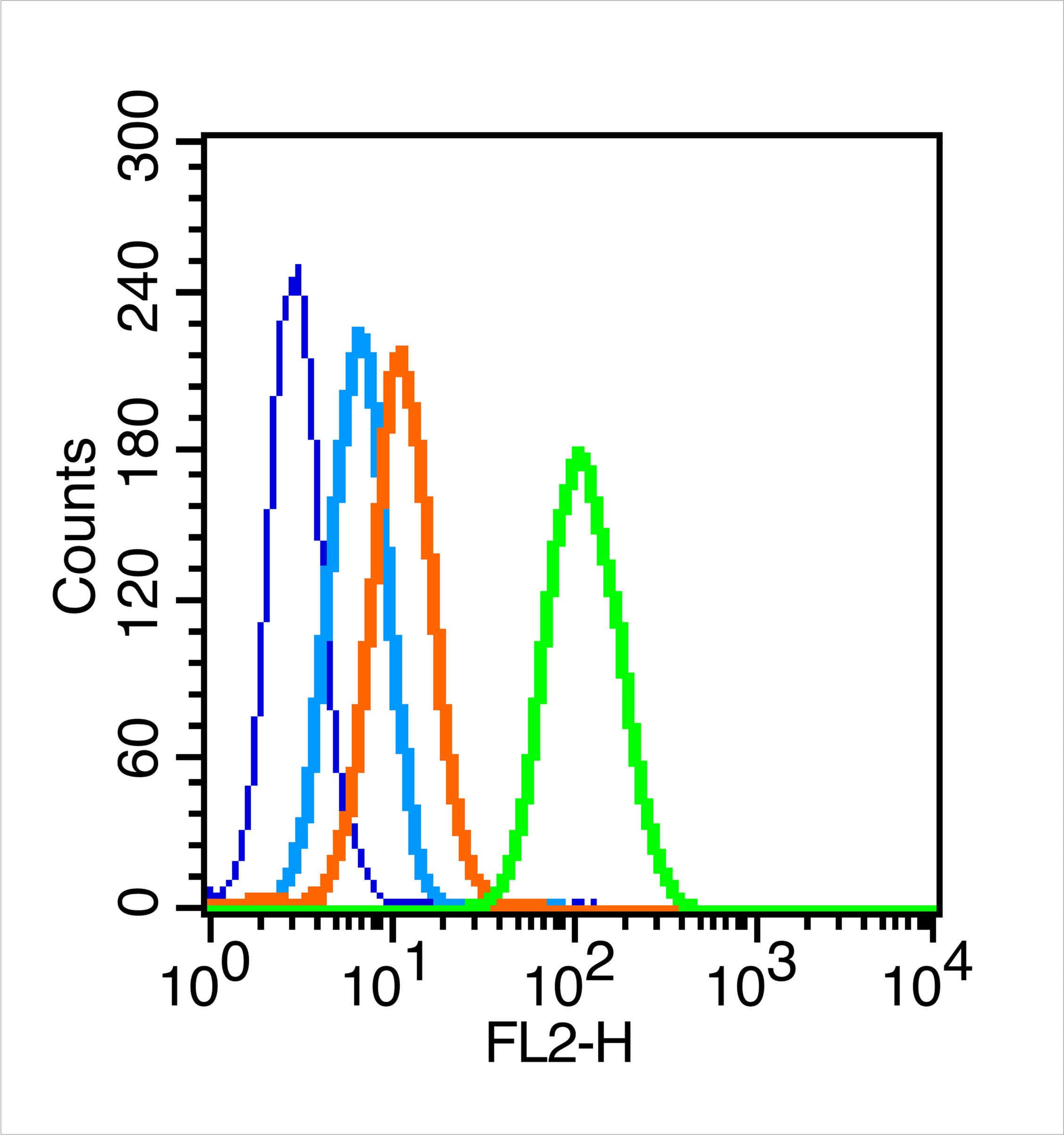 Blank control (blue line): MCF7 (blue).
Primary Antibody (green line): Rabbit Anti-Integrin Alpha V receptor antibody (SL2203R)
Dilution: 0.2μg /10^6 cells;
Isotype Control Antibody (orange line): Rabbit IgG .
Secondary Antibody (white blue line): Goat anti-rabbit IgG-PE
Dilution: 1μg /test.
Protocol
The cells were fixed with 70% methanol overnight at 4℃ . Cells stained with Primary Antibody for 30 min at room temperature. The cells were then incubated in 1 X PBS/2%BSA/10% goat serum to block non-specific protein-protein interactions followed by the antibody for 15 min at room temperature. The secondary antibody used for 40 min at room temperature. Acquisition of 20,000 events was performed.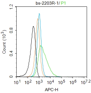 Blank control: MCF7.
Primary Antibody (green line): Rabbit Anti-CD51 antibody (SL2203R)
Dilution: 2μg /10^6 cells;
Isotype Control Antibody (orange line): Rabbit IgG .
Secondary Antibody : Goat anti-rabbit IgG-AF647
Dilution: 1μg /test.
Protocol
The cells were incubated in 5%BSA to block non-specific protein-protein interactions for 30 min at at room temperature .Cells stained with Primary Antibody for 30 min at room temperature. The secondary antibody used for 40 min at room temperature. Acquisition of 20,000 events was performed.
Cartpieces
Totalgoods,subtotals:¥Checkout
References (0)
No References
Bought notes(bought amounts latest0)
No one bought this product
User Comment(Total0User Comment Num)
- No comment


 +86 571 56623320
 +86 18668110335

