
Rabbit Anti-VEGFR3 antibody
Vascular endothelial growth factor receptor 3; VEGF Receptor 3; Tyrosine-protein kinase; Flt4; VEGFR3; AI323512; Chy; FLT4; FLT41; LMPH1A; LOC285682; PCL; VEGFR-3; fms-related tyrosine kinase 4; VGFR3_MOUSE.
View History [Clear]
Details
Product Name VEGFR3 Chinese Name vascular endothelial cell生长因子受体3抗体 Alias Vascular endothelial growth factor receptor 3; VEGF Receptor 3; Tyrosine-protein kinase; Flt4; VEGFR3; AI323512; Chy; FLT4; FLT41; LMPH1A; LOC285682; PCL; VEGFR-3; fms-related tyrosine kinase 4; VGFR3_MOUSE. literatures Research Area Tumour immunology Signal transduction Growth factors and hormones Kinases and Phosphatases The cell membrane受体 vascular endothelial cell Immunogen Species Rabbit Clonality Polyclonal React Species Human, Mouse, (predicted: Rat, ) Applications ELISA=1:5000-10000 IHC-P=1:100-500 IHC-F=1:100-500 Flow-Cyt=1μg /test ICC=1:100 IF=1:100-500 (Paraffin sections need antigen repair)
not yet tested in other applications.
optimal dilutions/concentrations should be determined by the end user.Theoretical molecular weight 151kDa Cellular localization The nucleus cytoplasmic The cell membrane Secretory protein Form Liquid Concentration 1mg/ml immunogen KLH conjugated synthetic peptide derived from human VEGFR-3: 901-1000/1298 <Cytoplasmic> Lsotype IgG Purification affinity purified by Protein A Buffer Solution 0.01M TBS(pH7.4) with 1% BSA, 0.03% Proclin300 and 50% Glycerol. Storage Shipped at 4℃. Store at -20 °C for one year. Avoid repeated freeze/thaw cycles. Attention This product as supplied is intended for research use only, not for use in human, therapeutic or diagnostic applications. PubMed PubMed Product Detail Vascular endothelial growth factors (VEGFs) are a family of closely related growth factors having a conserved pattern of eight cysteine esidues and sharing common VEGF receptors. VEGFs stimulate the proliferation of endothelial cells, induce angiogenesis, and increase vascular permeability in both large and small vessels. The mitogenic activity of VEGFs appears to be mediated by specific VEGF receptors. VEGF Receptor 3 is one of the five receptor tyrosine kinases (RTKs) (VEGF Receptor 1/Flt1, VEGF Receptor 2/KDR/Flk1, VEGF Receptor 3/Flt4, tie1 and tek/tie2) whose expression is almost exclusively restricted to endothelial cells
Function:
Tyrosine-protein kinase that acts as a cell-surface receptor for VEGFC and VEGFD, and plays an essential role in adult lymphangiogenesis and in the development of the vascular network and the cardiovascular system during embryonic development. Promotes proliferation, survival and migration of endothelial cells, and regulates angiogenic sprouting. Signaling by activated FLT4 leads to enhanced production of VEGFC, and to a lesser degree VEGFA, thereby creating a positive feedback loop that enhances FLT4 signaling. Modulates KDR signaling by forming heterodimers. Mediates activation of the MAPK1/ERK2, MAPK3/ERK1 signaling pathway, of MAPK8 and the JUN signaling pathway, and of the AKT1 signaling pathway. Phosphorylates SHC1. Mediates phosphorylation of PIK3R1, the regulatory subunit of phosphatidylinositol 3-kinase. Promotes phosphorylation of MAPK8 at 'Thr-183' and 'Tyr-185', and of AKT1 at 'Ser-473'.
Subunit:
Interacts with VEGFC and VEGFD. Monomer in the absence of bound VEGFC or VEGFD. Homodimer in the presence of bound VEGFC or VEGFD. Can also form a heterodimer with KDR. Interacts with PTPN14; the interaction is enhanced by stimulation with VEGFC. Interacts with CRK, GRB2, PTK2/FAK1, SHC1, PIK3R1 and PTPN11/SHP-2. Identified in a complex with SRC and ITGB1.
Subcellular Location:
Cell membrane; Single-pass type I membrane protein. Cytoplasm. Nucleus. Note=Ligand-mediated autophosphorylation leads to rapid internalization.
Isoform 1: Cell membrane; Single-pass type I membrane protein. Note=Ligand-mediated autophosphorylation leads to rapid internalization.
Isoform 2: Cell membrane; Single-pass type I membrane protein.
Isoform 3: Secreted. Cytoplasm.
Tissue Specificity:
Detected in endothelial cells (at protein level). Widely expressed. Detected in fetal spleen, lung and brain. Detected in adult liver, muscle, thymus, placenta, lung, testis, ovary, prostate, heart, and kidney.
Post-translational modifications:
Autophosphorylated on tyrosine residues upon ligand binding. Autophosphorylation occurs in trans, i.e. one subunit of the dimeric receptor phosphorylates tyrosine residues on the other subunit. Phosphorylation in response to H(2)O(2) is mediated by a process that requires SRC and PRKCD activity. Phosphorylation at Tyr-1068 is required for autophosphorylation at additional tyrosine residues. Phosphorylation at Tyr-1063 and Tyr-1337 is important for interaction with CRK and subsequent activation of MAPK8. Phosphorylation at Tyr-1230, Tyr-1231 and Tyr-1337 is important for interaction with GRB2 and subsequent activation of the AKT1 and MAPK1/ERK2 and/or MAPK3/ERK1 signaling pathways. In response to endothelial cell adhesion onto collagen, can also be phosphorylated in the absence of FLT4 kinase activity by SRC.
Similarity:
Belongs to the protein kinase superfamily. Tyr protein kinase family. CSF-1/PDGF receptor subfamily.
Contains 7 Ig-like C2-type (immunoglobulin-like) domains.
Contains 1 protein kinase domain.
SWISS:
P35916
Gene ID:
2324
Database links:Entrez Gene: 2324 Human
Entrez Gene: 14257 Mouse
Omim: 136352 Human
SwissProt: P35916 Human
SwissProt: P35917 Mouse
Unigene: 646917 Human
Unigene: 3291 Mouse
Unigene: 81043 Rat
VEGFR3又称FLt4主要在成熟组织的淋巴管endothelial cells上表达,VEGF-R3与淋巴管endothelial cells增殖和迁移有关,有刺激淋巴管新生的作用,目前多用于Tumour转移方面的研究。Product Picture 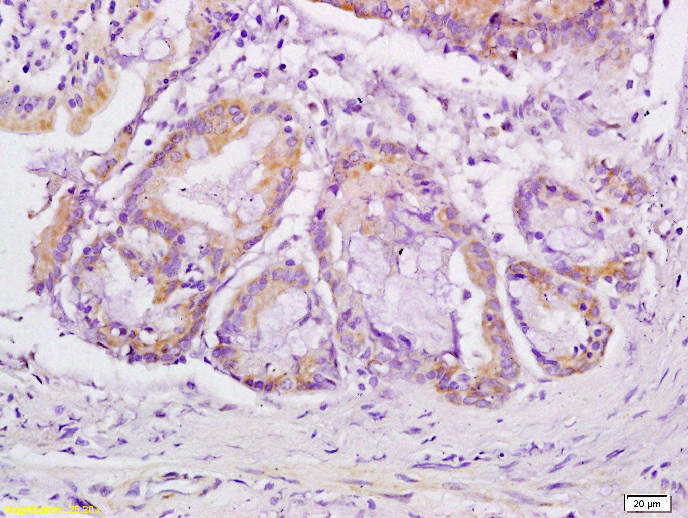 Tissue/cell: human gastric carcinoma;4% Paraformaldehyde-fixed and paraffin-embedded
Antigen retrieval: citrate buffer ( 0.01M, pH 6.0 ), Boiling bathing for 15min; Block endogenous peroxidase by 3% Hydrogen peroxide for 30min; Blocking buffer (normal goat serum) at 37℃ for 20 min
Incubation: Anti-VEGFR3 Polyclonal Antibody, Unconjugated(SL2202R) 1:200, overnight at 4°C, followed by conjugation to the secondary antibody and DAB staining
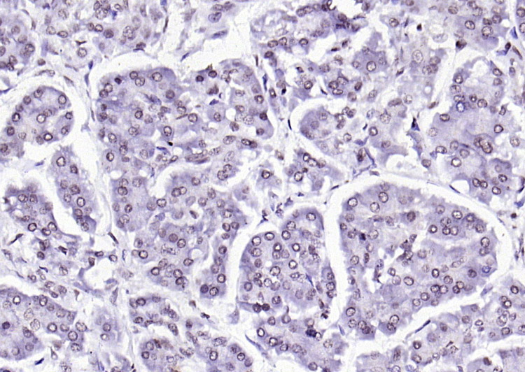 Paraformaldehyde-fixed, paraffin embedded (human pancreatic cancer); Antigen retrieval by boiling in sodium citrate buffer (pH6.0) for 15min; Block endogenous peroxidase by 3% hydrogen peroxide for 20 minutes; Blocking buffer (normal goat serum) at 37°C for 30min; Antibody incubation with (VEGFR3) Polyclonal Antibody, Unconjugated (bSL2202R) at 1:200 overnight at 4°C, followed by operating according to SP Kit(Rabbit) (sp-0023) instructionsand DAB staining.
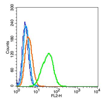 Blank control: A549(blue), the cells were fixed with 2% paraformaldehyde (10 min) and then permeabilized with ice-cold 90% methanol for 30 min on ice..
Isotype Control Antibody: Rabbit IgG(orange) ; Secondary Antibody: Goat anti-rabbit IgG-FITC(white blue), Dilution: 1:100 in 1 X PBS containing 0.5% BSA ; Primary Antibody Dilution: 1μl in 100 μL1X PBS containing 0.5% BSA(green). Cell: (mo)Splenocyte(2% BSA at 4°blocked for 30 minutes.1% Paraformaldehyde fixed for 30 minutes,0.1%TritonX-100 permeablized for 2 minutes).
Concentration:1:100.
Incubation: 40 minutes.
Host/Blank: Splenocytes.
Flow cytometric analysis of Rabbit Anti-VEGFR3 antibody (SL2202R)(green) compared with control in the absence of primary antibody (blue) followed by Splenocytes.
Secondary antibody: Goat Anti-rabbit IgG/FITC antibody (SL0295G-FITC).
Cartpieces
Totalgoods,subtotals:¥Checkout
References (0)
No References
Bought notes(bought amounts latest0)
No one bought this product
User Comment(Total0User Comment Num)
- No comment


 +86 571 56623320
 +86 18668110335

