
Rabbit Anti-PD-L1 antibody
CD274; B7 H; B7 H1; B7 homolog 1; B7-H1; B7H; B7H1; CD 274; CD274 antigen; CD274 molecule; MGC142294; MGC142296; OTTHUMP00000021029; PD L1; PD1L1_HUMAN; PD1L1_Mouse; PDCD1 ligand 1; PDCD1L1; PDCD1LG1; PDL 1; PDL1; Programmed cell death 1 ligand 1; Program
View History [Clear]
Details
Product Name PD-L1 Chinese Name 程序性死亡配体1(CD274)抗体 Alias CD274; B7 H; B7 H1; B7 homolog 1; B7-H1; B7H; B7H1; CD 274; CD274 antigen; CD274 molecule; MGC142294; MGC142296; OTTHUMP00000021029; PD L1; PD1L1_HUMAN; PD1L1_Mouse; PDCD1 ligand 1; PDCD1L1; PDCD1LG1; PDL 1; PDL1; Programmed cell death 1 ligand 1; Programmed death ligand 1; RGD1566211. Research Area Tumour Cell biology Signal transduction Apoptosis Immunogen Species Rabbit Clonality Polyclonal React Species Human, Mouse, (predicted: Rat, ) Applications WB=1:500-2000 Flow-Cyt=1ug/Test
not yet tested in other applications.
optimal dilutions/concentrations should be determined by the end user.Theoretical molecular weight 32kDa Cellular localization The cell membrane Form Liquid Concentration 1mg/ml immunogen KLH conjugated synthetic peptide derived from mouse PD-L1: 31-130/290 <Extracellular> Lsotype IgG Purification affinity purified by Protein A Buffer Solution 0.01M TBS(pH7.4) with 1% BSA, 0.03% Proclin300 and 50% Glycerol. Storage Shipped at 4℃. Store at -20 °C for one year. Avoid repeated freeze/thaw cycles. Attention This product as supplied is intended for research use only, not for use in human, therapeutic or diagnostic applications. PubMed PubMed Product Detail This gene encodes an immune inhibitory receptor ligand that is expressed by hematopoietic and non-hematopoietic cells, such as T cells and B cells and various types of tumor cells. The encoded protein is a type I transmembrane protein that has immunoglobulin V-like and C-like domains. Interaction of this ligand with its receptor inhibits T-cell activation and cytokine production. During infection or inflammation of normal tissue, this interaction is important for preventing autoimmunity by maintaining homeostasis of the immune response. In tumor microenvironments, this interaction provides an immune escape for tumor cells through cytotoxic T-cell inactivation. Expression of this gene in tumor cells is considered to be prognostic in many types of human malignancies, including colon cancer and renal cell carcinoma. Alternative splicing results in multiple transcript variants. [provided by RefSeq, Sep 2015]
Function:
Involved in the costimulatory signal, essential for T-cell proliferation and production of IL10 and IFNG, in an IL2-dependent and a PDCD1-independent manner. Interaction with PDCD1 inhibits T-cell proliferation and cytokine production.
Subunit:
Interacts with PDCD1.
Subcellular Location:
Isoform 1: Cell membrane; Single-pass type I membrane protein. Isoform 2: Endomembrane system; Single-pass type I membrane protein.
Tissue Specificity:
Highly expressed in the heart, skeletal muscle, placenta and lung. Weakly expressed in the thymus, spleen, kidney and liver. Expressed on activated T- and B-cells, dendritic cells, keratinocytes and monocytes.
Similarity:
Belongs to the immunoglobulin superfamily. BTN/MOG family.
Contains 1 Ig-like C2-type (immunoglobulin-like) domain.
Contains 1 Ig-like V-type (immunoglobulin-like) domain.
SWISS:
Q9EP73
Gene ID:
60533
Database links:Entrez Gene: 29126 Human
Entrez Gene: 60533 Mouse
Omim: 605402 Human
SwissProt: Q9NZQ7 Human
SwissProt: Q9EP73 Mouse
Unigene: 521989 Human
Unigene: 245363 Mouse
Unigene: 228198 Rat
程序性死亡配体1(B7-H1)可以促进epithelial cells的恶性转化,保护表皮细胞免于失巢凋亡,在Tumour的发生,进展和转归中发挥重要作用.
目前对CD274信号通路的进一步的研究必将为自身免疫性疾病、病毒感染性疾病和器官移植后排斥反应中T细胞介导的免疫应答调控提供了新的途径.B7-H1在癌症、类风湿、HIV感染等疾病中发现B7-H1有负调节作用。对B7-H1路径进行调控有助于自身免疫病及恶性Tumour的治疗。Product Picture 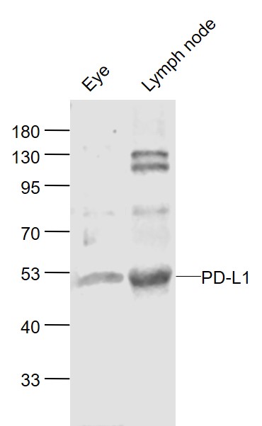 Sample:
Eye (Mouse) Lysate at 40 ug
Lymph node (Mouse) Lysate at 40 ug
Primary: Anti-PD-L1 (SL22022R) at 1/1000 dilution
Secondary: IRDye800CW Goat Anti-Rabbit IgG at 1/20000 dilution
Predicted band size: 48 kD
Observed band size: 50 kD
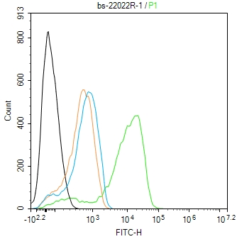 Blank control(black line):RAW264.7.
Primary Antibody (green line): Rabbit Anti-PD-L1 antibody (SL22022R)
Dilution:1ug/Test;
Secondary Antibody(white blue line): Goat anti-rabbit IgG-AF488
Dilution: 0.5ug/Test.
Isotype control(orange line): Normal Rabbit IgG
Protocol
The cells were fixed with 4% PFA (10min at room temperature)and then permeabilized with 90% ice-cold methanol for 20 min at -20℃, The cells were then incubated in 5%BSA to block non-specific protein-protein interactions for 30 min at room temperature .Cells stained with Primary Antibody for 30 min at room temperature. The secondary antibody used for 40 min at room temperature. Acquisition of 20,000 events was performed.
Cartpieces
Totalgoods,subtotals:¥Checkout
References (0)
No References
Bought notes(bought amounts latest0)
No one bought this product
User Comment(Total0User Comment Num)
- No comment


 +86 571 56623320
 +86 18668110335

