
Rabbit Anti-Cytokeratin 19 antibody
40 kDa keratin intermediate filament; CK 19; ck19; CK19; CK-19; KRT19; Cytokeratin19; Cytokeratin-19; k19; k1cs; Keratin 19; Keratin-19; Keratin type I 40 kD; Keratin type i 40kD; Keratin type I cytoskeletal 19; krt19; mgc15366; K1C19_HUMAN; Keratin, type
View History [Clear]
Details
Product Name Cytokeratin 19 Chinese Name 细胞角蛋白19抗体 Alias 40 kDa keratin intermediate filament; CK 19; ck19; CK19; CK-19; KRT19; Cytokeratin19; Cytokeratin-19; k19; k1cs; Keratin 19; Keratin-19; Keratin type I 40 kD; Keratin type i 40kD; Keratin type I cytoskeletal 19; krt19; mgc15366; K1C19_HUMAN; Keratin, type I cytoskeletal 19; Keratin, type I, 40 kd. literatures Research Area Tumour Stem cells Immunogen Species Rabbit Clonality Polyclonal React Species Human, Mouse, (predicted: Rat, Chicken, Dog, Pig, Cow, Horse, Rabbit, ) Applications WB=1:500-2000 ELISA=1:5000-10000 IHC-P=1:100-500 IHC-F=1:100-500 Flow-Cyt=1μg /test IF=1:100-500 (Paraffin sections need antigen repair)
not yet tested in other applications.
optimal dilutions/concentrations should be determined by the end user.Theoretical molecular weight 44kDa Cellular localization cytoplasmic The cell membrane Extracellular matrix Form Liquid Concentration 1mg/ml immunogen KLH conjugated synthetic peptide derived from human Cytokeratin 19.: 101-200/403 Lsotype IgG Purification affinity purified by Protein A Buffer Solution 0.01M TBS(pH7.4) with 1% BSA, 0.03% Proclin300 and 50% Glycerol. Storage Shipped at 4℃. Store at -20 °C for one year. Avoid repeated freeze/thaw cycles. Attention This product as supplied is intended for research use only, not for use in human, therapeutic or diagnostic applications. PubMed PubMed Product Detail The protein encoded by this gene is a member of the keratin family. The keratins are intermediate filament proteins responsible for the structural integrity of epithelial cells and are subdivided into cytokeratins and hair keratins. The type I cytokeratins consist of acidic proteins which are arranged in pairs of heterotypic keratin chains. Unlike its related family members, this smallest known acidic cytokeratin is not paired with a basic cytokeratin in epithelial cells. It is specifically expressed in the periderm, the transiently superficial layer that envelopes the developing epidermis. The type I cytokeratins are clustered in a region of chromosome 17q12-q21.
Function:
Involved in the organization of myofibers. Together with KRT8, helps to link the contractile apparatus to dystrophin at the costameres of striated muscle.
Subunit:
Heterotetramer of two type I and two type II keratins. Interacts with PNN and the actin-binding domain of DMD. Interacts with HCV core protein.
Tissue Specificity:
Expressed in a defined zone of basal keratinocytes in the deep outer root sheath of hair follicles. Also observed in sweat gland and mammary gland ductal and secretory cells, bile ducts, gastrointestinal tract, bladder urothelium, oral epithelia, esophagus, ectocervical epithelium (at protein level). Expressed in epidermal basal cells, in nipple epidermis and a defined region of the hair follicle. Also seen in a subset of vascular wall cells in both the veins and artery of human umbilical cord, and in umbilical cord vascular smooth muscle. Observed in muscle fibers accumulating in the costameres of myoplasm at the sarcolemma in structures that contain dystrophin and spectrin.
Similarity:
Belongs to the intermediate filament family.
SWISS:
P08727
Gene ID:
3880
Database links:Entrez Gene: 3880 Human
Entrez Gene: 16669 Mouse
Omim: 148020 Human
SwissProt: P08727 Human
SwissProt: P19001 Mouse
Unigene: 654568 Human
Unigene: 439699 Mouse
结构蛋白(Structural Proteins)
CK-19细胞角蛋白-19(又称CYFRa21-1)主要用于Tumour方面的研究,近年来大多用于消化系统恶性Tumour的鉴别,有学者认为:CK-19在胰腺Stem cells分化过程中有细胞角蛋白19的表达,把细胞角蛋白19作为胰腺Stem cells的Maker。阳性部位:主要在胞浆。Product Picture 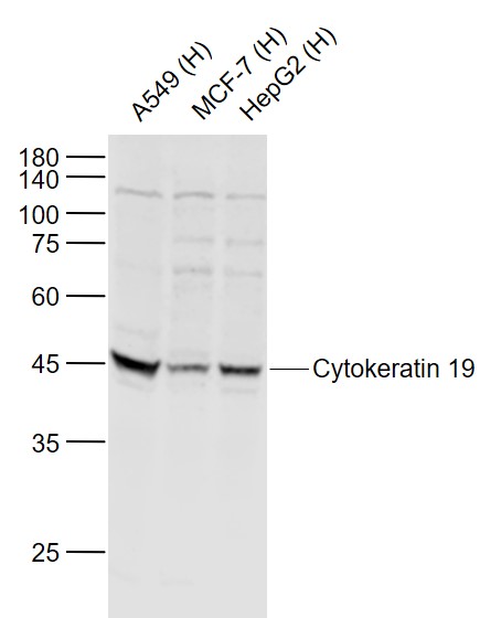 Sample:
Lane 1: A549 (Human) Cell Lysate at 30 ug
Lane 2: MCF-7 (Human) Cell Lysate at 30 ug
Lane 3: HepG2 (Human) Cell Lysate at 30 ug
Primary: Anti-Cytokeratin 19 (SL2190R) at 1/1000 dilution
Secondary: IRDye800CW Goat Anti-Rabbit IgG at 1/20000 dilution
Predicted band size: 42 kD
Observed band size: 42 kD
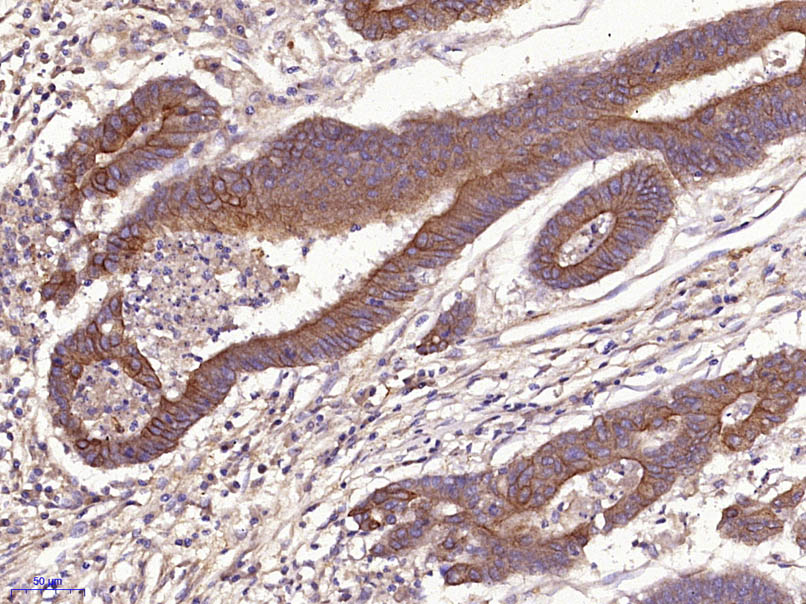 Paraformaldehyde-fixed, paraffin embedded (Human stomach carcinoma); Antigen retrieval by microwave in sodium citrate buffer (pH6.0) ; Block endogenous peroxidase by 3% hydrogen peroxide for 30 minutes; Blocking buffer (3% BSA) at RT for 30min; Antibody incubation with (Cytokeratin 19) Polyclonal Antibody, Unconjugated (SL2190R) at 1:400 overnight at 4℃, followed by conjugation to the secondary antibody (labeled with HRP)and DAB staining.
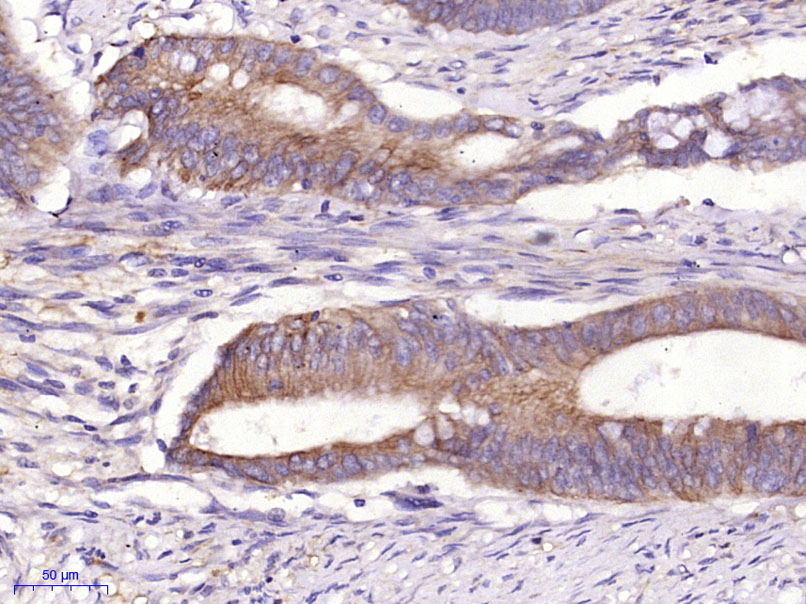 Paraformaldehyde-fixed, paraffin embedded (Human cervical carcinoma); Antigen retrieval by microwave in sodium citrate buffer (pH6.0) ; Block endogenous peroxidase by 3% hydrogen peroxide for 30 minutes; Blocking buffer (3% BSA) at RT for 30min; Antibody incubation with (Cytokeratin 19) Polyclonal Antibody, Unconjugated (SL2190R) at 1:400 overnight at 4℃, followed by conjugation to the secondary antibody (labeled with HRP)and DAB staining.
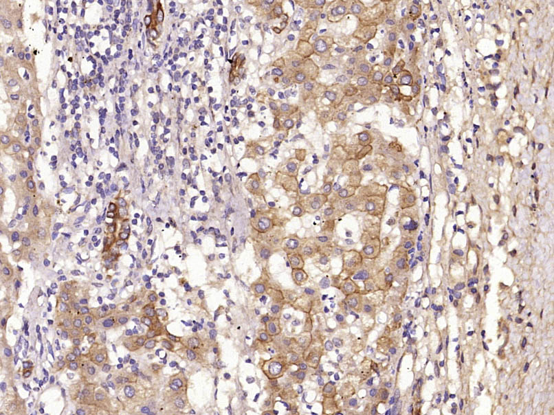 Paraformaldehyde-fixed, paraffin embedded (Human liver carcinoma); Antigen retrieval by microwave in sodium citrate buffer (pH6.0) ; Block endogenous peroxidase by 3% hydrogen peroxide for 30 minutes; Blocking buffer (3% BSA) at RT for 30min; Antibody incubation with (Cytokeratin 19) Polyclonal Antibody, Unconjugated (SL2190R) at 1:400 overnight at 4℃, followed by conjugation to the secondary antibody (labeled with HRP)and DAB staining.
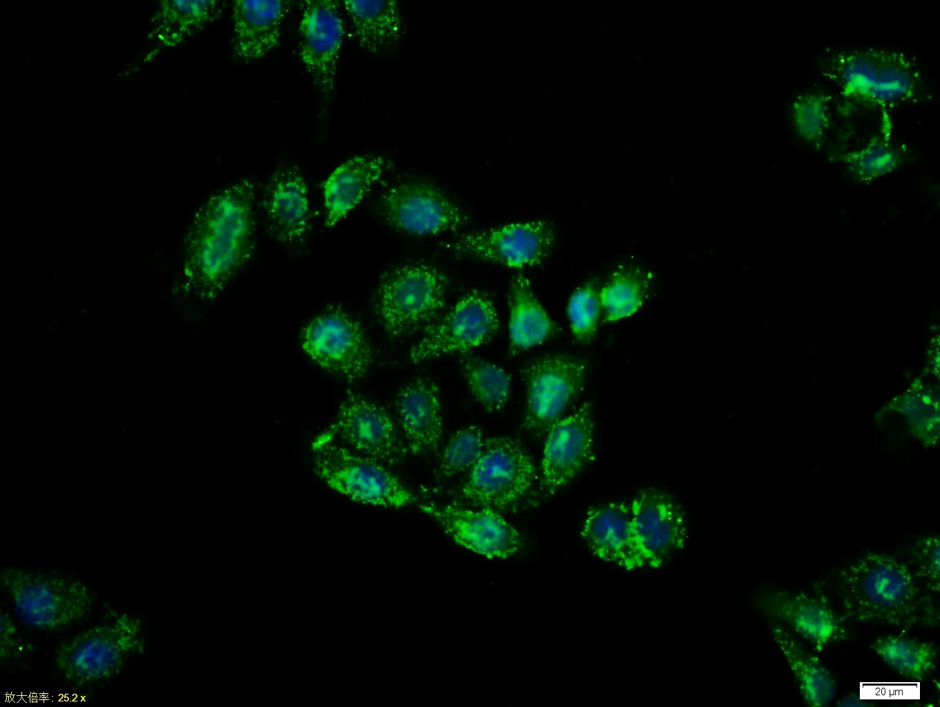 Tissue/cell:MCF7 cell; 4% Paraformaldehyde-fixed; Triton X-100 at room temperature for 20 min; Blocking buffer (normal goat serum, C-0005) at 37°C for 20 min; Antibody incubation with (Cytokeratin 19) polyclonal Antibody, Unconjugated (SL2190R) 1:100, 90 minutes at 37°C; followed by a FITC conjugated Goat Anti-Rabbit IgG antibody at 37°C for 90 minutes, DAPI (blue, C02-04002) was used to stain the cell nuclei.Tissue/cell:MCF7 cell; 4% Paraformaldehyde-fixed; Triton X-100 at room temperature for 20 min; Blocking buffer (normal goat serum, C-0005) at 37°C for 20 min; Antibody incubation with (Cytokeratin 19) polyclonal Antibody, Unconjugated (SL2190R) 1:100, 90 minutes at 37°C; followed by a FITC conjugated Goat Anti-Rabbit IgG antibody at 37°C for 90 minutes, DAPI (blue, C02-04002) was used to stain the cell nuclei.
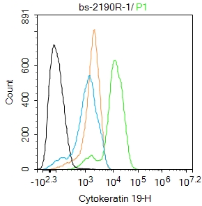 Blank control:MCF-7.
Primary Antibody (green line): Rabbit Anti-Cytokeratin 19 antibody (SL2190R)
Dilution: 1ug/Test;
Secondary Antibody : Goat anti-rabbit IgG-FITC
Dilution: 0.5ug/Test.
Protocol
The cells were fixed with 4% PFA (10min at room temperature)and then permeabilized with 90% ice-cold methanol for 20 min at -20℃.The cells were then incubated in 5%BSA to block non-specific protein-protein interactions for 30 min at room temperature .Cells stained with Primary Antibody for 30 min at room temperature. The secondary antibody used for 40 min at room temperature. Acquisition of 20,000 events was performed.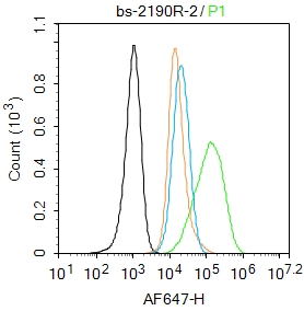 Blank control:A549.
Primary Antibody (green line): Rabbit Anti-Cytokeratin 19 antibody (SL2190R)
Dilution: 2μg /10^6 cells;
Isotype Control Antibody (orange line): Rabbit IgG .
Secondary Antibody : Goat anti-rabbit IgG-AF647
Dilution: 1μg /test.
Protocol
The cells were fixed with 4% PFA (10min at room temperature)and then permeabilized with 0.1% PBST for 20 min at room temperature. The cells were then incubated in 5%BSA to block non-specific protein-protein interactions for 30 min at room temperature .Cells stained with Primary Antibody for 30 min at room temperature. The secondary antibody used for 40 min at room temperature. Acquisition of 20,000 events was performed.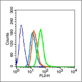 Blank control (blue line): Hela (fixed with 70% methanol (Overnight at 4℃) and then permeabilized with 90% ice-cold methanol for 20 min at -20℃).
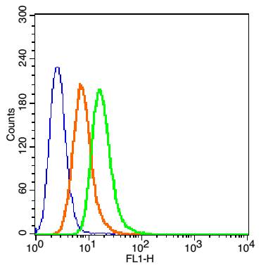
Primary Antibody (green line): Rabbit Anti-Cytokeratin 19 antibody (SL2190R),Dilution: 1μg /10^6 cells;
Isotype Control Antibody (orange line): Rabbit IgG .
Secondary Antibody (white blue line): Goat anti-rabbit IgG-PE,Dilution: 1μg /test.
 Blank control(blue): MCF7(fixed with 2% paraformaldehyde (10 min) and then permeabilized with ice-cold 90% methanol for 30 min on ice).
Primary Antibody: Rabbit Anti-Cytokeratin 19/FITC Conjugated antibody (SL2190R /FITC), Dilution: 1μg in 100 μL 1X PBS containing 0.5% BSA;
Isotype Control Antibody: Rabbit IgG/FITC(orange) ,used under the same conditions.
Cartpieces
Totalgoods,subtotals:¥Checkout
References (0)
No References
Bought notes(bought amounts latest0)
No one bought this product
User Comment(Total0User Comment Num)
- No comment


 +86 571 56623320
 +86 18668110335

