
Rabbit Anti-UMOD antibody
Uromucoid; ADMCKD2; FJHN; HNFJ; HNFJ1; MCKD2; medullary cystic kidney disease 2 (autosomal dominant); Tamm Horsfall glycoprotein; Tamm Horsfall urinary glycoprotein; Tamm-Horsfall urinary glycoprotein; THGP; THP; Umod; UROM_MOUSE; uromodulin (uromucoid, T
View History [Clear]
Details
Product Name UMOD Chinese Name 尿调节蛋白抗体 Alias Uromucoid; ADMCKD2; FJHN; HNFJ; HNFJ1; MCKD2; medullary cystic kidney disease 2 (autosomal dominant); Tamm Horsfall glycoprotein; Tamm Horsfall urinary glycoprotein; Tamm-Horsfall urinary glycoprotein; THGP; THP; Umod; UROM_MOUSE; uromodulin (uromucoid, Tamm-Horsfall glycoprotein); Uromodulin; Uromodulin, secreted form. literatures Research Area Cell biology immunology Immunogen Species Rabbit Clonality Polyclonal React Species (predicted: Cow, ) Applications ELISA=1:5000-10000 IHC-P=1:100-500 IHC-F=1:100-500 Flow-Cyt=1µg/Test IF=1:100-500 (Paraffin sections need antigen repair)
not yet tested in other applications.
optimal dilutions/concentrations should be determined by the end user.Theoretical molecular weight 61/65kDa Cellular localization The cell membrane Secretory protein Form Liquid Concentration 1mg/ml immunogen KLH conjugated synthetic peptide derived from mouse UMOD: 351-450/642 Lsotype IgG Purification affinity purified by Protein A Buffer Solution 0.01M TBS(pH7.4) with 1% BSA, 0.03% Proclin300 and 50% Glycerol. Storage Shipped at 4℃. Store at -20 °C for one year. Avoid repeated freeze/thaw cycles. Attention This product as supplied is intended for research use only, not for use in human, therapeutic or diagnostic applications. PubMed PubMed Product Detail The protein encoded by this gene is the most abundant protein in mammalian urine under physiological conditions. Its excretion in urine follows proteolytic cleavage of the ectodomain of its glycosyl phosphatidylinosital-anchored counterpart that is situated on the luminal cell surface of the loop of Henle. This protein may act as a constitutive inhibitor of calcium crystallization in renal fluids. Excretion of this protein in urine may provide defense against urinary tract infections caused by uropathogenic bacteria. Defects in this gene are associated with the renal disorders medullary cystic kidney disease-2 (MCKD2), glomerulocystic kidney disease with hyperuricemia and isosthenuria (GCKDHI), and familial juvenile hyperuricemic nephropathy (FJHN). Alternative splicing of this gene results in multiple transcript variants. [provided by RefSeq, Jul 2013].
Function:
Uromodulin: Functions in biogenesis and organization of the apical membrane of epithelial cells of the thick ascending limb of Henle's loop (TALH), where it promotes formation of complex filamentous gel-like structure providing the water barrier permeability. May serve as a receptor for binding and endocytosis for cytokines (IL-1, IL-2) and TNF. Facilitates neutrophil migration across renal epithelial (By similarity).
Uromodulin, secreted form: Secreted into urine after proteolytically cleaveage. Into the urine, may contribute to colloid osmotic pressure, retards passage of positively charged electrolytes, prevents urinary tract infection and modulates formation of supersaturated salts and their crystals.
Subcellular Location:
Apical cell membrane; Lipid-anchor, GPI-anchor (By similarity). Basolateral cell membrane; Lipid-anchor, GPI-anchor (By similarity). Cell projection, cilium membrane (By similarity). Note=Only a small fraction is sorts to the basolateral pole of tubular epithelial cells compared to apical localization (By similarity).
Uromodulin, secreted form: Secreted (By similarity).
Post-translational modifications:
N-glycosylated.
Similarity:
Contains 3 EGF-like domains.
Contains 1 ZP domain.
SWISS:
Q91X17
Gene ID:
22242
Database links:Entrez Gene: 7369 Human
Omim: 191845 Human
SwissProt: P07911 Human
Unigene: 654425 Human
Product Picture 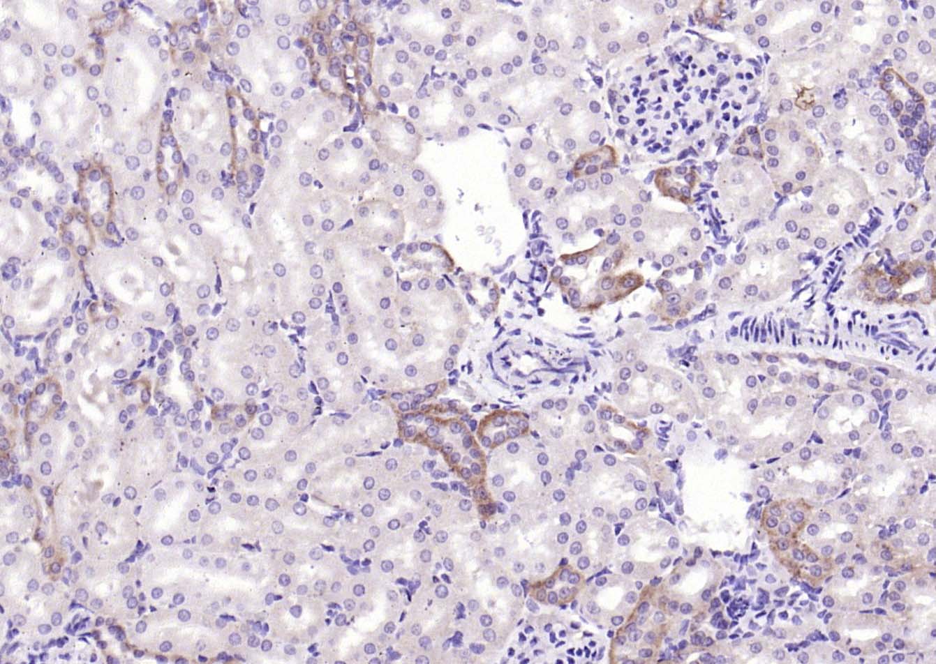 Paraformaldehyde-fixed, paraffin embedded (rat kidney); Antigen retrieval by boiling in sodium citrate buffer (pH6.0) for 15min; Block endogenous peroxidase by 3% hydrogen peroxide for 20 minutes; Blocking buffer (normal goat serum) at 37°C for 30min; Antibody incubation with (Uromucoid) Polyclonal Antibody, Unconjugated (SL2189R) at 1:2000 overnight at 4°C, followed by operating according to SP Kit(Rabbit) (sp-0023) instructionsand DAB staining.
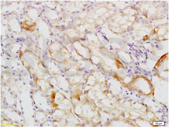 Tissue/cell: mouse kidney tissue; 4% Paraformaldehyde-fixed and paraffin-embedded;
Antigen retrieval: citrate buffer ( 0.01M, pH 6.0 ), Boiling bathing for 15min; Block endogenous peroxidase by 3% Hydrogen peroxide for 30min; Blocking buffer (normal goat serum,C-0005) at 37℃ for 20 min;
Incubation: Anti-MCKD2/UMOD Polyclonal Antibody, Unconjugated(SL2189R) 1:200, overnight at 4°C, followed by conjugation to the secondary antibody(SP-0023) and DAB(C-0010) staining
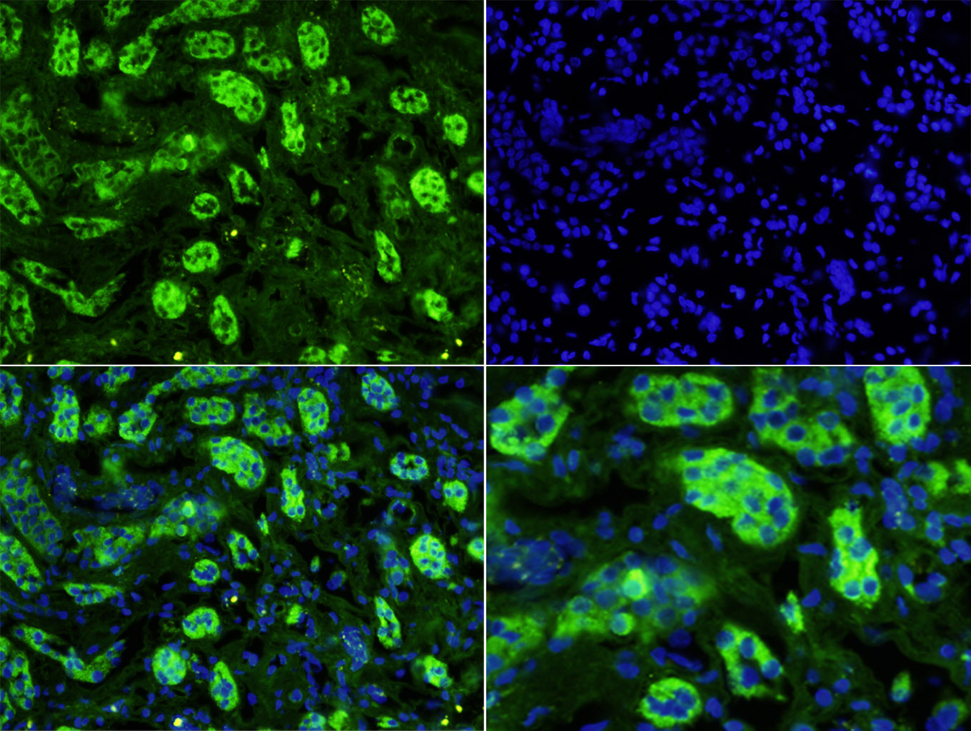 Tissue/cell: human kidney tissue;4% Paraformaldehyde-fixed and paraffin-embedded;
Antigen retrieval: citrate buffer ( 0.01M, pH 6.0 ), Boiling bathing for 15min; Blocking buffer (normal goat serum,C-0005) at 37℃ for 20 min;
Incubation: Anti-Uromucoid Polyclonal Antibody, Unconjugated(SL2189R) 1:200, overnight at 4°C; The secondary antibody was Goat Anti-Rabbit IgG, Cy3 conjugated(SL0295G-FITC)used at 1:200 dilution for 40 minutes at 37°C. DAPI(5ug/ml,blue,C-0033) was used to stain the cell nuclei
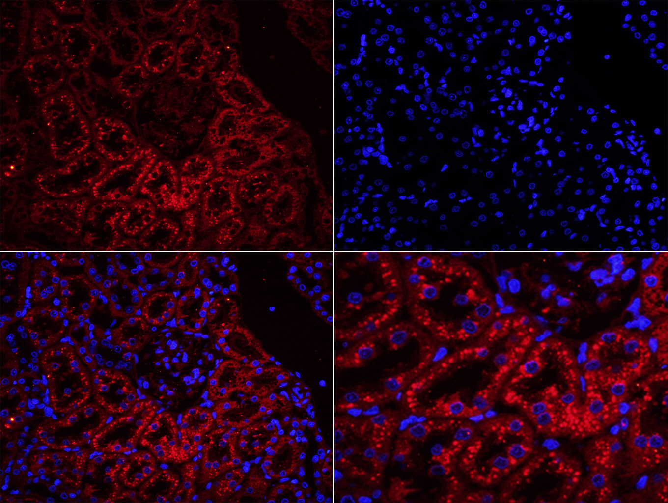 Tissue/cell: rat kidney tissue;4% Paraformaldehyde-fixed and paraffin-embedded;
Antigen retrieval: citrate buffer ( 0.01M, pH 6.0 ), Boiling bathing for 15min; Blocking buffer (normal goat serum,C-0005) at 37℃ for 20 min;
Incubation: Anti-Uromucoid Polyclonal Antibody, Unconjugated(SL2189R) 1:200, overnight at 4°C; The secondary antibody was Goat Anti-Rabbit IgG, Cy3 conjugated(SL0295G-Cy3)used at 1:200 dilution for 40 minutes at 37°C. DAPI(5ug/ml,blue,C-0033) was used to stain the cell nuclei
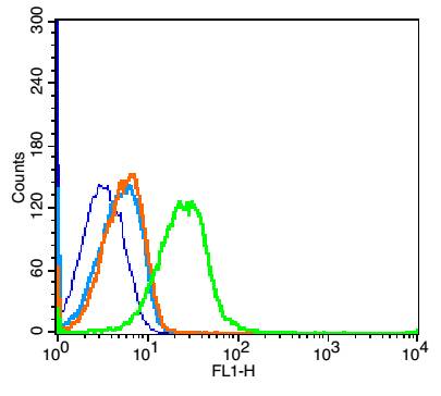 Blank control: Mouse kidney (blue).
Primary Antibody:Rabbit Anti-Coxsackie Adenovirus Receptor antibody (SL2189R,Green); Dilution: 1μg in 100 μL 1X PBS containing 0.5% BSA;
Isotype Control Antibody: Rabbit IgG(orange) ,used under the same conditions;
Secondary Antibody: Goat anti-rabbit IgG-FITC(white blue), Dilution: 1:200 in 1 X PBS containing 0.5% BSA.
Protocol
The cells were fixed with 2% paraformaldehyde for 10 min at 37℃. Primary antibody (SL2189R, 1μg /1x10^6 cells) were incubated for 30 min at room temperature, followed by 1 X PBS containing 0.5% BSA + 10% goat serum (1 hour) to block non-specific protein-protein interactions. Then the Goat Anti-rabbit IgG/FITC antibody was added into the blocking buffer mentioned above to react with the primary antibody at 1/200 dilution for 40 min at room temperature. Acquisition of 20,000 events was performed.
Cartpieces
Totalgoods,subtotals:¥Checkout
References (0)
No References
Bought notes(bought amounts latest0)
No one bought this product
User Comment(Total0User Comment Num)
- No comment


 +86 571 56623320
 +86 18668110335

