
Rabbit Anti-PARP1 antibody
ADP ribosyltransferase (NAD+; poly (ADP ribose) polymerase); ADP ribosyltransferase NAD+; ADPRT 1; ADPRT; ADPRT1; msPARP; NAD(+) ADP ribosyltransferase 1; pADPRT 1; pADPRT1; PARP 1; PARP; Poly (ADP ribose) polymerase 1; poly (ADP ribose) polymerase family
View History [Clear]
Details
Product Name PARP1 Chinese Name 多腺苷二磷酸多聚酶抗体(N端) Alias ADP ribosyltransferase (NAD+; poly (ADP ribose) polymerase); ADP ribosyltransferase NAD+; ADPRT 1; ADPRT; ADPRT1; msPARP; NAD(+) ADP ribosyltransferase 1; pADPRT 1; pADPRT1; PARP 1; PARP; Poly (ADP ribose) polymerase 1; poly (ADP ribose) polymerase family, member 1; Poly adenosine diphosphate ADP ribose polymerase; Poly ADP ribose polymerase 1; Poly ADP ribose polymerase family member 1; Poly ADP ribose synthetase 1; poly(ADP ribose) synthetase; poly(ADP ribosyl)transferase; Poly[ADP ribose] synthetase 1; PPOL; sPARP 1; sPARP1; PARP1_HUMAN. literatures Research Area Chromatin and nuclear signals Apoptosis Immunogen Species Rabbit Clonality Polyclonal React Species Human, Mouse, (predicted: Rat, Dog, Cow, ) Applications WB=1:500-2000 ELISA=1:5000-10000 IHC-P=1:100-500 IHC-F=1:100-500 Flow-Cyt=0.2μg/Test IF=1:100-500 (Paraffin sections need antigen repair)
not yet tested in other applications.
optimal dilutions/concentrations should be determined by the end user.Theoretical molecular weight 111kDa Cellular localization The nucleus Form Liquid Concentration 1mg/ml immunogen KLH conjugated synthetic peptide derived from human PARP: 201-300/1014 Lsotype IgG Purification affinity purified by Protein A Buffer Solution 0.01M TBS(pH7.4) with 1% BSA, 0.03% Proclin300 and 50% Glycerol. Storage Shipped at 4℃. Store at -20 °C for one year. Avoid repeated freeze/thaw cycles. Attention This product as supplied is intended for research use only, not for use in human, therapeutic or diagnostic applications. PubMed PubMed Product Detail This gene encodes a chromatin-associated enzyme, poly(ADP-ribosyl)transferase, which modifies various nuclear proteins by poly(ADP-ribosyl)ation. The modification is dependent on DNA and is involved in the regulation of various important cellular processes such as differentiation, proliferation, and tumor transformation and also in the regulation of the molecular events involved in the recovery of cell from DNA damage. In addition, this enzyme may be the site of mutation in Fanconi anemia, and may participate in the pathophysiology of type I diabetes. [provided by RefSeq, Jul 2008].
Function:
Involved in the base excision repair (BER) pathway, by catalyzing the poly(ADP-ribosyl)ation of a limited number of acceptor proteins involved in chromatin architecture and in DNA metabolism. This modification follows DNA damages and appears as an obligatory step in a detection/signaling pathway leading to the reparation of DNA strand breaks. Mediates the poly(ADP-ribosyl)ation of APLF and CHFR. Positively regulates the transcription of MTUS1 and negatively regulates the transcription of MTUS2/TIP150. With EEF1A1 and TXK, forms a complex that acts as a T-helper 1 (Th1) cell-specific transcription factor and binds the promoter of IFN-gamma to directly regulate its transcription, and is thus involved importantly in Th1 cytokine production.
Subunit:
Component of a base excision repair (BER) complex, containing at least XRCC1, PARP2, POLB and LRIG3. Homo- and heterodimer with PARP2. Interacts with PARP3, APTX and SRY. The SWAP complex consists of NPM1, NCL, PARP1 and SWAP70. Interacts with TIAM2 and ZNF423 (By similarity). Interacts (when poly-ADP-ribosylated) with CHD1L. Interacts with the DNA polymerase alpha catalytic subunit POLA1; this interaction functions as part of the control of replication fork progression. Interacts with EEF1A1, RNF4 and TXK.
Subcellular Location:
Mitochondrion outer membrane; Single-pass membrane protein.
Nucleus membrane; Single-pass membrane protein.
Endoplasmic reticulum membrane; Single-pass membrane protein.
Nucleus.
Post-translational modifications:
Phosphorylated by PRKDC and TXK. Phosphorylated upon DNA damage, probably by ATM or ATR.
Poly-ADP-ribosylated by PARP2. Poly-ADP-ribosylation mediates the recruitment of CHD1L to DNA damage sites.
S-nitrosylated, leading to inhibit transcription regulation activity.
Similarity:
Contains 1 BRCT domain.
Contains 1 PARP alpha-helical domain.
Contains 1 PARP catalytic domain.
Contains 2 PARP-type zinc fingers.
SWISS:
P09874
Gene ID:
142
Database links:Entrez Gene: 142 Human
Entrez Gene: 11545 Mouse
Omim: 173870 Human
SwissProt: P09874 Human
SwissProt: P11103 Mouse
Unigene: 177766 Human
Unigene: 277779 Mouse
Unigene: 11327 Rat
PARP(poly ADP-ribose polymerase)是DNA修复酶。
PARP是Apoptosis核心成员半胱胺酸蛋白酶(caspase)的切割底物。因此,它在DNA损伤修复与Apoptosis中发挥着重要作用。Anti-PARP p85 是特意的PARPp85片段的特异抗体,由caspase剪切116kDa完整分子而得到的。
PARP是存在于多数真核细胞中的一个多功能蛋白质翻译后修饰酶。它通过识别结构损伤的DNA片段而被激活,对聚腺苷二磷酸核糖聚合酶PARP被认为是DNA损伤的感受器。它还能对许多核蛋白进行聚腺苷二磷酸核糖基化。因此,在DNA损伤修复与Apoptosis中发挥着重要作用,端锚聚合酶在癌细胞端粒结构的调控机制中有重要作用。Product Picture 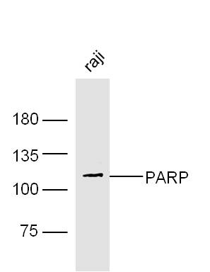 Sample:
Raji Cell (Human) Lysate at 30 ug
Primary: Anti- PARP (SL2138R) at 1/300 dilution
Secondary: IRDye800CW Goat Anti-Rabbit IgG at 1/20000 dilution
Predicted band size: 111 kD
Observed band size: 111 kD
 Sample:
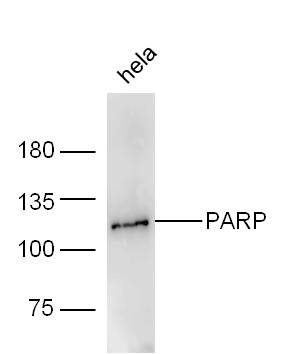
Hela Cell (Human) Lysate at 30 ug
Primary: Anti- PARP (SL2138R) at 1/300 dilution
Secondary: IRDye800CW Goat Anti-Rabbit IgG at 1/20000 dilution
Predicted band size: 111 kD
Observed band size: 111 kD
 Sample:
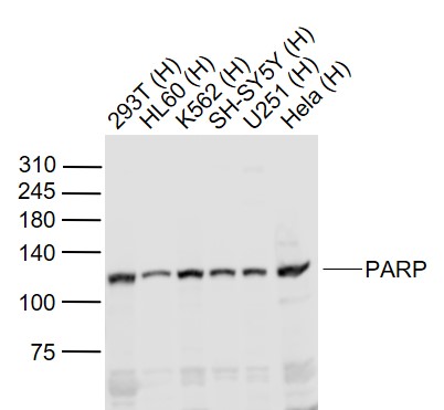
Lane 1: 293T (Human) Cell Lysate at 30 ug
Lane 2: HL60 (Human) Cell Lysate at 30 ug
Lane 3: K562 (Human) Cell Lysate at 30 ug
Lane 4: SH-SY5Y (Human) Cell Lysate at 30 ug
Lane 5: U251 (Human) Cell Lysate at 30 ug
Lane 6: Hela (Human) Cell Lysate at 30 ug
Primary: Anti-PARP (SL2138R) at 1/1000 dilution
Secondary: IRDye800CW Goat Anti-Rabbit IgG at 1/20000 dilution
Predicted band size: 115 kD
Observed band size: 115 kD
 Sample:
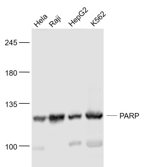
Hela(Human) Cell Lysate at 30 ug
Raji(Human) Cell Lysate at 30 ug
HepG2(Human) Cell Lysate at 30 ug
K562(Human) Cell Lysate at 30 ug
Primary: Anti- PARP (SL2138R) at 1/1000 dilution
Secondary: IRDye800CW Goat Anti-Rabbit IgG at 1/20000 dilution
Predicted band size: 111 kD
Observed band size: 111 kD
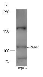 Protein:HepG2 lysate at 30ug;
Primary: Anti-PARP (SL2138R) at 1:300 dilution;
Secondary: HRP conjugated Goat-Anti-rabbit IgG(SL0295G-HRP) at 1: 5000 dilution;
Predicted band size:111 kD
Observed band size:111 kD
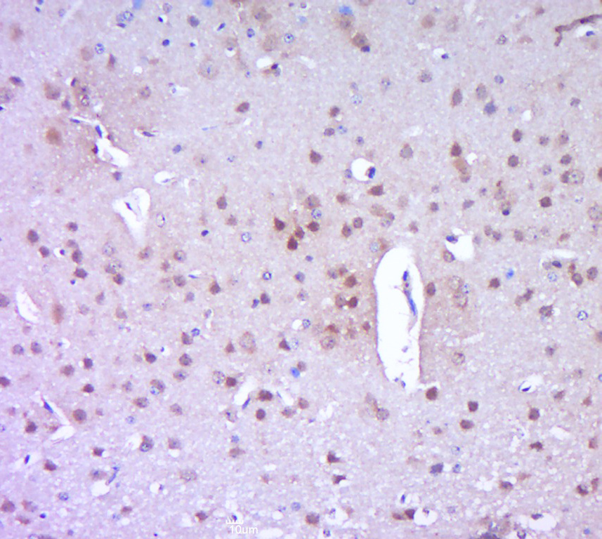 Paraformaldehyde-fixed, paraffin embedded (mouse brain tissue); Antigen retrieval by boiling in sodium citrate buffer (pH6.0) for 15min; Block endogenous peroxidase by 3% hydrogen peroxide for 20 minutes; Blocking buffer (normal goat serum) at 37°C for 30min; Antibody incubation with (PARP) Polyclonal Antibody, Unconjugated (SL2138R) at 1:400 overnight at 4°C, followed by a conjugated secondary (sp-0023) for 20 minutes and DAB staining.
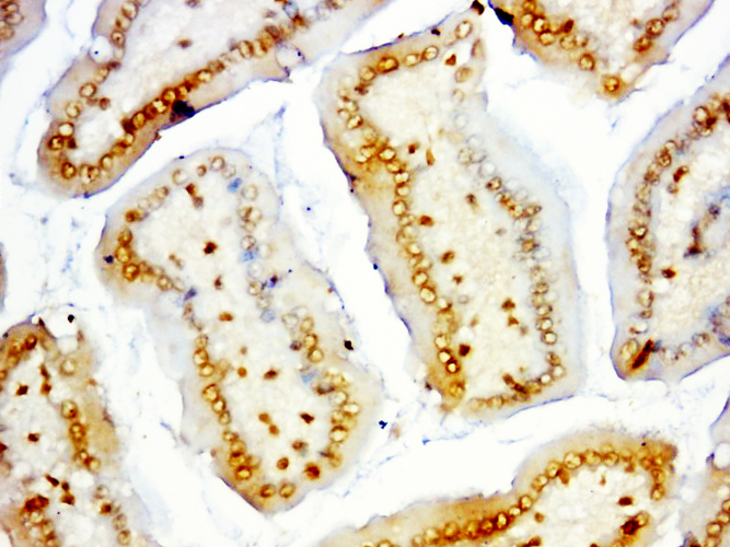 Paraformaldehyde-fixed, paraffin embedded (mouse intestines); Antigen retrieval by boiling in sodium citrate buffer (pH6.0) for 15min; Block endogenous peroxidase by 3% hydrogen peroxide for 20 minutes; Blocking buffer (normal goat serum) at 37°C for 30min; Antibody incubation with (PARP) Polyclonal Antibody, Unconjugated (SL2138R) at 1:500 overnight at 4°C, followed by a conjugated secondary (sp-0023) for 20 minutes and DAB staining.
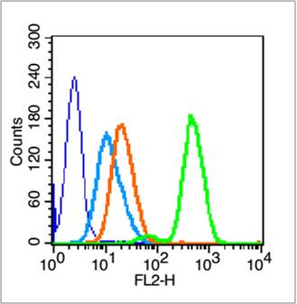 Blank control (blue line): HL60 cells (blue).
Primary Antibody (green line): Rabbit Anti- PARP antibody (SL2138R)
Dilution: 0.2μg /10^6 cells;
Isotype Control Antibody (orange line): Rabbit IgG .
Secondary Antibody (white blue line): Goat anti-rabbit IgG-PE
Dilution: 1μg /test.
Protocol
The cells were fixed with 70% methanol (Overnight at 4℃) and then permeabilized with 90% ice-cold methanol for 20 min at -20℃. Cells stained with Primary Antibody for 30 min at room temperature. The cells were then incubated in 1 X PBS/2%BSA/10% goat serum to block non-specific protein-protein interactions followed by the antibody for 15 min at room temperature. The secondary antibody used for 40 min at room temperature. Acquisition of 20,000 events was performed.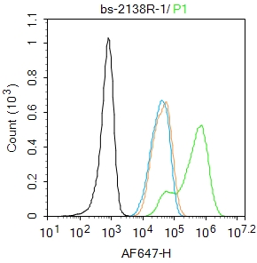 Blank control:293T.
Primary Antibody (green line): Rabbit Anti-PARP1 antibody (SL2138R)
Dilution: 1μg /10^6 cells;
Isotype Control Antibody (orange line): Rabbit IgG .
Secondary Antibody : Goat anti-rabbit IgG-AF647
Dilution: 1μg /test.
Protocol
The cells were fixed with 4% PFA (10min at room temperature)and then permeabilized with 90% ice-cold methanol for 20 min at -20℃.The cells were then incubated in 5%BSA to block non-specific protein-protein interactions for 30 min at room temperature .Cells stained with Primary Antibody for 30 min at room temperature. The secondary antibody used for 40 min at room temperature. Acquisition of 20,000 events was performed.
Cartpieces
Totalgoods,subtotals:¥Checkout
References (0)
No References
Bought notes(bought amounts latest0)
No one bought this product
User Comment(Total0User Comment Num)
- No comment


 +86 571 56623320
 +86 18668110335

