
Rabbit Anti-Yellow fever virus envelope glycoprotein E antibody
Envelope protein E; Genome polyprotein; polyprotein [Yellow fever virus]; polyprotein YFV; POLG_YEFV1; YFVgp1; YFVgp1 polyprotein precursor [ Yellow fever virus ]; Yellow fever virus (strain 17D vaccine) (YFV).
View History [Clear]
Details
Product Name Yellow fever virus envelope glycoprotein E Chinese Name 黄热病毒包膜glycoprotein抗体 Alias Envelope protein E; Genome polyprotein; polyprotein [Yellow fever virus]; polyprotein YFV; POLG_YEFV1; YFVgp1; YFVgp1 polyprotein precursor [ Yellow fever virus ]; Yellow fever virus (strain 17D vaccine) (YFV). Research Area immunology Bacteria and viruses Immunogen Species Rabbit Clonality Polyclonal Applications WB=1:500-2000 ELISA=1:5000-10000
not yet tested in other applications.
optimal dilutions/concentrations should be determined by the end user.Theoretical molecular weight 54/375kDa Cellular localization cytoplasmic The cell membrane Secretory protein Form Liquid Concentration 1mg/ml immunogen KLH conjugated synthetic peptide derived from Yellow fever virus envelope glycoprotein E: 601-700/3411 Lsotype IgG Purification affinity purified by Protein A Buffer Solution 0.01M TBS(pH7.4) with 1% BSA, 0.03% Proclin300 and 50% Glycerol. Storage Shipped at 4℃. Store at -20 °C for one year. Avoid repeated freeze/thaw cycles. Attention This product as supplied is intended for research use only, not for use in human, therapeutic or diagnostic applications. PubMed PubMed Product Detail Envelope protein E binding to host cell surface receptor is followed by virus internalization through clathrin-mediated endocytosis. Envelope protein E is subsequently involved in membrane fusion between virion and host late endosomes. Synthesized as a homodimer with prM which acts as a chaperone for envelope protein E. After cleavage of prM, envelope protein E dissociate from small envelope protein M and homodimerizes.
Function:
Capsid protein C self-assembles to form an icosahedral capsid about 30 nm in diameter. The capsid encapsulates the genomic RNA.
prM acts as a chaperone for envelope protein E during intracellular virion assembly by masking and inactivating envelope protein E fusion peptide. prM is matured in the last step of virion assembly, presumably to avoid catastrophic activation of the viral fusion peptide induced by the acidic pH of the trans-Golgi network. After cleavage by host furin, the pr peptide is released in the extracellular medium and small envelope protein M and envelope protein E homodimers are dissociated.
Envelope protein E binding to host cell surface receptor is followed by virus internalization through clathrin-mediated endocytosis. Envelope protein E is subsequently involved in membrane fusion between virion and host late endosomes. Synthesized as a homodimer with prM which acts as a chaperone for envelope protein E. After cleavage of prM, envelope protein E dissociate from small envelope protein M and homodimerizes.
Non-structural protein 1 is involved in virus replication and regulation of the innate immune response.
Non-structural protein 2A may be involved viral RNA replication and capsid assembly (Potential).
Non-structural protein 2B is a required cofactor for the serine protease function of NS3.
Serine protease NS3 displays three enzymatic activities: serine protease, NTPase and RNA helicase. NS3 serine protease, in association with NS2B, performs its autocleavage and cleaves the polyprotein at dibasic sites in the cytoplasm: C-prM, NS2A-NS2B, NS2B-NS3, NS3-NS4A, NS4A-2K and NS4B-NS5. NS3 RNA helicase binds RNA and unwinds dsRNA in the 3' to 5' direction (By similarity).
Non-structural protein 4A induces host endoplasmic reticulum membrane rearrangements leading to the formation of virus-induced membranous vesicles hosting the dsRNA and polymerase, functioning as a replication complex. NS4A might also regulate the ATPase activity of the NS3 helicase (By similarity).
Peptide 2k functions as a signal peptide for NS4B and is required for the interferon antagonism activity of the latter.
Non-structural protein 4B inhibits interferon (IFN)-induced host STAT1 phosphorylation and nuclear translocation, thereby preventing the establishment of cellular antiviral state by blocking the IFN-alpha/beta pathway (By similarity).
RNA-directed RNA polymerase NS5 replicates the viral (+) and (-) genome, and performs the capping of genomes in the cytoplasm. NS5 methylates viral RNA cap at guanine N-7 and ribose 2'-O positions. Besides its role in genome replication, also prevents the establishment of cellular antiviral state by blocking the interferon-alpha/beta (IFN-alpha/beta) signaling pathway
Subunit:
Capsid protein C forms homodimers. prM and envelope protein E form heterodimers in the endoplasmic reticulum and Golgi. In immature particles, there are 60 icosaedrally organized trimeric spikes on the surface. Each spike consists of three heterodimers of envelope protein M precursor (prM) and envelope protein E. NS1 forms homodimers as well as homohexamers when secreted. NS1 may interact with NS4A. NS3 and NS2B form a heterodimer. NS3 is the catalytic subunit, whereas NS2B strongly stimulates the latter, acting as a cofactor. In the absence of the NS2B, NS3 protease is unfolded and inactive. NS3 interacts with unphosphorylated NS5; this interaction stimulates NS5 guanylyltransferase activity.
Subcellular Location:
Capsid protein C: Virion (Potential).
Peptide pr: Secreted.
Small envelope protein M: Virion membrane; Multi-pass membrane protein (Potential). Host endoplasmic reticulum membrane; Multi-pass membrane protein (Potential).
Envelope protein E: Virion membrane; Multi-pass membrane protein (Potential). Host endoplasmic reticulum membrane; Multi-pass membrane protein (Potential).
Non-structural protein 1: Secreted. Host endoplasmic reticulum membrane; Peripheral membrane protein; Lumenal side.
Non-structural protein 2A-alpha: Host endoplasmic reticulum membrane; Multi-pass membrane protein (Potential).
Non-structural protein 2A: Host endoplasmic reticulum membrane; Multi-pass membrane protein (Potential).
Serine protease subunit NS2B: Host endoplasmic reticulum membrane; Peripheral membrane protein; Cytoplasmic side.
Serine protease NS3: Host endoplasmic reticulum membrane; Peripheral membrane protein; Cytoplasmic side. Note=Remains non-covalently associated to NS3 protease.
Non-structural protein 4A: Host endoplasmic reticulum membrane; Multi-pass membrane protein. Note=Located in RE-associated vesicles hosting the replication complex.
Non-structural protein 4B: Host endoplasmic reticulum membrane; Multi-pass membrane protein.
RNA-directed RNA polymerase NS5: Host endoplasmic reticulum membrane; Peripheral membrane protein; Cytoplasmic side. Host nucleus. Note=Located in RE-associated vesicles hosting the replication complex.
Post-translational modifications:
Specific enzymatic cleavages in vivo yield mature proteins. The nascent protein C contains a C-terminal hydrophobic domain that act as a signal sequence for translocation of prM into the lumen of the ER. Mature protein C is cleaved at a site upstream of this hydrophobic domain by NS3. prM is cleaved in post-Golgi vesicles by a host furin, releasing the mature small envelope protein M, and peptide pr. Non-structural protein 2A-alpha, a C-terminally truncated form of non-structural protein 2A, results from partial cleavage by NS3. Specific enzymatic cleavages in vivo yield mature proteins Peptide 2K acts as a signal sequence and is removed from the N-terminus of NS4B by the host signal peptidase in the ER lumen. Signal cleavage at the 2K-4B site requires a prior NS3 protease-mediated cleavage at the 4A-2K site (By similarity).
RNA-directed RNA polymerase NS5 is phosphorylated on serines residues. This phosphorylation may trigger NS5 nuclear localization.
Envelope protein E and non-structural protein 1 are N-glycosylated.
Similarity:
In the N-terminal section; belongs to the class I-like SAM-binding methyltransferase superfamily. mRNA cap 0-1 NS5-type methyltransferase family.
Contains 1 helicase ATP-binding domain.
Contains 1 helicase C-terminal domain.
Contains 1 mRNA cap 0-1 NS5-type MT domain.
Contains 1 peptidase S7 domain.
Contains 1 RdRp catalytic domain.
SWISS:
P03314
Gene ID:
1502173
Product Picture 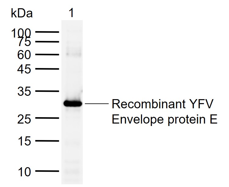 Sample:
Lane 1: Recombinant YFV Envelope protein E, Trx & His(SL41285P-10ng)
Primary: Anti-Yellow fever virus envelope glycoprotein E (SL2041R) at 1/1000 dilution
Secondary: IRDye800CW Goat Anti-Rabbit IgG at 1/20000 dilution
Predicted band size: 54/375 kDa
Observed band size: 30 kDa
Cartpieces
Totalgoods,subtotals:¥Checkout
Bought notes(bought amounts latest0)
No one bought this product
User Comment(Total0User Comment Num)
- No comment


 +86 571 56623320
 +86 18668110335

