
Rabbit Anti-Phospho-MYPT1 (Thr696)antibody
Myosin Phosphatase (phospho T696); Myosin Phosphatase (phospho Thr696); p-MYPT1(Thr696); Myosin Phosphatase; M130; MBS; MGC133042; Myosin phosphatase target subunit 1; Myosin phosphatase targeting subunit 1; MYPT 1; MYPT1; PPP1R12A; Protein phosphatase 1
View History [Clear]
Details
Product Name Phospho-MYPT1 (Thr696) Chinese Name 磷酸化肌球蛋白磷酸酶抗体 Alias Myosin Phosphatase (phospho T696); Myosin Phosphatase (phospho Thr696); p-MYPT1(Thr696); Myosin Phosphatase; M130; MBS; MGC133042; Myosin phosphatase target subunit 1; Myosin phosphatase targeting subunit 1; MYPT 1; MYPT1; PPP1R12A; Protein phosphatase 1 regulatory inhibitor subunit 12A; Protein phosphatase 1 regulatory subunit 12A; Protein phosphatase myosin binding subunit; MYPT1_HUMAN; Myosin phosphatase-targeting subunit 1; Protein phosphatase myosin-binding subunit. literatures Research Area Tumour immunology Signal transduction Apoptosis transcriptional regulatory factor Immunogen Species Rabbit Clonality Polyclonal React Species Human, (predicted: Mouse, Rat, Chicken, Dog, Cow, Horse, Rabbit, ) Applications ELISA=1:5000-10000 IHC-P=1:100-500 IHC-F=1:100-500 Flow-Cyt=1ug/Test ICC=1:100 IF=1:100-500 (Paraffin sections need antigen repair)
not yet tested in other applications.
optimal dilutions/concentrations should be determined by the end user.Theoretical molecular weight 113kDa Cellular localization cytoplasmic Form Liquid Concentration 1mg/ml immunogen KLH conjugated synthesised phosphopeptide derived from human MYPT1 around the phosphorylation site of Thr696: RS(p-T)QG Lsotype IgG Purification affinity purified by Protein A Buffer Solution Preservative: 15mM Sodium Azide, Constituents: 1% BSA, 0.01M PBS, pH 7.4 Storage Shipped at 4℃. Store at -20 °C for one year. Avoid repeated freeze/thaw cycles. Attention This product as supplied is intended for research use only, not for use in human, therapeutic or diagnostic applications. PubMed PubMed Product Detail Myosin phosphatase regulates the interaction of actin and myosin downstream of the guanosine triphosphatase Rho. The small guanosine triphosphatase Rho is implicated in myosin light chain (MLC) phosphorylation, which results in contraction of smooth muscle and interaction of actin and myosin in non muscle cells. The guanosine triphosphate (GTP) bound, active form of RhoA (GTP.RhoA) specifically interacted with the myosin binding subunit (MBS) of myosin phosphatase, which regulates the extent of phosphorylation of MLC. Rho associated kinase (Rho kinase), which is activated by GTP. RhoA, phosphorylated MBS and consequently inactivated myosin phosphatase. Overexpression of RhoA or activated RhoA in NIH 3T3 cells increased phosphorylation of MBS and MLC. Therefore Rho appears to inhibit myosin phosphatase through the action of Rho kinase.
Function:
Key regulator of protein phosphatase 1C (PPP1C). Mediates binding to myosin. As part of the PPP1C complex, involved in dephosphorylation of PLK1. Capable of inhibiting HIF1AN-dependent suppression of HIF1A activity.
Subunit:
Interacts (when phosphorylated at Ser-445, Ser-472 and Ser-910) with 14-3-3. Interacts with ROCK1 and ROCK2. Interacts with isoform 1 and isoform 2 of ZIPK/DAPK3. Interacts with RAF1. Interacts with HIF1AN.
Subcellular Location:
Cytoplasm. Note=Along actomyosin filaments and stress fibers.
Tissue Specificity:
Expressed in striated muscles, specifically in type 2a fibers (at protein level).
Post-translational modifications:
Phosphorylated by CIT (Rho-associated kinase). Phosphorylated cooperatively by ROCK1 and CDC42BP on Thr-696. Phosphorylated on upon DNA damage, probably by ATM or ATR. In vitro, phosphorylation of Ser-695 by PKA and PKG appears to prevent phosphorylation of the inhibitory site Thr-696, probably mediated by PRKG1. Phosphorylation at Ser-445, Ser-472 and Ser-910 by NUAK1 promotes interaction with 14-3-3, leading to inhibit interaction with myosin light chain MLC2, preventing dephosphorylation of MLC2. May be phosphorylated at Thr-696 by DMPK; may inhibit the myosin phosphatase activity. Phosphorylated at Ser-473 by CDK1 during mitosis, creating docking sites for the POLO box domains of PLK1. Subsequently, PLK1 binds and phosphorylates PPP1R12A.
Similarity:
Contains 6 ANK repeats.
SWISS:
O14974
Gene ID:
4659
Database links:Entrez Gene: 4659 Human
Entrez Gene: 17931 Mouse
Omim: 602021 Human
SwissProt: O14974 Human
SwissProt: Q9DBR7 Mouse
Unigene: 49582 Human
Unigene: 422959 Mouse
Unigene: 482714 Mouse
Unigene: 162937 Rat
Product Picture 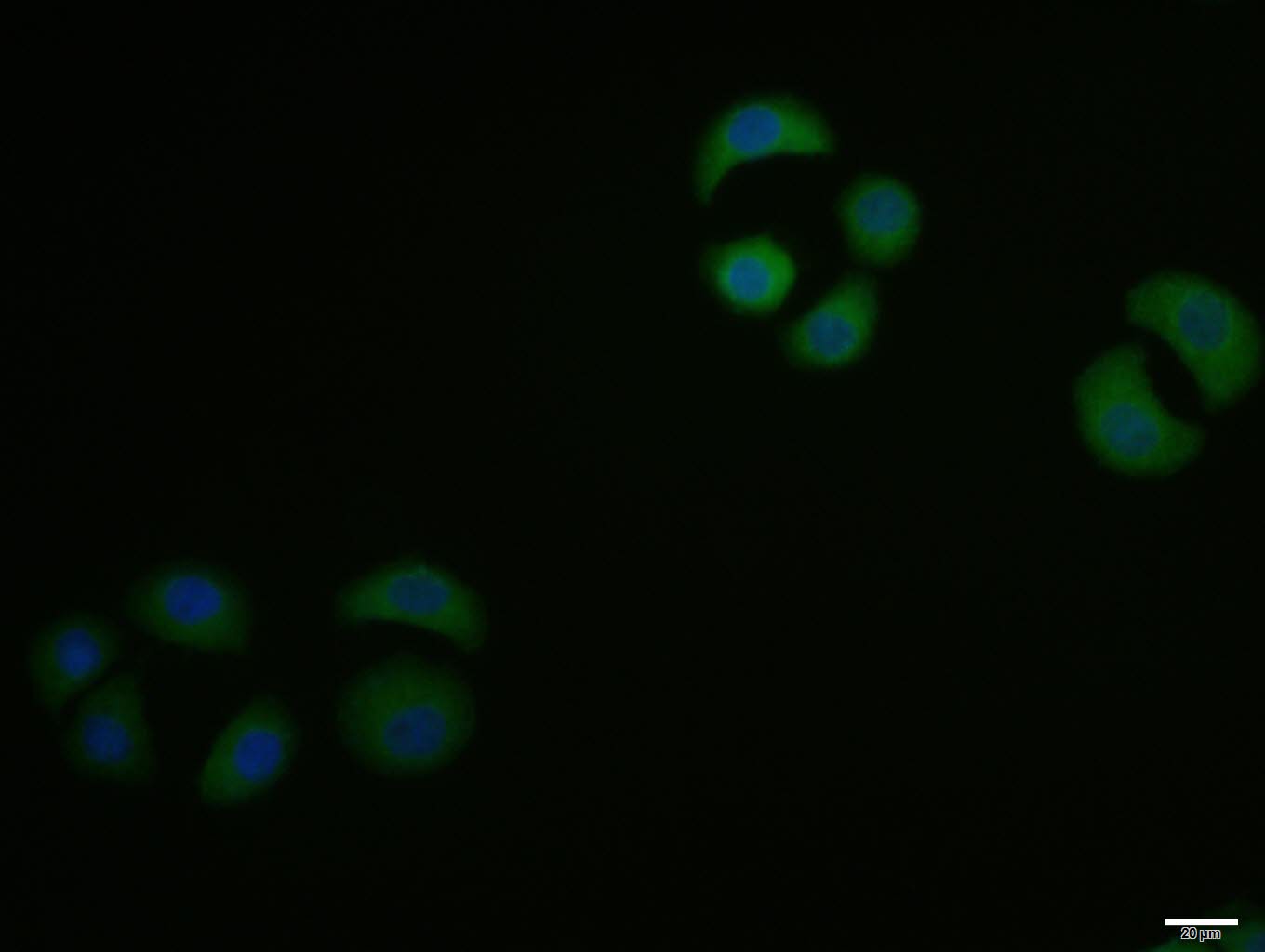 HepG2 cell; 4% Paraformaldehyde-fixed; Triton X-100 at room temperature for 20 min; Blocking buffer (normal goat serum, C-0005) at 37°C for 20 min; Antibody incubation with (Phospho-MYPT1 (Thr696)) polyclonal Antibody, Unconjugated (SL20264R) 1:100, 90 minutes at 37°C; followed by a conjugated Goat Anti-Rabbit IgG antibody at 37°C for 90 minutes, DAPI (blue, C02-04002) was used to stain the cell nuclei.
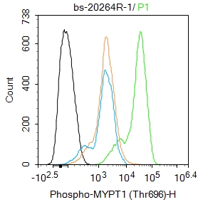 Blank control(black line):HepG2.
Primary Antibody (green line): Rabbit Anti-Phospho-MYPT1 (Thr696) antibody (SL20264R)
Dilution:1ug/Test;
Secondary Antibody : Goat anti-rabbit IgG-AF488
Dilution: 0.5ug/Test.
Negative control(white blue line): PBS
Isotype control(orange line): Normal Rabbit IgG
Protocol
The cells were fixed with 4% PFA (10min at room temperature)and then permeabilized with 90% ice-cold methanol for 20 min at -20℃.The cells were then incubated in 5%BSA to block non-specific protein-protein interactions for 30 min at room temperature .Cells stained with Primary Antibody for 30 min at room temperature. The secondary antibody used for 40 min at room temperature. Acquisition of 20,000 events was performed.
Cartpieces
Totalgoods,subtotals:¥Checkout
Bought notes(bought amounts latest0)
No one bought this product
User Comment(Total0User Comment Num)
- No comment


 +86 571 56623320
 +86 18668110335

