
Rabbit Anti-Phospho-FoxO1 (Ser319)antibody
FOXO1A (phospho S319); p-FOXO1A (phospho S319); FKHR(Phospho-Ser319); Forkhead box protein O1; Afxh; AI876417; FKHR; Fkhr1; Foxo1a; Forkhead; FKH 1; FKH1; FKH1; FKHR; FKHR; Forkhead (Drosophila) homolog 1 (rhabdomyosarcoma); Forkhead (Drosophila) homolog
View History [Clear]
Details
Product Name Phospho-FoxO1 (Ser319) Chinese Name 磷酸化叉头蛋白家族1(Ser319)抗体 Alias FOXO1A (phospho S319); p-FOXO1A (phospho S319); FKHR(Phospho-Ser319); Forkhead box protein O1; Afxh; AI876417; FKHR; Fkhr1; Foxo1a; Forkhead; FKH 1; FKH1; FKH1; FKHR; FKHR; Forkhead (Drosophila) homolog 1 (rhabdomyosarcoma); Forkhead (Drosophila) homolog 1 (rhabdomyosarcoma); Forkhead box O1; Forkhead box protein O1; Forkhead box protein O1A; Forkhead in rhabdomyosarcoma; Forkhead, Drosophila, homolog of, in rhabdomyosarcoma; FOXO1; FOXO1; FOXO1_HUMAN; FOXO1A. literatures Product Type Phosphorylated anti Research Area Tumour immunology Signal transduction transcriptional regulatory factor Kinases and Phosphatases Immunogen Species Rabbit Clonality Polyclonal React Species Human, Mouse, Rat, (predicted: Chicken, Dog, Pig, Cow, Horse, ) Applications WB=1:500-2000 ELISA=1:5000-10000 IHC-P=1:100-500 IHC-F=1:100-500 IF=1:100-500 (Paraffin sections need antigen repair)
not yet tested in other applications.
optimal dilutions/concentrations should be determined by the end user.Theoretical molecular weight 72kDa Cellular localization The nucleus cytoplasmic Form Liquid Concentration 1mg/ml immunogen KLH conjugated synthesised phosphopeptide derived from human FoxO1 around the phosphorylation site of Ser319: TS(p-S)NA Lsotype IgG Purification affinity purified by Protein A Buffer Solution Preservative: 15mM Sodium Azide, Constituents: 1% BSA, 0.01M PBS, pH 7.4 Storage Shipped at 4℃. Store at -20 °C for one year. Avoid repeated freeze/thaw cycles. Attention This product as supplied is intended for research use only, not for use in human, therapeutic or diagnostic applications. PubMed PubMed Product Detail This gene belongs to the forkhead family of transcription factors which are characterized by a distinct forkhead domain. The specific function of this gene has not yet been determined; however, it may play a role in myogenic growth and differentiation. Translocation of this gene with PAX3 has been associated with alveolar rhabdomyosarcoma. [provided by RefSeq].
Function:
Transcription factor which acts as a regulator of cell responses to oxidative stress. In the presence of KIRT1, mediates down-regulation of cyclin D1 and up-regulation of CDKN1B levels which are required for cell transition from proliferative growth to quiescence. Triggers death of postmitotic neurons when phosphorylated by CDK1. Activates transcription of PMAIP1.
Subunit:
Interacts with LRPPRC. Interacts with SIRT1 and this interaction requires the presence of KRIT1. Interacts with NLK. Binds to CDK1 and 14-3-3 proteins.
Subcellular Location:
Cytoplasm. Nucleus. Note=Shuttles between cytoplasm and nucleus. Translocates to the nucleus upon oxidative stress induced phosphorylation at Ser-212 by STK4/MST1. Translocates to the nucleus upon phosphorylation of Thr-24, Ser-256 and Ser-322 by SGK1.
Tissue Specificity:
Ubiquitous.
Post-translational modifications:
Phosphorylated by AKT1; insulin-induced. Phosphorylated by NLK, which inhibits transcriptional activity and promotes nuclear export (By similarity). IGF1 rapidly induces phosphorylation of Ser-256, Thr-24, and Ser-319. Phosphorylation of Ser-256 decreases DNA-binding activity and promotes the phosphorylation of Thr-24, and Ser-319, permitting phosphorylation of Ser-322 and Ser-325, probably by CK1, leading to nuclear exclusion and loss of function. Phosphorylation of Ser-329 is independent of IGF1 and leads to reduced function. Phosphorylated upon DNA damage, probably by ATM or ATR. Phosphorylation of Ser-249 by CDK1 disrupts 14-3-3 proteins binding and thereby promotes FOXO1 nuclear accumulation and subsequent transcription activation and cell death. Phosphorylated by STK4/MST1 on Ser-212 upon oxidative stress. Phosphorylated on Thr-24, Ser-256 and Ser-322 by SGK1 resulting in its translocation from the nucleus to the cytoplasm.
DISEASE:
Defects in FOXO1 are a cause of rhabdomyosarcoma type 2 (RMS2) [MIM:268220]. It is a form of rhabdomyosarcoma, a highly malignant tumor of striated muscle derived from primitive mesenchimal cells and exhibiting differentiation along rhabdomyoblastic lines. Rhabdomyosarcoma is one of the most frequently occurring soft tissue sarcomas and the most common in children. It occurs in four forms: alveolar, pleomorphic, embryonal and botryoidal rhabdomyosarcomas. Note=Chromosomal aberrations involving FOXO1 are found in rhabdomyosarcoma. Translocation (2;13)(q35;q14) with PAX3; translocation t(1;13)(p36;q14) with PAX7. The resulting protein is a transcriptional activator.
Similarity:
Contains 1 fork-head DNA-binding domain.
SWISS:
Q12778
Gene ID:
2308
Database links:Entrez Gene: 2308 Human
Entrez Gene: 56458 Mouse
Omim: 136533 Human
SwissProt: Q12778 Human
SwissProt: Q9R1E0 Mouse
Unigene: 370666 Human
Unigene: 29891 Mouse
Unigene: 116108 Rat
Product Picture 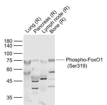 Sample:
Lane 1: Lung (Rat) Lysate at 40 ug
Lane 2: Pancreas (Rat) Lysate at 40 ug
Lane 3: Lymph node (Rat) Lysate at 40 ug
Lane 4: Bone (Rat) Lysate at 40 ug
Primary: Anti-Phospho-FoxO1 (Ser319) (SL20095R) at 1/1000 dilution
Secondary: IRDye800CW Goat Anti-Rabbit IgG at 1/20000 dilution
Predicted band size: 78 kD
Observed band size: 75 kD
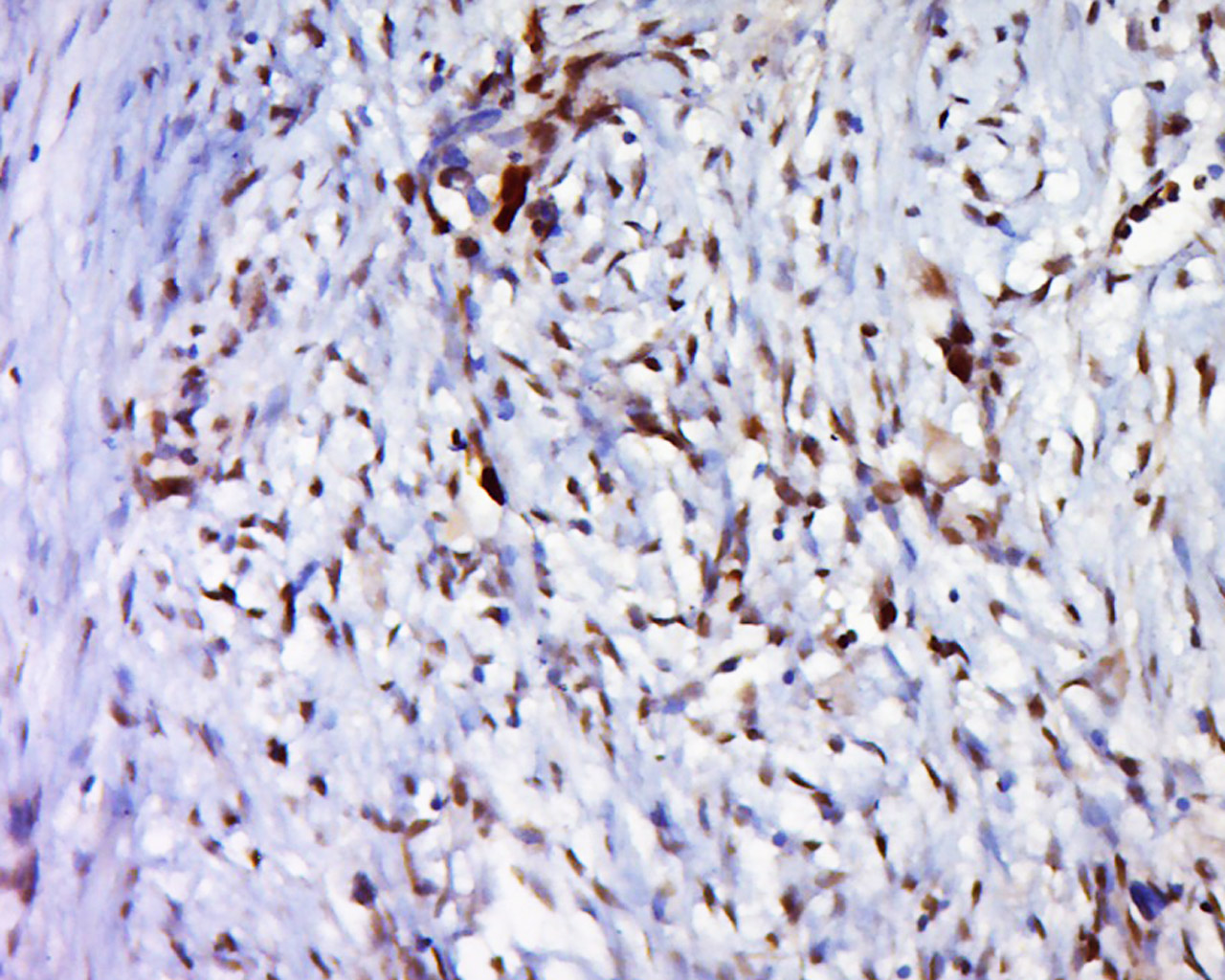 Tissue/cell: human cervical carcinoma; 4% Paraformaldehyde-fixed and paraffin-embedded;
Antigen retrieval: citrate buffer ( 0.01M, pH 6.0 ), Boiling bathing for 15min; Block endogenous peroxidase by 3% Hydrogen peroxide for 30min; Blocking buffer (normal goat serum,C-0005) at 37℃ for 20 min;
Incubation: Anti-Phospho-FoxO1(Ser319)Polyclonal Antibody, Unconjugated(SL20095R) 1:500, overnight at 4°C, followed by conjugation to the secondary antibody(SP-0023) and DAB(C-0010) staining
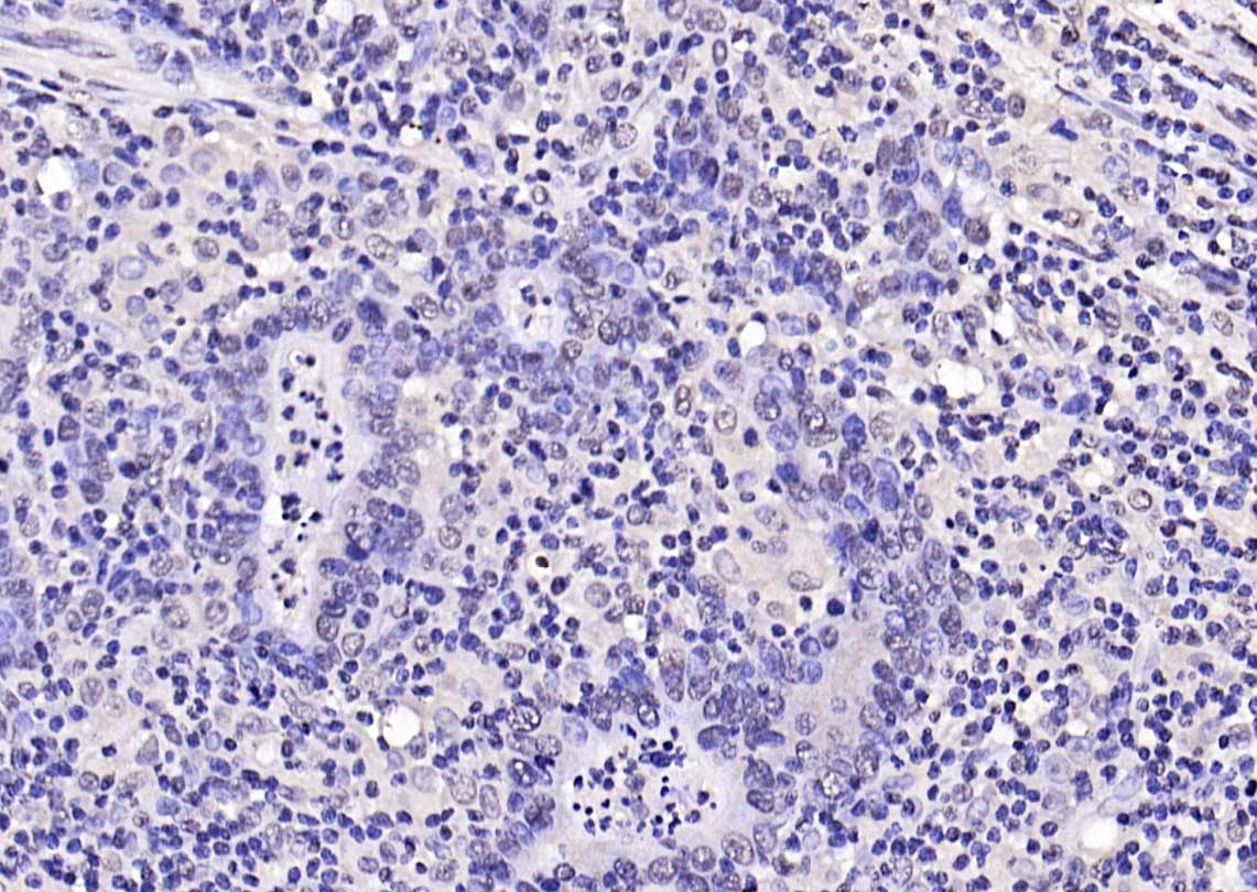 Paraformaldehyde-fixed, paraffin embedded (human gastric carcinoma); Antigen retrieval by boiling in sodium citrate buffer (pH6.0) for 15min; Block endogenous peroxidase by 3% hydrogen peroxide for 20 minutes; Blocking buffer (normal goat serum) at 37°C for 30min; Antibody incubation with (Phospho-FoxO1 (Ser319)) Polyclonal Antibody, Unconjugated (SL20095R) at 1:200 overnight at 4°C, followed by operating according to SP Kit(Rabbit) (sp-0023) instructionsand DAB staining.
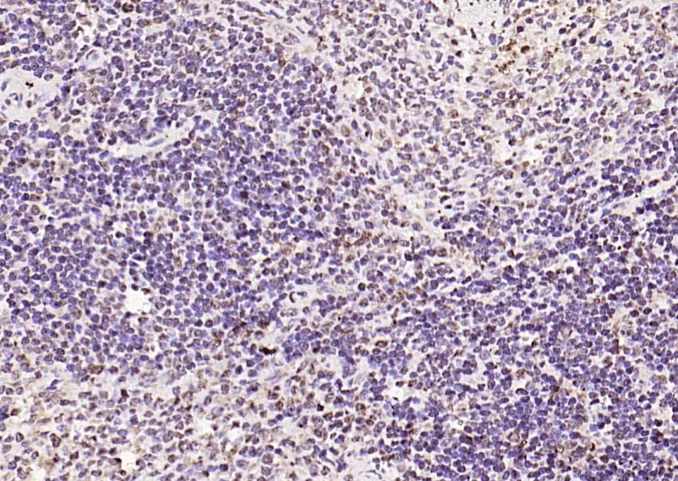 Paraformaldehyde-fixed, paraffin embedded (rat spleen); Antigen retrieval by boiling in sodium citrate buffer (pH6.0) for 15min; Block endogenous peroxidase by 3% hydrogen peroxide for 20 minutes; Blocking buffer (normal goat serum) at 37°C for 30min; Antibody incubation with (Phospho-FoxO1 (Ser319)) Polyclonal Antibody, Unconjugated (SL20095R) at 1:200 overnight at 4°C, followed by operating according to SP Kit(Rabbit) (sp-0023) instructionsand DAB staining.
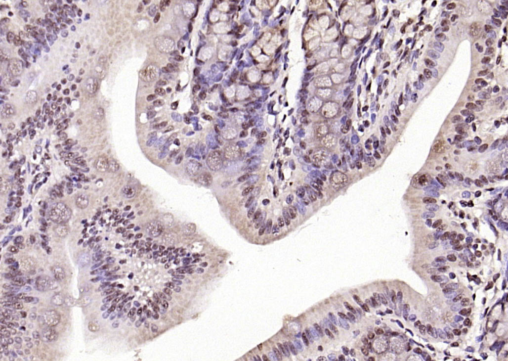 Paraformaldehyde-fixed, paraffin embedded (rat colon); Antigen retrieval by boiling in sodium citrate buffer (pH6.0) for 15min; Block endogenous peroxidase by 3% hydrogen peroxide for 20 minutes; Blocking buffer (normal goat serum) at 37°C for 30min; Antibody incubation with (Phospho-FoxO1 (Ser319)) Polyclonal Antibody, Unconjugated (SL20095R) at 1:200 overnight at 4°C, followed by operating according to SP Kit(Rabbit) (sp-0023) instructionsand DAB staining.
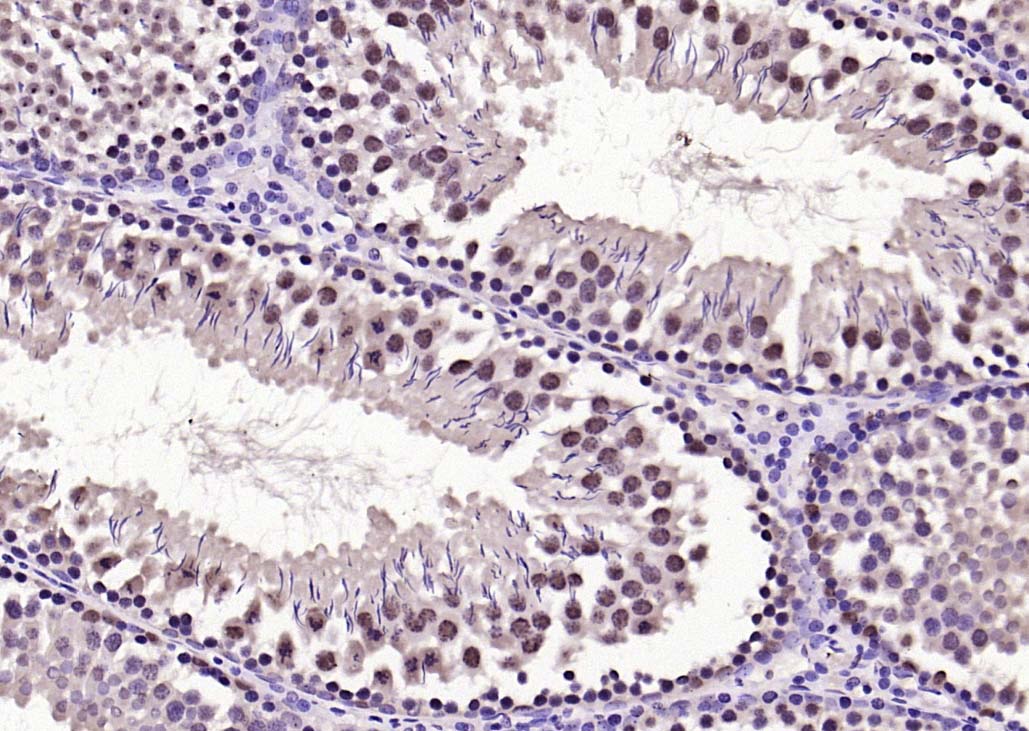 Paraformaldehyde-fixed, paraffin embedded (rat testis); Antigen retrieval by boiling in sodium citrate buffer (pH6.0) for 15min; Block endogenous peroxidase by 3% hydrogen peroxide for 20 minutes; Blocking buffer (normal goat serum) at 37°C for 30min; Antibody incubation with (Phospho-FoxO1 (Ser319)) Polyclonal Antibody, Unconjugated (SL20095R) at 1:200 overnight at 4°C, followed by operating according to SP Kit(Rabbit) (sp-0023) instructionsand DAB staining.
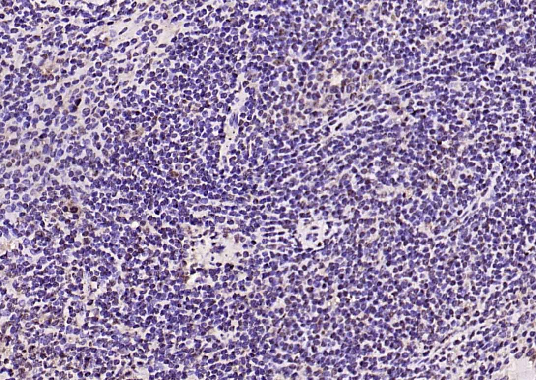 Paraformaldehyde-fixed, paraffin embedded (mouse spleen); Antigen retrieval by boiling in sodium citrate buffer (pH6.0) for 15min; Block endogenous peroxidase by 3% hydrogen peroxide for 20 minutes; Blocking buffer (normal goat serum) at 37°C for 30min; Antibody incubation with (Phospho-FoxO1 (Ser319)) Polyclonal Antibody, Unconjugated (SL20095R) at 1:200 overnight at 4°C, followed by operating according to SP Kit(Rabbit) (sp-0023) instructionsand DAB staining.
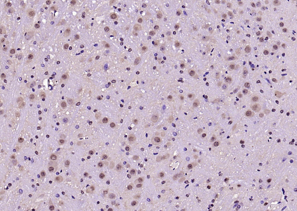 Paraformaldehyde-fixed, paraffin embedded (mouse brain); Antigen retrieval by boiling in sodium citrate buffer (pH6.0) for 15min; Block endogenous peroxidase by 3% hydrogen peroxide for 20 minutes; Blocking buffer (normal goat serum) at 37°C for 30min; Antibody incubation with (Phospho-FoxO1 (Ser319)) Polyclonal Antibody, Unconjugated (SL20095R) at 1:200 overnight at 4°C, followed by operating according to SP Kit(Rabbit) (sp-0023) instructionsand DAB staining.
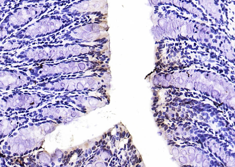 Paraformaldehyde-fixed, paraffin embedded (mouse colon); Antigen retrieval by boiling in sodium citrate buffer (pH6.0) for 15min; Block endogenous peroxidase by 3% hydrogen peroxide for 20 minutes; Blocking buffer (normal goat serum) at 37°C for 30min; Antibody incubation with (Phospho-FoxO1 (Ser319)) Polyclonal Antibody, Unconjugated (SL20095R) at 1:200 overnight at 4°C, followed by operating according to SP Kit(Rabbit) (sp-0023) instructionsand DAB staining.
 Paraformaldehyde-fixed, paraffin embedded (mouse liver); Antigen retrieval by boiling in sodium citrate buffer (pH6.0) for 15min; Block endogenous peroxidase by 3% hydrogen peroxide for 20 minutes; Blocking buffer (normal goat serum) at 37°C for 30min; Antibody incubation with (Phospho-FoxO1 (Ser319)) Polyclonal Antibody, Unconjugated (SL20095R) at 1:200 overnight at 4°C, followed by operating according to SP Kit(Rabbit) (sp-0023) instructionsand DAB staining.
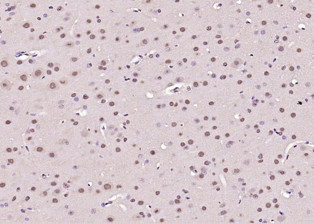 Paraformaldehyde-fixed, paraffin embedded (rat brain); Antigen retrieval by boiling in sodium citrate buffer (pH6.0) for 15min; Block endogenous peroxidase by 3% hydrogen peroxide for 20 minutes; Blocking buffer (normal goat serum) at 37°C for 30min; Antibody incubation with (Phospho-FoxO1 (Ser319)) Polyclonal Antibody, Unconjugated (SL20095R) at 1:200 overnight at 4°C, followed by operating according to SP Kit(Rabbit) (sp-0023) instructionsand DAB staining.
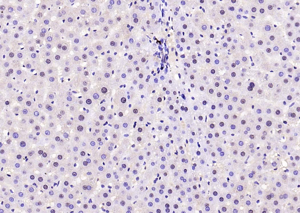 Paraformaldehyde-fixed, paraffin embedded (rat liver); Antigen retrieval by boiling in sodium citrate buffer (pH6.0) for 15min; Block endogenous peroxidase by 3% hydrogen peroxide for 20 minutes; Blocking buffer (normal goat serum) at 37°C for 30min; Antibody incubation with (Phospho-FoxO1 (Ser319)) Polyclonal Antibody, Unconjugated (SL20095R) at 1:200 overnight at 4°C, followed by operating according to SP Kit(Rabbit) (sp-0023) instructionsand DAB staining.
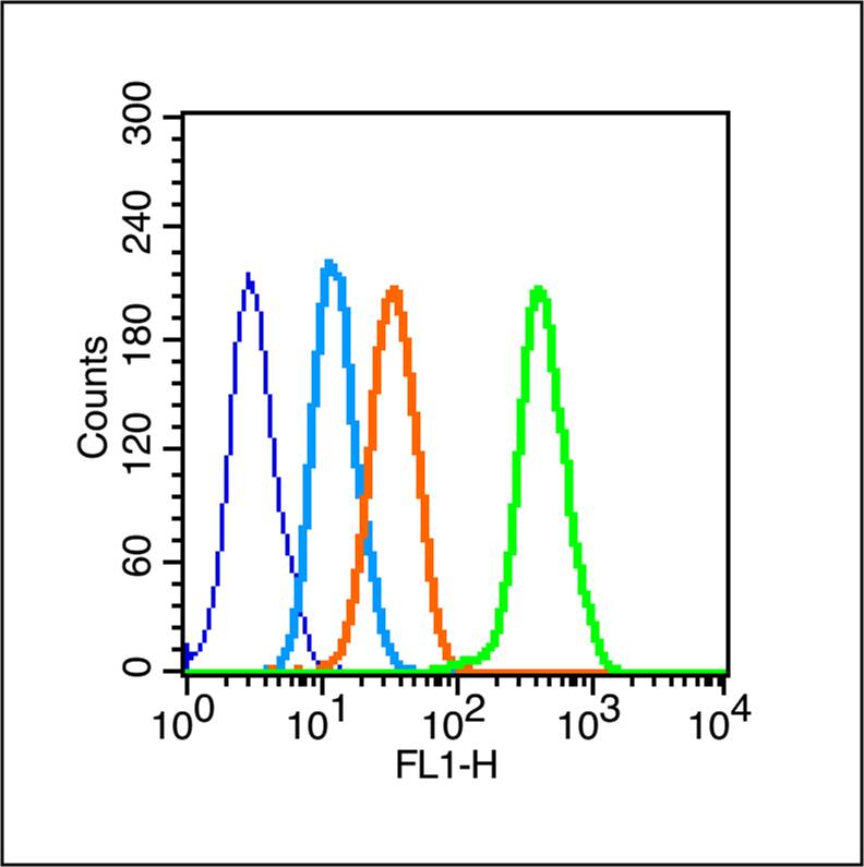 Blank control (Black line): Raji (Black).
Primary Antibody (green line): Rabbit Anti-Phospho-FoxO1 (Ser319) antibody (SL20095R)
Dilution: 1μg /10^6 cells;
Isotype Control Antibody (orange line): Rabbit IgG .
Secondary Antibody (white blue line): Goat anti-rabbit IgG-PE
Dilution: 1μg /test.
Protocol
The cells were fixed with 4% PFA (10 min) and then permeabilized with 90% ice-cold methanol for 20 min on ice. Cells stained with Primary Antibody for 30 min at room temperature. The cells were then incubated in 1 X PBS/2%BSA/10% goat serum to block non-specific protein-protein interactions followed by the antibody for 15 min at room temperature. The secondary antibody used for 40 min at room temperature. Acquisition of 20,000 events was performed.
Cartpieces
Totalgoods,subtotals:¥Checkout
Partial purchase records(bought amounts latest0)
No one bought this product
User Comment(Total0User Comment Num)
- No comment


 +86 571 56623320


