
Rabbit Anti-VAMP1 antibody
Vesicle Associated Membrane Protein 1; DKFZp686H12131; SYB 1; SYB1; Synaptobrevin 1; Synaptobrevin1; Synaptobrevin-1; VAMP 1; VAMP-1; Vesicle associated membrane protein 1; Vesicle associated membrane protein 1 synaptobrevin 1; VAMP1_HUMAN; Vesicle-associ
View History [Clear]
Details
Product Name VAMP1 Chinese Name VAMP1囊泡相关膜蛋白1抗体 Alias Vesicle Associated Membrane Protein 1; DKFZp686H12131; SYB 1; SYB1; Synaptobrevin 1; Synaptobrevin1; Synaptobrevin-1; VAMP 1; VAMP-1; Vesicle associated membrane protein 1; Vesicle associated membrane protein 1 synaptobrevin 1; VAMP1_HUMAN; Vesicle-associated membrane protein 1. Research Area Tumour immunology Neurobiology Signal transduction Mitochondrion Immunogen Species Rabbit Clonality Polyclonal React Species Human, Mouse, Rat, (predicted: Chicken, Dog, Pig, Cow, Horse, Rabbit, Guinea Pig, ) Applications ELISA=1:5000-10000 IHC-P=1:100-500 IHC-F=1:100-500 IF=1:100-500 (Paraffin sections need antigen repair)
not yet tested in other applications.
optimal dilutions/concentrations should be determined by the end user.Theoretical molecular weight 13kDa Cellular localization cytoplasmic The cell membrane Mitochondrion Form Liquid Concentration 1mg/ml immunogen KLH conjugated synthetic peptide derived from human VAMP-1: 51-118/118 <Cytoplasmic> Lsotype IgG Purification affinity purified by Protein A Buffer Solution 0.01M TBS(pH7.4) with 1% BSA, 0.03% Proclin300 and 50% Glycerol. Storage Shipped at 4℃. Store at -20 °C for one year. Avoid repeated freeze/thaw cycles. Attention This product as supplied is intended for research use only, not for use in human, therapeutic or diagnostic applications. PubMed PubMed Product Detail Synaptobrevins/VAMPs, syntaxins, and the 25-kD synaptosomal-associated protein SNAP25 are the main components of a protein complex involved in the docking and/or fusion of synaptic vesicles with the presynaptic membrane. VAMP1 is a member of the vesicle-associated membrane protein (VAMP)/synaptobrevin family. Multiple alternative splice variants that encode proteins with alternative carboxy ends have been described, but the full-length nature of some variants has not been defined. [provided by RefSeq, Jul 2008].
Function:
Involved in the targeting and/or fusion of transport vesicles to their target membrane.
Subunit:
Interacts with VAPA and VAPB.
Subcellular Location:
Isoform 1: Cytoplasmic vesicle, secretory vesicle, synaptic vesicle membrane; Single-pass type IV membrane protein. Cell junction, synapse, synaptosome.
Isoform 2: Cytoplasmic vesicle membrane; Single-pass type IV membrane protein. Cell junction, synapse, synaptosome.
Isoform 3: Mitochondrion outer membrane; Single-pass type IV membrane protein.
Tissue Specificity:
Nervous system, skeletal muscle and adipose tissue.
Similarity:
Belongs to the synaptobrevin family.
Contains 1 v-SNARE coiled-coil homology domain.
SWISS:
P23763
Gene ID:
6843
Database links:Entrez Gene: 6843 Human
Entrez Gene: 22317 Mouse
Omim: 185880 Human
SwissProt: P23763 Human
SwissProt: Q62442 Mouse
Unigene: 20021 Human
Unigene: 32321 Mouse
Unigene: 31977 Rat
Product Picture 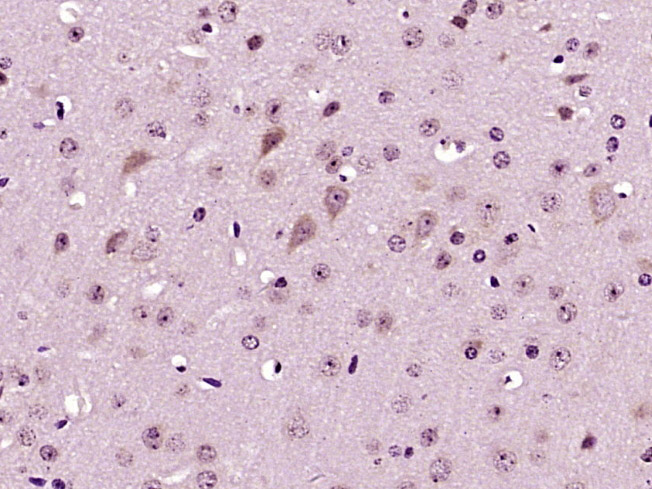 Paraformaldehyde-fixed, paraffin embedded (mouse brain tissue); Antigen retrieval by boiling in sodium citrate buffer (pH6.0) for 15min; Block endogenous peroxidase by 3% hydrogen peroxide for 20 minutes; Blocking buffer (normal goat serum) at 37°C for 30min; Antibody incubation with (VAMP1) Polyclonal Antibody, Unconjugated (SL1950R) at 1:400 overnight at 4°C, followed by operating according to SP Kit(Rabbit) (sp-0023) instructionsand DAB staining.
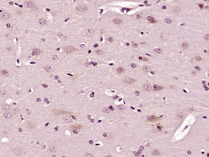 Paraformaldehyde-fixed, paraffin embedded (rat brain tissue); Antigen retrieval by boiling in sodium citrate buffer (pH6.0) for 15min; Block endogenous peroxidase by 3% hydrogen peroxide for 20 minutes; Blocking buffer (normal goat serum) at 37°C for 30min; Antibody incubation with (VAMP1) Polyclonal Antibody, Unconjugated (SL1950R) at 1:400 overnight at 4°C, followed by operating according to SP Kit(Rabbit) (sp-0023) instructionsand DAB staining.
 Blank control:Molt-4.
Primary Antibody (green line): Rabbit Anti-VAMP1 antibody (SL1950)
Dilution: 0.2μg /10^6 cells;
Isotype Control Antibody (orange line): Rabbit IgG .
Secondary Antibody : Goat anti-rabbit IgG-PE
Dilution: 0.2μg /test.
Protocol
The cells were fixed with 4% PFA (10min at room temperature)and then permeabilized with 20% PBST for 20 min at-20℃. The cells were then incubated in 5%BSA to block non-specific protein-protein interactions for 30 min at at room temperature .Cells stained with Primary Antibody for 30 min at room temperature. The secondary antibody used for 40 min at room temperature. Acquisition of 20,000 events was performed.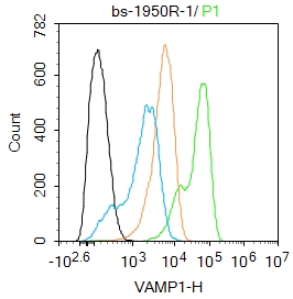 Blank control(black line):U87MG.
Primary Antibody (green line): Rabbit Anti-VAMP1 antibody (SL1950R)
Dilution:1ug/Test;
Secondary Antibody : Goat anti-rabbit IgG-AF488
Dilution: 0.5ug/Test.
Negative control(white blue line): PBS
Isotype control(orange line): Normal Rabbit IgG
Protocol
The cells were fixed with 4% PFA (10min at room temperature)and then permeabilized with 90% ice-cold methanol for 20 min at -20℃.The cells were then incubated in 5%BSA to block non-specific protein-protein interactions for 30 min at room temperature .Cells stained with Primary Antibody for 30 min at room temperature. The secondary antibody used for 40 min at room temperature. Acquisition of 20,000 events was performed.
Cartpieces
Totalgoods,subtotals:¥Checkout
References (0)
No References
Bought notes(bought amounts latest0)
No one bought this product
User Comment(Total0User Comment Num)
- No comment


 +86 571 56623320
 +86 18668110335

