
Rabbit Anti-PDCD1LG2 antibody
PD L2; PD-L2;PD1L2_HUMAN; B7 dendritic cell molecule; B7-DC; B7DC; bA574F11.2; Btdc; Butyrophilin B7 DC; Butyrophilin B7-DC; Butyrophilin B7DC; CD 273; CD273; CD273 antigen; MGC142238; MGC142240; PD 1 ligand 2; PD-1 ligand 2; PD1 ligand 2; PDCD 1 ligand
View History [Clear]
Details
Product Name PDCD1LG2 Chinese Name 程序性死亡配体2(CD273)抗体 Alias PD L2; PD-L2; PD1L2_HUMAN; B7 dendritic cell molecule; B7-DC; B7DC; bA574F11.2; Btdc; Butyrophilin B7 DC; Butyrophilin B7-DC; Butyrophilin B7DC; CD 273; CD273; CD273 antigen; MGC142238; MGC142240; PD 1 ligand 2; PD-1 ligand 2; PD1 ligand 2; PDCD 1 ligand 2; PDCD1 ligand 2; PDCD1L2; PDL 2; PDL2; Programmed cell death 1 ligand 2; Programmed death ligand 2. literatures Research Area immunology Immunogen Species Rabbit Clonality Polyclonal React Species Human, (predicted: Mouse, Rat, Cow, ) Applications ELISA=1:5000-10000 Flow-Cyt=1ug/Test ICC=1:100
not yet tested in other applications.
optimal dilutions/concentrations should be determined by the end user.Theoretical molecular weight 29kDa Detection molecular weight 42kDa Cellular localization The cell membrane Secretory protein Form Liquid Concentration 1mg/ml immunogen KLH conjugated synthetic peptide derived from human PDCD1LG2: 41-150/273 Lsotype IgG Purification affinity purified by Protein A Buffer Solution 0.01M TBS(pH7.4) with 1% BSA, 0.03% Proclin300 and 50% Glycerol. Storage Shipped at 4℃. Store at -20 °C for one year. Avoid repeated freeze/thaw cycles. Attention This product as supplied is intended for research use only, not for use in human, therapeutic or diagnostic applications. PubMed PubMed Product Detail Involved in negative regulation of activated T cell proliferation; negative regulation of interferon-gamma production; and negative regulation of interleukin-10 production. Predicted to be located in plasma membrane. Predicted to be active in external side of plasma membrane. Biomarker of pulmonary tuberculosis. [provided by Alliance of Genome Resources, Nov 2021]
Function:
Involved in the costimulatory signal, essential for T-cell proliferation and IFNG production in a PDCD1-independent manner. Interaction with PDCD1 inhibits T-cell proliferation by blocking cell cycle progression and cytokine production.
Subunit:
Interacts with PDCD1.
Subcellular Location:
Isoform 3: Secreted (Probable).
Isoform 2: Endomembrane system; Single-pass type I membrane protein.
Isoform 1: Cell membrane; Single-pass type I membrane protein.
Tissue Specificity:
Highly expressed in heart, placenta, pancreas, lung and liver and weakly expressed in spleen, lymph nodes and thymus.
Similarity:
Belongs to the immunoglobulin superfamily. BTN/MOG family.
Contains 1 Ig-like C2-type (immunoglobulin-like) domain.
Contains 1 Ig-like V-type (immunoglobulin-like) domain.
SWISS:
Q9BQ51
Gene ID:
80380
Database links:Entrez Gene: 80380 Human
Omim: 605723 Human
SwissProt: Q9BQ51 Human
Product Picture  HepG2 cell; 4% Paraformaldehyde-fixed; Triton X-100 at room temperature for 20 min; Blocking buffer (normal goat serum, C-0005) at 37°C for 20 min; Antibody incubation with (PDCD1LG2) polyclonal Antibody, Unconjugated (SL1868R) 1:100, 90 minutes at 37°C; followed by a conjugated Goat Anti-Rabbit IgG antibody at 37°C for 90 minutes, DAPI (blue, C02-04002) was used to stain the cell nuclei.
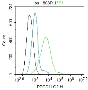 Blank control:HepG2.
Primary Antibody (green line): Rabbit Anti-PDCD1LG2 antibody (SL1868R)
Dilution: 1ug/Test;
Secondary Antibody (white blue line) : Goat anti-rabbit IgG-AF488
Dilution: 0.5ug/Test.
Isotype control(orange line):Normal Rabbit IgG
Protocol
The cells were incubated in 5%BSA to block non-specific protein-protein interactions for 30 min at room temperature .Cells stained with Primary Antibody for 30 min at room temperature. The secondary antibody used for 40 min at room temperature. Acquisition of 20,000 events was performed.
Cartpieces
Totalgoods,subtotals:¥Checkout
Bought notes(bought amounts latest0)
No one bought this product
User Comment(Total0User Comment Num)
- No comment


 +86 571 56623320
 +86 18668110335

