
Rabbit Anti-PD-1 antibody
Programmed cell death protein 1; CD279; CD279 antigen; hPD 1; hPD-1; hSLE1; PD 1; PD1; PDCD 1; PDCD1; PDCD1_HUMAN; Programmed cell death 1; Protein PD 1; Protein PD-1; SLEB2; Systemic lupus erythematosus susceptibility 2.
View History [Clear]
Details
Product Name PD-1 Chinese Name 程序性死亡1(CD279)抗体 Alias Programmed cell death protein 1; CD279; CD279 antigen; hPD 1; hPD-1; hSLE1; PD 1; PD1; PDCD 1; PDCD1; PDCD1_HUMAN; Programmed cell death 1; Protein PD 1; Protein PD-1; SLEB2; Systemic lupus erythematosus susceptibility 2. literatures Research Area Tumour Cell biology immunology Apoptosis Immunogen Species Rabbit Clonality Polyclonal React Species Human, Mouse, Rat, (predicted: Cow, Horse, Rabbit, ) Applications WB=1:500-2000 ELISA=1:5000-10000 IHC-P=1:100-500 IHC-F=1:100-500 Flow-Cyt=1μg /test IF=1:100-500 (Paraffin sections need antigen repair)
not yet tested in other applications.
optimal dilutions/concentrations should be determined by the end user.Theoretical molecular weight 32kDa Detection molecular weight 55-60 kDa Cellular localization The cell membrane Form Liquid Concentration 1mg/ml immunogen KLH conjugated synthetic peptide derived from human PD-1: 201-288/288 Lsotype IgG Purification affinity purified by Protein A Buffer Solution 0.01M TBS(pH7.4) with 1% BSA, 0.03% Proclin300 and 50% Glycerol. Storage Shipped at 4℃. Store at -20 °C for one year. Avoid repeated freeze/thaw cycles. Attention This product as supplied is intended for research use only, not for use in human, therapeutic or diagnostic applications. PubMed PubMed Product Detail Programmed cell death protein 1 (PDCD1) is an immune-inhibitory receptor expressed in activated T cells; it is involved in the regulation of T-cell functions, including those of effector CD8+ T cells. In addition, this protein can also promote the differentiation of CD4+ T cells into T regulatory cells. PDCD1 is expressed in many types of tumors including melanomas, and has demonstrated to play a role in anti-tumor immunity. Moreover, this protein has been shown to be involved in safeguarding against autoimmunity, however, it can also contribute to the inhibition of effective anti-tumor and anti-microbial immunity. [provided by RefSeq, Aug 2020]
Function:
Inhibitory cell surface receptor involved in the regulation of T-cell function during immunity and tolerance. Upon ligand binding, inhibits T-cell effector functions in an antigen-specific manner. Possible cell death inducer, in association with other factors.
Subunit:
Monomer.
Subcellular Location:
Membrane; Single-pass type I membrane protein.
Tissue Specificity:
Ta,Ba,Ma,Thy
DISEASE:
Systemic lupus erythematosus 2 (SLEB2) [MIM:605218]: A chronic, relapsing, inflammatory, and often febrile multisystemic disorder of connective tissue, characterized principally by involvement of the skin, joints, kidneys and serosal membranes. It is of unknown etiology, but is thought to represent a failure of the regulatory mechanisms of the autoimmune system. The disease is marked by a wide range of system dysfunctions, an elevated erythrocyte sedimentation rate, and the formation of LE cells in the blood or bone marrow. {ECO:0000269|PubMed:12402038}. Note=Disease susceptibility is associated with variations affecting the gene represented in this entry.
Similarity:
Contains 1 Ig-like V-type (immunoglobulin-like) domain.
SWISS:
Q15116
Gene ID:
5133
Database links:Entrez Gene: 5133 Human
Entrez Gene: 18566 Mouse
Omim: 600244 Human
SwissProt: Q15116 Human
SwissProt: Q02242 Mouse
Unigene: 158297 Human
Unigene: 5024 Mouse
Unigene: 105023 Rat
Product Picture 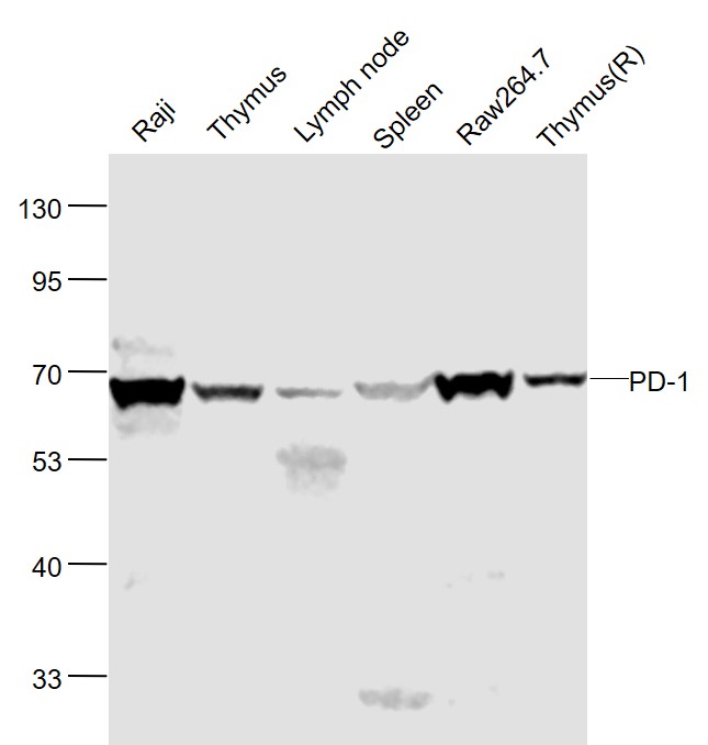 Sample:
Raji(Human) Cell Lysate at 30 ug
Thymus(Mouse) Lysate at 40 ug
Lymph node(Mouse) Lysate at 40 ug
Spleen(Mouse) Lysate at 40 ug
Raw264.7(Mouse) Cell Lysate at 40 ug
Thymus (Rat) Lysate at 40 ug
Primary: Anti-PD-1 (SL1867R) at 1/1000 dilution
Secondary: IRDye800CW Goat Anti-Rabbit IgG at 1/20000 dilution
Predicted band size: 55 kD
Observed band size: 58 kD
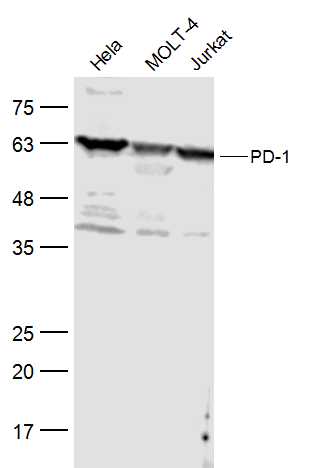 Sample:
Hela(Human) Cell Lysate at 30 ug
MOLT-4(Human) Cell Lysate at 30 ug
Jurkat(Human) Cell Lysate at 30 ug
Primary: Anti-PD-1 (SL1867R) at 1/500 dilution
Secondary: IRDye800CW Goat Anti-Rabbit IgG at 1/20000 dilution
Predicted band size: 32 kD
Observed band size: 55 kD
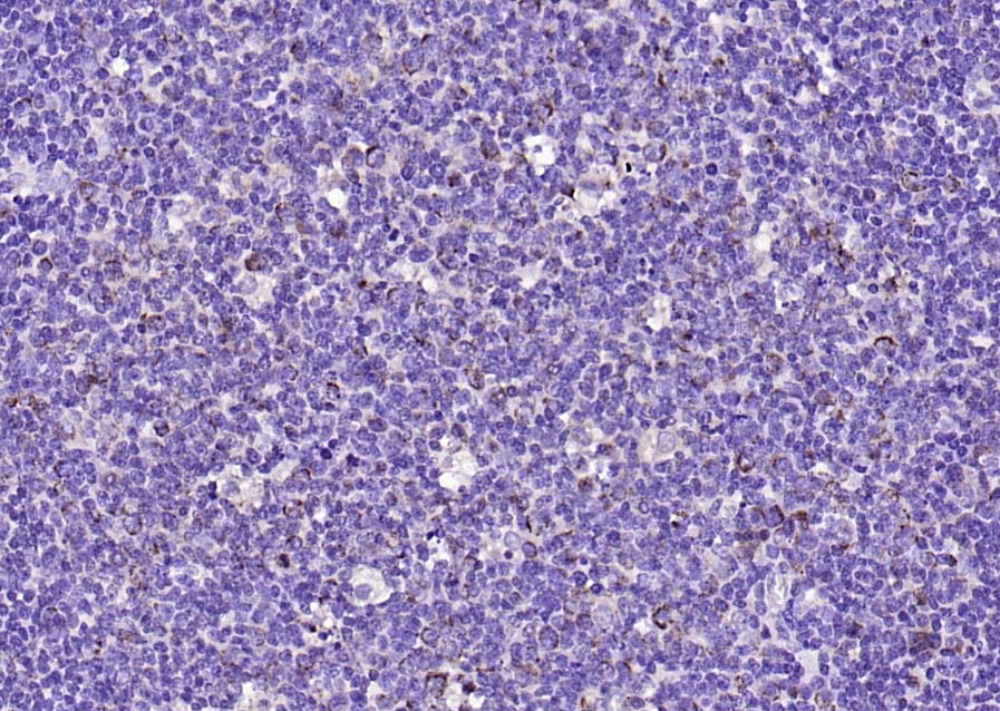 Paraformaldehyde-fixed, paraffin embedded (human tonsil); Antigen retrieval by boiling in sodium citrate buffer (pH6.0) for 15min; Block endogenous peroxidase by 3% hydrogen peroxide for 20 minutes; Blocking buffer (normal goat serum) at 37°C for 30min; Antibody incubation with (PD-1 ) Polyclonal Antibody, Unconjugated (SL1867R) at 1:200 overnight at 4°C, followed by operating according to SP Kit(Rabbit) (sp-0023) instructionsand DAB staining.
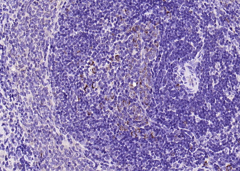 Paraformaldehyde-fixed, paraffin embedded (rat spleen); Antigen retrieval by boiling in sodium citrate buffer (pH6.0) for 15min; Block endogenous peroxidase by 3% hydrogen peroxide for 20 minutes; Blocking buffer (normal goat serum) at 37°C for 30min; Antibody incubation with (PD-1 ) Polyclonal Antibody, Unconjugated (SL1867R) at 1:200 overnight at 4°C, followed by operating according to SP Kit(Rabbit) (sp-0023) instructionsand DAB staining.
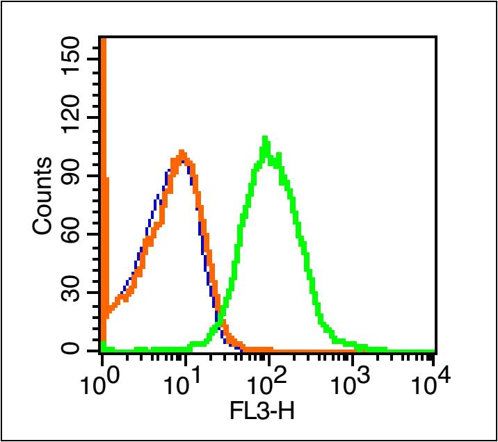 Blank control (blue line): Mouse spleen cells(blue).
Primary Antibody (green line): Rabbit Anti-PD-1/PE-CY7 Conjugated antibody (SL1867R-PE-CY7)
Dilution: 1μg /10^6 cells;
Isotype Control Antibody (orange line): Rabbit IgG-PE-CY7 .
Protocol
The cells were fixed with 70% ice-cold methanol overnight at 4℃ . The cells were then incubated in 1 X PBS/2%BSA/10% goat serum to block non-specific protein-protein interactions followed by the antibody for 15 min at room temperature. Cells stained with Primary Antibody for 30 min at room temperature.Acquisition of 20,000 events was performed.
Cartpieces
Totalgoods,subtotals:¥Checkout
References (0)
No References
Bought notes(bought amounts latest0)
No one bought this product
User Comment(Total0User Comment Num)
- No comment


 +86 571 56623320
 +86 18668110335

