
Rabbit Anti-DC-SIGNR1/CD209b antibody
SIGN Related 1;CD209 antigen like protein B; CD209b antigen; DC SIGN related protein 1; DC SIGNR1; OtB7.
View History [Clear]
Details
Product Name DC-SIGNR1/CD209b Chinese Name CD209b抗体 Alias SIGN Related 1; CD209 antigen like protein B; CD209b antigen; DC SIGN related protein 1; DC SIGNR1; OtB7. Research Area Cell biology Cell Surface Molecule lymphocyte t-lymphocyte Immunogen Species Rabbit Clonality Polyclonal React Species Mouse, Applications WB=1:500-2000 ELISA=1:5000-10000 IHC-P=1:100-500 IHC-F=1:100-500 ICC=1:100-500 IF=1:100-500 (Paraffin sections need antigen repair)
not yet tested in other applications.
optimal dilutions/concentrations should be determined by the end user.Theoretical molecular weight 37kDa Cellular localization The cell membrane Form Liquid Concentration 1mg/ml immunogen KLH conjugated synthetic peptide derived from mouse CD209b: 51-150/325 <Extracellular> Lsotype IgG Purification affinity purified by Protein A Buffer Solution 0.01M TBS(pH7.4) with 1% BSA, 0.03% Proclin300 and 50% Glycerol. Storage Shipped at 4℃. Store at -20 °C for one year. Avoid repeated freeze/thaw cycles. Attention This product as supplied is intended for research use only, not for use in human, therapeutic or diagnostic applications. PubMed PubMed Product Detail Antigen-presenting cells are localized in essentially every tissue, where they operate at the interface of innate and acquired immunity by capturing pathogens and presenting pathogen-derived peptides to T cells. Dendritic cells capture antigens or viruses in peripheral tissue and transport them to lymphoid organs, an event that induces cellular T cell responses. The mouse CD209 family of cell adhesion receptors consists of CD209a (also known as DC-SIGN), CD209b, CD209c, CD209d, CD209e, CD209f and CD209g, all of which function to mediate the endocytosis and subsequent degradation of pathogens within lysosomal compartments. There are two human CD209 proteins, designated DC-SIGN and DC-SIGNR, which function in a similar manner to the mouse proteins.
Function:
SIGN R1 is a specific marker for the identification of macrophage subpopulations present in the marginal zone of spleen (the so-called marginal zone macrophages (MZM)), in the lymph node medulla, and in some strains, in the peritoneal cavity. MZM of the spleen are involved in the clearance of polysaccharides. Mouse SIGN R1 is a C type lectin, like DC SIGN which is expressed on Human dendritic cells (DCs). However, Mmouse SIGN R1 itself is not expressed on DCs. SIGN R1 exists in an aggregated form, resistant to dissociation into monomers upon boiling in SDS under reducing conditions. SIGN R1 mediates the uptake of encapsulated organisms and may be an important mediator for the uptake of microbes in both spleen and lymph node, particularly through the recognition of microbial polysaccharides.
Subcellular Location:
Membrane Single-pass type II membrane protein
Tissue Specificity:
Expressed in skin, spleen and lung, probably in a subset of dendritic cells. Detected in spleen extrafollicular paracortical areas including the red pulp and marginal zones, and at lower levels, in the follicular area. Detected in skin suprabasal areas adjacent to the epidermis and in epidermal cell layer.
Similarity:
Contains 1 C-type lectin domain.
SWISS:
Q8CJ91
Gene ID:
69165
Database links:Entrez Gene: 69165 Mouse
SwissProt: Q8CJ91 Mouse
Product Picture 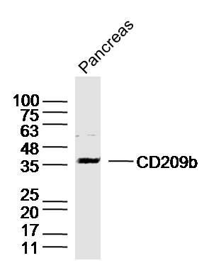 Sample:Pancreas (Mouse) Lysate at 40 ug
Primary: Anti-DC-SIGNR1/CD209b(SL17488R)at 1/300 dilution
Secondary: IRDye800CW Goat Anti-Rabbit IgG at 1/20000 dilution
Predicted band size: 37kD
Observed band size: 37kD
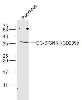 Sample:
Pancreas (Mouse) Lysate at 40 ug
Primary: Anti-DC-SIGNR1/CD209b(SL17488R) at 1/300 dilution
Secondary: IRDye800CW Goat Anti-Rabbit IgG at 1/20000 dilution
Predicted band size: 37 kD
Observed band size: 37 kD
Cartpieces
Totalgoods,subtotals:¥Checkout
Bought notes(bought amounts latest0)
No one bought this product
User Comment(Total0User Comment Num)
- No comment


 +86 571 56623320
 +86 18668110335

